166
KISEP Case Report J Kor Soc Echo 2005;13:166-168
간폐증후군
연세대학교 의과대학 내과학교실 이 지 혁・하 종 원
The Hepatopulmonary Syndrome
Jihyuk Rhee, MD and Jong-Won Ha, MD, PhD
Department of Internal Medicine, Yonsei University College of Medicine, Seoul, Korea
간폐증후군은 간질환, 폐환기의 이상으로 야기되는 동맥 저산소혈증, 그리고 광범위한 폐혈관 확장의 3징으로 정의된다. 특히 폐 모세혈관의 비정상적 확장에 의한 폐동맥과 폐정맥의 직접적인 교통은 우-좌 단락을 야기하며 이로 인한 동맥 저산소증은 호흡 곤란의 원인이 된다. 조영 심초음파는 비침습적으로 이러한 우-좌 단락을 확인 및 진단할 수 있는 유일한 방법으로 사료된다. 본 증례는 호흡 곤란을 호소하는 63세의 간경변 환자에서 조영 심초음파를 이용하여 간폐증후군을 진단한 증례이다. 중심 단어:간폐증후군・조영 심초음파・우-좌 단락.
Introduction
The hepatopulmonary syndrome (HPS) is defined as the triad of liver disease, pulmonary gas exchange abnormalities leading to arterial deoxygenation, and widespread pulmon-ary vascular dilatation.1) Although both acute and chronic
liver diseases have been associated with HPS, most comm-only it is associated with cirrhosis. Portal hypertension seems to be the predominant factor related to this syndrome.2) The
hallmark of pulmonary vascular changes in HPS is dilated vessels at the precapillary and capillary level and direct arteriovenous communications. This causes right-to-left shun-ting of blood flow, mismatch between ventilation and per-fusion, and diffusion limitation. Contrast transthoracic echo-cardiography may be the only diagnostic test to demonstrate right-to-left shunt in these patients.3) It is performed by
intravenous injection of agitated saline as an echocardiogra-phic contrast, while visualizing the atria. In the case of HPS, contrast appears in the left atrium 2 to 5 s after it is seen in the right atrium.
Case Report
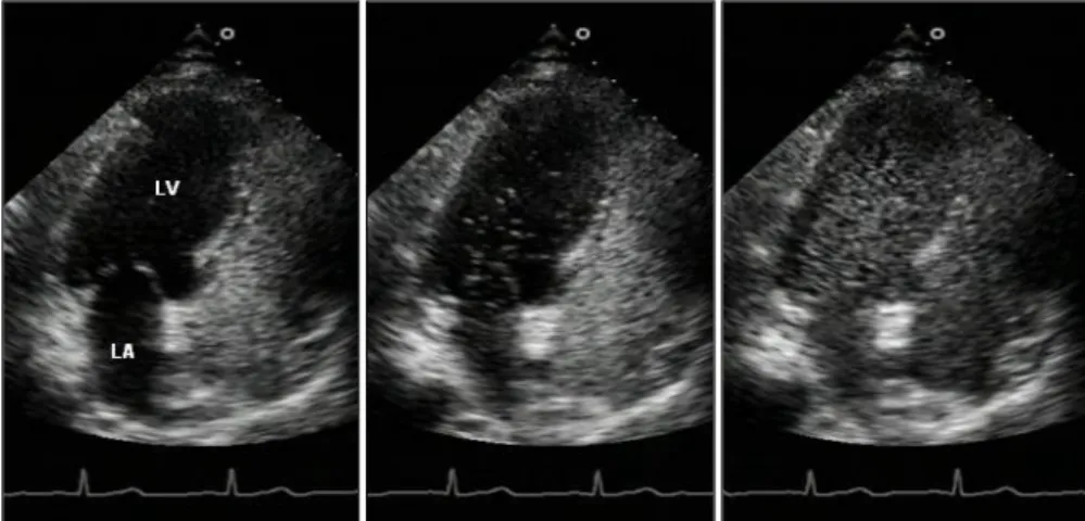
A 63-year-old woman was evaluated for progressively worsening exertional dyspnea. Her past medical history was unremarkable except liver cirrhosis for 10 years. The elec-trocardiogram revealed sinus rhythmwith early transition in V1. Pulmonary function test was normal. Transthoracic echo-cardiography showed borderline enlarged left atrium but nor-mal sized other cardiac chambers with nornor-mal left ventri-cular systolic function (ejection fraction 68%). HPS was suspected because of a long history of liver cirrhosis. Contrast echocardiography was performed by intravenous injection of hand-agitated saline while visualizing the atria. Contrast appears in the left atrium 3 to 5 seconds after it is seen in the right atrium, suggestive of pulmonary arteriovenous fistula (Fig. 1).
Discussion
HPS is defined as the clinical triad of advanced liver disease, arterial deoxygenation and intrapulmonary vascular dilatation.1) HPS is a well-recognized complication of liver
cirrhosis, but its pathogenesis is not completely understood. It is one of pathophysiologic mechanism that pulmonary ca-pillary is dilated upto 500 μm (normal range:8-15 μm) and the vascular resistance reduced, so abnormal hyperdy-논문접수일:2005년 11월 21일 심사완료일:2005년 12월 21일 교신저자:하종원, 120-752 서울 서대문구 신촌동 134번지 연세대학교 의과대학 내과학교실 전화:(02) 2228-8460・전송:(02) 393-2041 E-mail:[email protected]
・Jihyuk Rhee, et alJihyuk Rhee, et al:The Hepatopulmonary SyndromeJihyuk Rhee, et alJihyuk Rhee, et al
167 namic pulmonary circulation results in shunting of deoxy-dized venous blood into systemic arterial network. Excessive pulmonary nitric oxide production seems to be one of the factors that contribute to the intrapulmonary vascular dilata-tion. Other mediators such as endothelin-1 and the heme oxygenase-1/carbon monoxide system have recently been found to be important contributors.4)
The prevalence of HPS among patients with liver cirrhosis has been reported to be between 5.3 and 17.5%.5)6)
HPS may also occur in liver diseases such as acute viral hepatitis and fulminant hepatic failure.7)8) A few cases with
noncirrhotic portal hypertension complicated by HPS has also been re-ported.9)10)
The major clinical manifestations are arterial hypoxemia, clubbed fingers and spider nevi. Orthodeoxia is also the char-acteristic clinical feature.
For the detection of intrapulmonary vascular dilatations and shunting, contrast echocardiography is the method of choice because it is of easy execution and it can differentiate between the intrapulmonary and intracardiac communica-tions.3)11) In the case of HPS, contrast appears in the left
atrium 2 to 5 s after it is seen in the right atrium, while si-multaneously or within 2 heart beats in intracardiac shunt. Our case was also approved by hand-agitated saline contrast echocardiography.
99m Technetium macroaggregated albumin (Tc-99m MAA) lung perfusion scan can further specify the diagnosis of HPS and quantify the magnitude of shunting. On the other hand, Koksal and collegues have reported that high resolu-tion computed tomography (HRCT) may be helpful in the diagnosis of HPS by demonstrating the dilated peripheral pul-monary vessels or increased pulpul-monary artery to bronchus
ratios in patients with liver disease and hypoxemia.12)
Despite of multifarious trial, no clearly effective medical treatments have been found. Although liver transplantation seems feasible to reverse this situation,11) it is associated with increased postoperative morbidity and mortality. Swanson et al reported that preoperative arterial oxygen tension of 50 mmHg or less and Tc-99m MAA shunt fractions of 20% or more are strong predictors of postoperative mortality that can be used to stratify patients with better outcome.13)
There-fore, early detection of HPS can be related with reduction of morbidity and postoperative mortality.
KEY WORDS:Hepatopulmonary syndrome・Contrast ec-hocardiography・Right-to-left shunt.
REFERENCES
1) Rodriguez-Roisin R, Agusti AGN, Roca J. The hepatopulmonary
syndrome: new name, old complexities. Thorax 1992;47:897-902.
2) Kaymakoglu S, Kahraman T, Kudat H, Demir K, Cakaloglu Y, Ad-alet I, Dincer D, Besisik F, Boztas GB, Ozen AB, Mungan Z, Okten A. Hepatopulmonary Syndrome in Noncirrhotic Portal Hypertensive
Patients. Digestive Diseases and Sciences 2003 Mar;48(3):556-60.
3) Krowka MJ, Tajik AJ, Dickson ER, Wiesner RH, Cortese DA.
Intra-pulmonary vascular dilatations (IPVD) in liver transplant candida-tes: screening by two-dimensional contrast-enhanced echocardiogra-phy. Chest 1990;97:1165-70.
4) Fallon MB. Mechanisms of pulmonary vascular complications of
liver disease: hepatopulmonary syndrome. J Clin Gastroenterol 2005 Apr;39(4 Suppl 2):S138-42.
5) Kennedy KC, Knudson RJ. Exercise-aggravated hypoxemia and
orthodeoxia in cirrhosis. Chest 1977;72:305-9.
6) Abrams GA, Jaffe CC, Hoffer PB, Binder HJ, Fallon MB.
Diagno-stic utility of contrast echocardiography and lung perfusion scan in patients with hepatopulmonary syndrome. Gastroenterology 1995; 109:1283-8.
7) Regev A, Yeshurun M, Rodriguez M, Sagie A, Neff GW, Molina EG, Schiff ER. Transient hepatopulmonary syndrome in a patient with
acute hepatitis A. J Viral Hep 2001;8:83-6.
8) Scott VL, Dodson SF, Kang Y. The hepatopulmonary syndrome.
Surg Clin North Am 1999;79:23-41.
9) Marchand EJ, De Jesus M, Biascoechea R. Cyanotic syndrome of
Fig. 1. Contrast
echocardiogra-phy in hepatopulmonary syndro-me. Delayed opacification of the left-sided heart (5 beats after the opacification of the right-sided heart in this figure) was showed during contrast echocardiogra-phy using hand-agitated saline.
J Kor Soc Echo 2005;13:166-168
168
portal hypertension in hepatosplenic schistosomiasis and portal cir-rhosis. Am J Cardiol 1962;10:496-506.
10) Babbs C, Warnes TW, Haboubi NY. Noncirrhotic portal
hyperten-sion with hypoxaemia. Gut 1988;29:129-31.
11) Lima B, Martinelli A, Franca AV. Hepatopulmonary syndrome:
pa-thogenesis, diagnosis and treatment. Arq Gastroenterol 2004 Oct-Dec;41(4):250-8.
12) Koksal D, Kacar S, Koksal AS, Tufekcioglu O, Kucukay F, Okten S,
Sasmaz N, Arda K, Sahin B. Evaluation of Intrapulmonary Vascular
Dilatations with High-Resolution Computed Thorax Tomography in Patients with Hepatopulmonary Syndrome. J Clin Gastroenterol 2006 Jan;40(1):77-83.
13) Swanson KL, Wiesner RH, Krowka MJ. Natural history of
hepa-topulmonary syndrome: Impact of liver transplantation. Hepatology 2005 May;41(5):1122-9.