Litholytic agents as an alternative treatment
modality in patients with biliary dyspepsia
Young Min Kim, MD
a, Sung Ill Jang, MD, PhD
a,∗, Jae Hee Cho, MD, PhD
a,b, Dong Hee Koh, MD, PhD
c,∗,
Chang-Il Kwon, MD, PhD
d, Tae Hoon Lee, MD, PhD
e, Seok Jeong, MD, PhD
f, Dong Ki Lee, MD, PhD
aAbstract
Biliary dyspepsia presents as biliary colic in the absence of explanatory structural abnormalities. Causes include gallbladder dyskinesia, sphincter of Oddi dysfunction, biliary tract sensitivity, microscopic sludges, and duodenal hypersensitivity. However, no consensus treatment guideline exists for biliary dyspepsia. We investigated the effects of medical treatments on biliary dyspepsia. We retrospectively reviewed the electronic medical records of 414 patients who had biliary pain and underwent cholescintigraphy from 2008 to 2018. We enrolled patients who received litholytic agents and underwent follow-up scans after medical treatment. We divided the patients into the GD group (biliary dyspepsia with reduced gallbladder ejection fraction [GBEF]) and the NGD group (biliary dyspepsia with normal GBEF). We compared pre- and post-treatment GBEF and symptoms.
Among 57 patients enrolled, 40 (70.2%) patients had significant GBEF improvement post-treatment, ranging from 34.4±22.6% to 53.8±26.8% (P<.001). In GD group (n=35), 28 patients had GBEF improvement after medical treatment, and value of GBEF significantly improved from 19.5±11.0 to 47.9±27.3% (P<.001). In NGD group (n=22), 12 patients had GBEF improvement after medical treatment, but value of GBEF did not have significant change. Most patients (97.1% in GD group and 81.8% in NGD group) had improved symptoms after medical treatment. No severe complication was reported during treatment period.
Litholytic agents improved biliary colic in patients with biliary dyspepsia. Therefore, these agents present an alternative treatment modality for biliary dyspepsia with or without gallbladder dyskinesia. Notably, biliary colic in patients with gallbladder dyskinesia resolved after normalization of the GBEF. Further prospective and large-scale mechanistic studies are warranted.
Abbreviations: CNU= combination of chenodeoxycholic acid and ursodeoxycholic acid, DISIDA = technetium-99m diisopropyl iminodiacetic acid cholescintigraphy, GB= gallbladder, GBEF = gallbladder ejection fraction, GD = gallbladder dyskinesia, NGD = non-gallbladder dyskinesia, UDCA= ursodeoxycholic acid.
Keywords:biliary dyspepsia, gallbladder dyskinesia, litholytic agent
1. Introduction
Biliary dyspepsia is defined as the presence of biliary pain with no organic, systemic, or metabolic cause.[1] The causes of biliary
dyspepsia include gallbladder dyskinesia (GD), sphincter of Oddi dysfunction, biliary tract sensitivity, microscopic sludges, duodenal hypersensitivity, and parasitic infestation.[2] Among these causes, GD, also referred to as functional gallbladder (GB) disorder or GB dysfunction, is a rare functional disorder defined
as biliary pain in the absence of gallstone or other structural pathology. The Rome IV criteria describe in detail the diagnostic criteria for biliary pain in the epigastrium and/or right upper quadrant.[3]The diagnosis of biliary pain due to GD is supported by a low gallbladder ejection fraction (GBEF) on GB scintigraphy and normal laboratory test results.[3,4]The prevalence of GD has increased due to the introduction of diagnostic modalities such as GB scintigraphy.[5]Several studies have examined the prevalence Editor: Raffaele Pezzilli.
This work was funded by the National Research Foundation, Ministry of Science and ICT of Korea (NRF-2018R1C1B5045210) and a faculty research grant from the Yonsei University College of Medicine (6-2017-0082).
The authors have no conflicts of interest to disclose.
The datasets generated during and/or analyzed during the current study are available from the corresponding author on reasonable request. a
Department of Internal Medicine, Gangnam Severance Hospital, Yonsei University College of Medicine, Seoul,b
Department of Internal Medicine, Gachon University College of Medicine, Gil Medical Center, Incheon,c
Department of Internal Medicine, Hallym University Dongtan Sacred Heart Hospital, Hallym University College of Medicine,d
Digestive Disease Center, CHA Bundang Medical Center, CHA University, Seongnam,e
Department of Internal Medicine, Soonchunhyang University College of Medicine, Cheonan Hospital, Cheonan,f
Department of Internal Medicine, Inha University School of Medicine, Incheon, South Korea. ∗
Correspondence: Sung Ill Jang, Department of Internal Medicine, Gangnam Severance Hospital, Yonsei University College of Medicine, 211 Eonjuro, Gangnam-gu, Seoul 06273, Korea (e-mail: [email protected]); Dong Hee Koh, Division of Gastroenterology, Department of Internal Medicine, Hallym University Dongtan Sacred Heart Hospital, 7 Keunjaebong-gil, Hwaseong 18450, Korea (e-mail: [email protected]).
Copyright© 2020 the Author(s). Published by Wolters Kluwer Health, Inc.
This is an open access article distributed under the terms of the Creative Commons Attribution-Non Commercial License 4.0 (CCBY-NC), where it is permissible to download, share, remix, transform, and buildup the work provided it is properly cited. The work cannot be used commercially without permission from the journal. How to cite this article: Kim YM, Jang SI, Cho JH, Koh DH, Kwon CI, Lee TH, Jeong S, Lee DK. Litholytic agents as an alternative treatment modality in patients with biliary dyspepsia. Medicine 2020;99:34(e21698).
Received: 18 February 2020 / Received infinal form: 29 June 2020 / Accepted: 3 July 2020 http://dx.doi.org/10.1097/MD.0000000000021698
Observational Study
Medicine
of biliary dyspepsia. One group reported on the prevalence of biliary pain without gallstone in Italy; 2.0% of the enrolled patients had biliary dyspepsia,[6] and the prevalence of GD
differed according to sex. Other large-population studies showed that GD was more common in females (20.7%) than in males (7.6%).[7,8]
Medical and surgical therapies can be used to treat biliary dyspepsia according to its cause, but no consensus treatment guideline has been established. A previous study of the efficacy of cholecystectomy for biliary dyskinesia showed that patients who received cholecystectomy exhibited significant symptom im-provement compared with nonsurgical patients.[9]
However, surgery as the primary treatment method for GD is burdensome for elderly patients and those with comorbidities. In such patients, medical treatment for GD is preferred. However, data on the effect of medical treatment on GD are scarce.
We investigated the effects of medical treatments on biliary dyspepsia, with a focus on 2 litholytic agents. The primary outcomes were increased GBEF and symptom improvement after medical treatment.
2. Materials and methods
2.1. Patients
In this retrospective study, we reviewed the electronic medical records of 414 patients who presented with biliary pain and underwent technetium-99m diisopropyl iminodiacetic acid (DISIDA) cholescintigraphy between July 2008 and August 2018, in 6 tertiary hospitals. All reviewed patients were≥18 years and of Korean. Exclusion criteria were patients with no post-treatment DISIDA scan; an organic lesion causing biliary pain; no medical treatment; abnormal laboratory test results (serum levels of aspartate transaminase/alanine transaminase, bilirubin, gam-ma-glutamyl transferase, amylase, or lipase); acute or episodic symptoms; and history of incomplete electronic medical records. In addition, patients who had impaired mobility, renal insufficiency, electrolyte imbalance, and history of abdominal surgery were also excluded.
The study protocol conformed to the ethical guidelines of the World Medical Association Declaration of Helsinki and was approved by the Institutional Review Board of Gangnam Severance Hospital (IRB No: 3-2019-0244). Informed consent was not required because this study was a retrospective analysis of existing administrative and clinical data.
2.2. Data collection
Data were retrospectively extracted from the electronic medical records of 6 tertiary hospitals. The data included demographic, clinical, laboratory, and imaging parameters, and thefindings of upper gastrointestinal endoscopies performed at the time of treatment initiation.
Demographic data included age, sex, body mass index (BMI), and personal and medical histories. Imaging and GBEF data were collected from initial and follow-up DISIDA scans. Imaging parameters included abdominal ultrasonography, abdominal computed tomography, magnetic resonance imaging, and endoscopic ultrasonography. Regarding litholytic agents, we collected data on the type, duration, and side effects of medication. We analyzed data from patients who received 2 litholytic agents: Ursa (ursodeoxycholic acid [UDCA];
Daewoong Pharm. Co. Ltd., Seoul, South Korea) and CNU (magnesium trihydrate salt of chenodeoxycholic acid and ursodeoxycholic acid [CNU]; Myungmoon Pharm. Co., Seoul, South Korea).
2.3. DISIDA scanning
All enrolled patients underwent pre- and post-treatment DISIDA scans, during which GBEF values were measured and recorded. The method used for DISIDA scanning is described in detail below.[10]
The DISIDA scan was performed after the patient had fasted overnight. Each subject was given 8 mCi DISIDA intravenously under a large-field-of-view gamma camera. Serial hepatobiliary analogue images were obtained at 5, 10, 20, 30, 45, and 60 minutes after the injection or until the GB was adequatelyfilled. Immediately after completion of the filling phase, the patients drank 200 mL milk containing about 13 g fat (130 kcal). Analogue images were recorded 30 minutes after ingestion of the milk. The GBEF was derived by calculating the counts in the GB before and 30 minutes after ingestion of the milk. Background regions over the liver were also generated and subtracted from the GB counts to derive the net GB count.
2.4. Definitions
Biliary dyspepsia defined as biliary colic without any organic, systemic, or metabolic origin. Biliary colic was steady pain in the right upper quadrant and/or epigastric area, lasting for at least 30 minutes according to Rome IV criteria.[4]To exclude patients,
who have organic, systemic, and metabolic origin, we reviewed the result of laboratory and imaging parameters, and thefindings of upper gastrointestinal endoscopies. These enrolled patients, who had biliary dyspepsia, were divided into 2 groups according to the results of the initial DISIDA scans. Patients who had reduced initial GBEFs (< 38%) were allocated to the GD group. Patients who had normal initial GBEFs (≥ 38%) were allocated to the non-gallbladder dyskinesia (NGD) group.
After medical treatment, we evaluated the GBEFs and symptoms of the patients. The GBEF was evaluated by follow-up DISIDA scan and classified as showing improvement, no change, or deterioration. The improvement of symptoms after treatment was also evaluated.
2.5. Statistical analysis
Continuous variables are reported as means±standard devia-tions. We used Studentt test or the Wilcoxon rank-sum test to compare continuous variables between the groups. Categorical variables are reported as numbers and percentages and were compared by Chi-squared test or Fisher exact test. Statistical analysis was performed using the SPSS version 23.0 (IBM Corp., Armonk, NY). Two-tailed P values<.05 were considered to indicate statistical significance.
3. Results
3.1. Patients
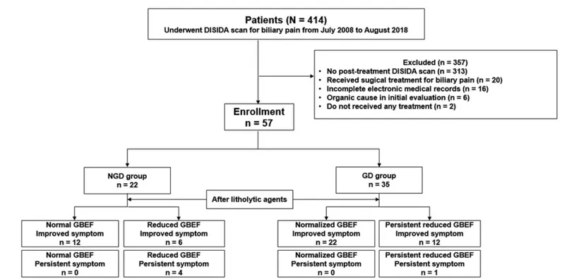
In total, 414 patients underwent DISIDA scans for biliary pain between July 2008, and August 2018. Among these patients, 6 were excluded because their biliary pain was determined to be of
organic cause. The remaining 408 patients were diagnosed with biliary dyspepsia. Among them, we excluded those with the following characteristics:
(1) lack of post-treatment DISIDA scan (n=313) (2) receipt of surgical treatment for biliary pain (n=20) (3) incompleteness of electronic medical records (n=16) (4) receipt of no treatment (n=2). As a result, 57 patients were
enrolled in this study (Fig. 1)
3.2. Baseline characteristics
The mean age of the participants was 46.4±15.9 years, and females were predominant (71.9%). The average BMI was 23.3± 5.0 kg/m2, and 10 (17.5%) patients were obese according to the
Asia-Pacific criteria. Three patients were current smokers, and 9 patients had histories of alcohol consumption. Two and 8 patients had diabetes mellitus and hypertension, respectively (Table 1).
Table 2 shows the litholytic agents prescribed for biliary dyspepsia. Among the patients, 19 (33.3%) received CNU and 38 (66.7%) received UDCA. The mean treatment duration was 15.6±20.1 months.
3.3. Result of DISIDA scan
On initial DISIDA scans, the mean pre-treatment GBEF was 34.4 ±22.6%, and 35 (61.4%) patients had GBEFs<38% (Table 1). Therefore, 35 patients were diagnosed with GD, and the remaining 22 patients were allocated to the NGD group. Mean time interval between initial and follow-up DISIDA scan was 8.6 ±6.8 months.
The quantitative changes in the GBEF are shown in Table 3. For all patients, the initial mean GBEF (34.4±22.6%) was significantly lower than that after medical treatment (53.8 ± 26.8%;P<.001). In addition, UDCA significantly increased the GBEF from 29.4±18.8% to 51.2±26.4% (P<.001) for all Figure 1. Study flow chart. We enrolled in this retrospective study 57 patients with biliary pain who underwent cholescintigraphy. DISIDA, technetium-99m diisopropyl iminodiacetic acid cholescintigraphy; GB, gallbladder; GBEF, gallbladder ejection fraction; NGD, biliary pain with normal gallbladder ejection fraction; GD, biliary pain with reduced gallbladder ejection fraction.
Table 1
Baseline characteristics of the study population.
Variables All patients (N=57)
Age (yr, mean±SD) 46.4±15.9
Male (n, %) 16 (28.1) BMI (kg/m2, mean±SD) 23.3±5.0 Current smoker (n, %) 3 (5.3) Alcohol history (n, %) 9 (15.8) DM (n, %) 2 (3.5) Hypertension (n, %) 8 (14.0)
Initial DISIDA scan
Pretreatment GBEF (%, mean±SD) 34.4±22.6 GD diagnoses based on pretreatment GBEF (n, %) 35 (61.4)
BMI=body mass index, DISIDA=technetium-99m diisopropyl iminodiacetic acid cholescintigraphy, DM= diabetes mellitus, GB=gallbladder, GBEF=gallbladder ejection fraction, GD=gallbladder dyskinesia.
Table 2
Litholytic agents prescribed to the study population.
Variables All patients (N=57)
Litholytic agents (n, %)
CNU 19 (33.3)
UDCA 38 (66.7)
Duration of medication (months, mean±SD) 15.6±20.1 Improved biliary pain (n, %) 52 (91.2)
GD group 34/35 (97.1)
NGD group 18/22 (81.8)
Complications
Diarrhea 1 (1.8)
Constipation 1 (1.8)
CNU=combination of chenodeoxycholic acid and ursodeoxycholic acid, GD =gallbladder dyskinesia, NGD=non-gallbladder dyskinesia, UDCA =ursodeoxycholic acid.
patients. Patients receiving CNU treatment also showed a significant difference in the GBEF (P=.107). Figure 2 (A) and (B) are pre- and post-treatment GBEF dot plots for all patients. In the GD group, the mean pre-treatment GBEF (19.5%± 11.0%) was significantly lower than that after medical treatment (47.9±27.3%, P<.001; Table 3). In patients with GD, CNU and UDCA significantly increased the GBEF (Fig. 2D). CNU treatment increased the GBEF from 21.4±11.8% to 46.4± 30.5% (P=.035; Fig. 2E), and UDCA treatment increased the GBEF from 18.9±10.8% to 48.4±26.8% (P<.001; Fig. 2F). The GBEF did not change significantly in the NGD group (P=.401; Table 3, Fig. 2C).
Figure 3 shows representative DISIDA scans of a treatment-responsive patient with GD (A) before and (B) after CNU treatment. This patient’s GBEF was 20.1% before treatment and 85.0% after treatment.
Figure 2. Pre- and post-treatment gallbladder ejection fractions. (A) Dot plot and (B) paired dot plot for all patients. Dot plots for the NGD group (C) and GD group (D). Dot plots for the patients in the GD group who received CNU (E) and UDCA (F).∗P< .05. GBEF, gallbladder ejection fraction; NGD, biliary pain with normal gallbladder ejection fraction; GD, biliary pain with reduced gallbladder ejection fraction.
Table 3
Comparison of pre- and post-treatment GBEFs.
Pretreatment Post-treatment P-value All patients (n=57) 34.4±22.6 53.8±26.8 <.001 CNU (n=19) 44.5±26.4 58.9±27.5 .107 UDCA (n=38) 29.4±18.8 51.2±26.4 <.001 GD group (n=35) 19.5±11.0 47.9±27.3 <.001 CNU (n=9) 21.4±11.8 46.4±30.5 .035 UDCA (n=26) 18.9±10.8 48.4±26.8 <.001 NGD group (n=22) 58.1±14.4 63.1±23.6 .401 CNU (n=10) 65.3±15.9 70.2±19.7 .548 UDCA (n=12) 52.1±10.2 57.2±25.7 .533
CNU=combination of chenodeoxycholic acid and ursodeoxycholic acid, GB=gallbladder, GBEF= gallbladder ejection fraction, GD=gallbladder dyskinesia, NGD =non-gallbladder dyskinesia, UDCA = ursodeoxycholic acid.
3.4. Symptom improvement after medical treatment Biliary pain symptoms improved in 52 (91.2%) patients (97.1% in the GD group, 81.8% in the NGD group). The rate of symptom improvement was significantly greater in the GD group than in the NGD group (P<.001).
Among the 35 patients with GD, all 22 patients with normalized GBEFs after medical treatment experienced improve-ment of their biliary pain. Of the 13 patients who had persistent GD (as indicated by their GBEFs) despite medical treatment, 12 patients had improved symptoms and 1 patient had persistent symptoms (Fig. 1).
Two patients had side effects while using UDCA (diarrhea and constipation, respectively). However, these side effects were not serious enough to warrant discontinuation of the treatment.
4. Discussion
We determined that litholytic agents improved symptoms of biliary dyspepsia. In addition, patients with GD experienced significant improvement in symptoms and the GBEF after medical treatment. Although the mechanism of GD is unclear, researchers have made plausible suggestions. First, a narrowed cystic duct can induce incomplete GB emptying, eventually causing chronic cholecystitis and biliary pain.[11]Another group reported an association between the cystic duct, rather than the common bile duct or sphincter of Oddi, and GB dysfunction.[12] Second, microlithiasis is associated with GD. One previous study compared GB emptying among 3 groups (healthy controls, patients with gallstones, and patients with microlithiasis).[13]The GBEF was significantly lower in patients with microlithiasis than Figure 3. Representative DISIDA scans of a patient with GD with improved GBEF. DISIDA scans (A) before medical treatment (GBEF =20.1%) and (B) after medical treatment (GBEF=85.0%). DISIDA, technetium-99m diisopropyl iminodiacetic acid cholescintigraphy; GB, gallbladder; GD, gallbladder dyskinesia; GBEF, gallbladder ejection fraction.
in control patients. Beyond these potential mechanisms, several factors, such as prostaglandin E2, are also associated with the pathogenesis of GD.[4,14,15]
Among these factors, we hypothesized that UDCA might have an effect on dissolution of microlithiasis, thereby, improved GD. However, further analysis of bile from these patients is warranted to evaluate the effect of medical treatment with UDCA on micro-lithiasis, and UDCA was shown to correct this bile abnormality.
We used a GBEF cutoff value for GD of 38%. No cut-off value for GD has been established, although values of 35% to 50% have been set in previous studies.[16,17] Further research to establish uniform cutoff value for GD should be performed. However, our study will be meaningful to compare incidence of GD and result of our study with other studies, which use cutoff value as 38%.
In patients with GD in this study, the proportion of symptom improvement (97.1%) was not consistent with that of GBEF normalization (80.0%). Whether this effect was due to medical treatment is not certain, because no GBEF cut-off value has been evaluated formally or established.[4] Patients with GD may respond to medical treatments, such as UDCA, but the sample for this study was too small for such an evaluation. Patients who had biliary pain and normal GBEFs experienced significant improve-ment in pain symptoms. Interestingly, the GBEF was not significantly changed after treatment. The mechanism of symptom improvement was not clear because of the heterogene-ity of the NGD group. The causes of biliary dyspepsia in the NGD group could include sphincter of Oddi dysfunction, biliary tract sensitivity, and microscopic gallstones or sludges.[18,19]
Our study has several strengths over previous studies. First, biliary pain and GBEF values were required to diagnose GD. Most previous studies that aimed to evaluate treatment effects focused solely on symptom improvement. Symptoms can be subjective, even when evaluated using a pain scale. We evaluated the effects of medication on not only symptoms but also the GBEF, a more objective and reliable measure. Second, our study enrolled patients with GD and NGD. Many patients visit outpatient clinics because of biliary pain located in the epigastrium and/or right upper quadrant. Physicians perform various tests, including laboratory and imaging tests, as part of their initial evaluation. When the results of these tests are normal, many clinicians suspect functional gastrointestinal disorder or irritable bowel syndrome. A previous study showed that the evaluation of GB function is necessary to exclude GD in patients who present with biliary pain.[20]Our study confirms this finding in a larger number of patients.
This study has several limitations. First, this study was retrospective and sample size was too small. When patients have biliary colic, DISIDA scan is not a routine test in our clinic. Moreover, patients tend to not be performed follow-up DISIDA scan when they recover from symptoms. Because of this small sample size, it is hard to analyze control group. Only 2 patients did not receive any treatment. Prospective and large-scale study is warranted. Second, we evaluated symptoms by retrospectively reviewing patients’ electronic medical records. Further investiga-tion of symptoms using a method such as a visual analogue scale is needed because the pain evaluation was subjective. Third, we evaluated the effects of litholytic agents, but did not determine whether medical treatment is superior to surgical treatment. Further study of medical and surgical treatments is thus warranted. Fourth, according to the Rome IV criteria, GD is not the only condition that induces biliary pain; other conditions
(e.g., sphincter of Oddi dysfunction and duodenal hypersensitiv-ity) can also induce biliary pain.[21]Because we reviewed medical records that included DISIDA scans, we were unable to diagnose these other conditions. For example, manometry for the diagnosis of sphincter of Oddi dysfunction is not a routine test. Therefore, the NGD group was heterogeneous in terms of the causes of biliary dyspepsia.
In conclusion, the litholytic agents UDCA and CNU modulated the GBEF and induced symptom improvement in patients with biliary dyspepsia. In addition, the complication rate of medical treatment was low. Therefore, treatment with litholytic agents may be an alternative to surgical treatment for biliary dyspepsia, especially in patients for whom surgery is contraindicated. Notably, biliary pain resolved after normalization of the GBEF in patients with GD.
Author contributions
Critical revision of the manuscript: S.I.J., D.H.K., S.J., D.K.L. Data acquisition, analysis, and interpretation: Y.M.K., S.I.J., D.
H.K., J.H.C., C.I.K., T.H.L., S.J., D.K.L. Drafting of the manuscript: Y.M.K., S.I.J.
Important intellectual content: S.I.J., D.H.K., J.H.C., C.I.K., T.H. L., S.J., D.K.L.
Statistical analysis: Y.M.K., S.I.J. Study concept and design: Y.M.K., S.I.J. Study supervision: S.I.J., D.H.K., D.K.L.
Technical, or material support: Y.M.K., J.H.C., C.I.K., T.H.L.
References
[1] Shaffer E. Acalculous biliary pain: new concepts for an old entity. Dig Liver Dis 2003;35(Suppl 3):S20–5.
[2] Behar J, Corazziari E, Guelrud M, et al. Functional gallbladder and sphincter of Oddi disorders. Gastroenterology 2006;130:1498–509. [3] Stanghellini V, Chan FK, Hasler WL, et al. Gastroduodenal Disorders.
Gastroenterology 2016;150:1380–92.
[4] Cotton PB, Elta GH, Carter CR, et al. Gallbladder and Sphincter of Oddi Disorders. Gastroenterology 2016;150:1420–9.
[5] Tabet J, Anvari M. Laparoscopic cholecystectomy for gallbladder dyskinesia: clinical outcome and patient satisfaction. Surg Laparosc Endosc Percutan Tech 1999;9:382–6.
[6] Barbara L, Sama C, Morselli Labate AM, et al. A population study on the prevalence of gallstone disease: the Sirmione Study. Hepatology 1987;7:913–7.
[7] The epidemiology of gallstone disease in Rome, Italy. Part I. Prevalence data in men. The Rome Group for Epidemiology and Prevention of Cholelithiasis (GREPCO). Hepatology 1988;8:904–6.
[8] Rome Group for the Epidemiology and Prevention of Cholelithiasis (GREPCO) Prevalence of gallstone disease in an Italian adult female population. Am J Epidemiol 1984;119:796–805.
[9] Yost F, Margenthaler J, Presti M, et al. Cholecystectomy is an effective treatment for biliary dyskinesia. Am J Surg 1999;178:462–5. [10] DiBaise JK, Richmond BK, Ziessman HA, et al.
Cholecystokinin-cholescintigraphy in adults: consensus recommendations of an interdis-ciplinary panel. Clin Gastroenterol Hepatol 2012;9:376–84.
[11] Yap L, Wycherley AG, Morphett AD, et al. Acalculous biliary pain: cholecystectomy alleviates symptoms in patients with abnormal cholescintigraphy. Gastroenterology 1991;101:786–93.
[12] Ruffolo TA, Sherman S, Lehman GA, et al. Gallbladder ejection fraction and its relationship to sphincter of Oddi dysfunction. Dig Dis Sci 1994;39:289–92.
[13] Sharma BC, Agarwal DK, Dhiman RK, et al. Bile lithogenicity and gallbladder emptying in patients with microlithiasis: effect of bile acid therapy. Gastroenterology 1998;115:124–8.
[14] Alcon S, Morales S, Camello PJ, et al. Contribution of different phospholipases and arachidonic acid metabolites in the response of gallbladder smooth muscle to cholecystokinin. Biochem Pharmacol 2002;64:1157–67.
[15] Pozo MJ, Camello PJ, Mawe GM. Chemical mediators of gallbladder dysmotility. Curr Med Chem 2004;11:1801–12.
[16] Paajanen H, Miilunpohja S, Joukainen S, et al. Role of quantitative cholescintigraphy for planning laparoscopic cholecystectomy in patients with gallbladder dyskinesia and chronic abdominal pain. Surg Laparosc Endosc Percutan Tech 2009;19:16–9.
[17] Goussous N, Maqsood H, Spiegler E, et al. HIDA scan for functional gallbladder disorder: ensure that you know how the scan was done. Hepatobiliary Pancreat Dis Int 2017;16: 197–201.
[18] Varadarajulu S, Hawes R. Key issues in sphincter of Oddi dysfunction. Gastrointest Endosc Clin N Am 2003;13:671–94.
[19] Corazziari E, Shaffer EA, Hogan WJ, et al. Functional disorders of the biliary tract and pancreas. Gut 1999;45(Suppl 2):II48–54.
[20] Jung SW, Joo MS, Choi HC, et al. Epigastric symptoms of gallbladder dyskinesia mistaken for functional dyspepsia: retrospective observation-al study. Medicine (Bobservation-altimore) 2017;96:e6702.
[21] Drossman DA. Functional gastrointestinal disorders: history, patho-physiology, clinical features and Rome IV. Gastroenterology 2016; 150:1262–79.