with a Moxifloxacin-Containing Regimen
Won-Jung Koh,aGoohyeon Hong,aSu-Young Kim,aByeong-Ho Jeong,aHye Yun Park,aKyeongman Jeon,aO Jung Kwon,a Seung-Heon Lee,bChang Ki Kim,bSung Jae Shinc
Division of Pulmonary and Critical Care Medicine, Department of Medicine, Samsung Medical Center, Sungkyunkwan University School of Medicine, Seoul, South Koreaa ; Korean Institute of Tuberculosis, Chungbuk, South Koreab
; Department of Microbiology, Yonsei University College of Medicine, Seoul, South Koreac
Moxifloxacin (MXF) has in vitro and in vivo activity against Mycobacterium avium complex (MAC) in experimental models. However, no data are available concerning its treatment effect in patients with MAC lung disease. The aim of this study was to evaluate the clinical efficacy of an MXF-containing regimen for the treatment of refractory MAC lung disease. Patients with MAC lung disease who were diagnosed between January 2002 and December 2011 were identified from our hospital database. We identified 41 patients who received MXF for >4 weeks for the treatment of refractory MAC lung disease. A total of 41 pa-tients were treated with an MXF-containing regimen because of a persistent positive culture after at least 6 months of clarithro-mycin-based standardized antibiotic therapy. The median duration of antibiotic therapy before MXF administration was 410 days (interquartile range [IQR], 324 to 683 days). All patients had culture-positive sputum when MXF treatment was initiated. The median duration of MXF administration was 332 days (IQR, 146 to 547 days). The overall treatment success rate was 29% (12/41), and the median time to sputum conversion was 91 days (IQR, 45 to 190 days). A positive sputum acid-fast-bacillus smear at the start of treatment with MXF-containing regimens was an independent predictor of an unfavorable microbiological re-sponse. Our results indicate that MXF may improve treatment outcomes in about one-third of patients with persistently culture-positive MAC lung disease who fail to respond to clarithromycin-based standardized antibiotic treatment. Prospective studies are required to assess the clinical efficacy of MXF treatment for refractory MAC lung disease.
P
ulmonary disease caused by nontuberculous mycobacteria (NTM) appears to be increasing worldwide (1–6). Mycobacte-rium avium complex (MAC), consisting of MycobacteMycobacte-rium avium and Mycobacterium intracellulare, is the most common etiologic agent in lung disease caused by NTM (1,2). A major therapeutic advance in the treatment of MAC lung disease was the introduc-tion of newer macrolides such as clarithromycin (CLR) and azithromycin, which have in vitro and clinical activity against MAC disease (1,2). Macrolides, together with ethambutol (EMB) and rifampin (RIF), are the cornerstones of MAC therapy (1,2). However, the treatment success rate for MAC lung disease is un-satisfactory. Macrolide-based therapy results in the successful eradication of an MAC pulmonary infection in only 60 to 80% of cases (7–10). Many patients fail to respond to treatment, relapse, or develop CLR-resistant MAC disease after receiving macrolide-based therapy (7–10).Moxifloxacin (MXF) is an 8-methoxy fluoroquinolone with better in vitro activity than older quinolones against MAC (11). Murine experimental infection models showed that MXF exhib-ited favorable activities against MAC in vitro and in vivo (12,13). Although MXF is not formally recommended for the treatment of MAC lung disease, it has been frequently prescribed in routine clinical practice, partly as a result of the unsatisfactory rate of response to recommended first-line regimens (14). However, no data are available concerning its effect on MAC lung disease. The aim of this study was to evaluate the clinical efficacy of an MXF-containing regimen for the treatment of MAC lung disease. MATERIALS AND METHODS
Patients. Consecutive patients with MAC lung disease who were diag-nosed between January 2002 and December 2011 were identified from the NTM Registry of Samsung Medical Center (a 1,961-bed referral hospital
in Seoul, South Korea) (10,15,16). During this 10-year period, 913 pa-tients were diagnosed with MAC lung disease. All of the papa-tients met the diagnostic criteria for NTM lung disease according to American Thoracic Society guidelines (1). Of these patients, 494 (54%) began long-term an-tibiotic treatment on 31 December 2011. Ultimately, we identified pa-tients who received MXF forⱖ4 weeks for the treatment of refractory MAC lung disease. The Institutional Review Board at our institution ap-proved this retrospective study; informed consent for the use of patient medical data was waived.
Antibiotic treatment. All of the patients with MAC lung disease who began antibiotic therapy received standardized combination oral antibi-otic therapy (1). For most patients, the regimen included CLR at 1,000 mg/day, EMB at 15 mg/kg/day, and RIF at 450 mg/day (body weight⬍ 50 kg) or 600 mg/day (body weightⱖ 50 kg). Streptomycin was administered intramuscularly in 13 (32%) patients with severe fibrocavitary disease or CLR-resistant MAC lung disease before MXF treatment.
The indication of MXF-containing antibiotic treatment for refractory MAC lung disease in this study was a persistent positive culture after at least 6 months of CLR-based standardized antibiotic therapy. All patients had culture-positive sputum when MXF treatment was initiated. A daily dose of 400 mg of MXF was prescribed. Sputum conversion was defined as three consecutive negative cultures; the time of conversion was defined as the date of the first negative culture. Treatment success was defined as negative cultures for the infecting MAC strains for 12 months or longer
Received 12 November 2012 Returned for modification 24 December 2012 Accepted 26 February 2013
Published ahead of print 11 March 2013
Address correspondence to Won-Jung Koh, [email protected]. W.-J.K. and G.H. contributed equally to this article.
Copyright © 2013, American Society for Microbiology. All Rights Reserved. doi:10.1128/AAC.02281-12
after the initiation of MXF therapy (17). We excluded two patients who received an adjunctive surgery with less than 2 months of MXF therapy, because the treatment effect of MXF could not be evaluated.
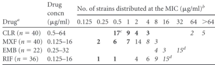
Drug susceptibility tests. Drug susceptibility testing was performed at the Korean Institute of Tuberculosis. The MICs of CLR, RIF, EMB, and MXF were determined using the broth microdilution method as de-scribed by the Clinical and Laboratory Standards Institute (CLSI) (18). The drug concentration ranges for tested drugs were as follows: for CLR, 0.5 to 64g/ml; for MXF, 0.125 to 16 g/ml; for EMB, 0.25 to 32 g/ml; and for RIF, 0.125 to 16g/ml. MAC isolates with an MIC ⱕ 8 g/ml were regarded as susceptible to CLR, while those with an MICⱖ 32 g/ml were regarded as resistant. An isolate with an MICⱕ 1 g/ml was defined as susceptible to MXF, while that with an MICⱖ 4 g/ml was defined as resistant (18). Breakpoints for the susceptibility and resistance of MAC to RIF and EMB have not been defined by the CLSI. In this study, isolates were considered resistant if the RIF or EMB MIC wasⱖ8 g/ml (19).
Statistical analysis. All data are presented as medians and interquar-tile ranges (IQR) for continuous variables and as numbers (percentages) for categorical variables. Categorical variables were analyzed using Pear-son’s2test or Fisher’s exact test. Multivariable logistic regression analysis
was performed to assess the effects of independent factors on the final treatment outcome. Variables with a P valueⱕ 0.2 by univariable analysis were considered for multivariable analysis (20). All P values were two sided, with P⬍ 0.05 considered to be statistically significant. All of the statistical analyses were performed using PASW 18.0 (SPSS Inc., Chicago, IL); a two-sided P⬍ 0.05 was considered significant.
RESULTS
Clinical characteristics of the patients. A total of 41 patients (28
males and 13 females; median age, 65 years [IQR, 56 to 71 years]) with MAC lung disease who were treated using an MXF-contain-ing regimen were included in the study. The median body mass
index was 19.5 kg/m2(IQR, 18.2 to 21.2 kg/m2). None of the
patients were positive for HIV infection. The baseline character-istics of the patients are summarized inTable 1.
The etiologic agents included M. avium in 15 patients (37%) and M. intracellulare in 26 patients (63%). A total of 29 patients (71%) had a positive acid-fast-bacillus (AFB) smear at the time of MXF treatment initiation. A total of 19 patients (46%) had the fibrocavitary form of MAC lung disease, 20 patients (49%) had the nodular bronchiectatic form, and 2 patients (5%) had an unclas-sifiable form.
Drug susceptibility test results were available for 40 patients (98%). The MAC isolates recovered from seven patients (18%) showed resistance to CLR, while 11 isolates (28%) were resistant to MXF (Table 2).
MXF-containing antibiotic treatment. All patients were
treated with an MXF-containing regimen because of a persistent positive culture after at least 6 months of CLR-based standardized antibiotic therapy. The median duration of CLR-based antibiotic therapy before MXF administration was 410 days (IQR, 324 to 683 days). Streptomycin was continuously administered in eight pa-tients and newly administered in five papa-tients with the MXF-con-taining regimen; the median duration of streptomycin use was 159 days (IQR, 78 to 248 days) in these patients.
The duration of MXF administration was a median of 332 days (IQR, 146 to 547 days) in 41 patients. However, MXF was discon-tinued during antibiotic therapy in 33 patients. The reasons for discontinuation were a persistent positive culture despite the ad-ministration of MXF (n⫽ 21; median, 413 days; IQR, 172 to 682 days), adverse effects associated with MXF such as a gastrointesti-nal disturbance or skin rash (n⫽ 10; median, 62 days; IQR, 36 to 237 days), death related to MAC lung disease (n⫽ 2; medians, 106 and 156 days), and other (n⫽ 1; median, 98 days).
Treatment outcomes. Of 41 patients with MAC lung disease
with persistent positive cultures after at least 6 months of CLR-based antibiotic therapy, 12 became sputum negative after MXF therapy. The median time to sputum conversion was 91 days (IQR, 45 to 190 days). Among these 12 patients with treatment success, eight patients received MXF to the end of antibiotic treat-ment for a median of 521 days (IQR, 415 to 598 days) and the median time to sputum conversion was 79 days (IQR, 45 to 190 days). Although MXF was discontinued in four patients after a median of 87 days (IQR, 45 to 125 days), sputum culture con-verted to negative after a median of 91 days (IQR, 49 to 119 days). Therefore, the overall treatment success rate was 29% (12/41) and TABLE 1 Baseline characteristics of 41 patients with Mycobacterium
avium complex lung disease who were treated with a moxifloxacin-containing antibiotic regimen
Patient characteristica Valuesb
Age, yr 65 (56–71)
Male 28 (68)
Body mass index, kg/m2 19.5 (18.2–21.2)
Current or ex-smoker 19 (46)
History of previous tuberculosis 29 (71) Pulmonary function test
FEV1 84% (65–94%) FVC 75% (63–90%) FEV1/FVC 80% (69–89%) Etiologic organism Mycobacterium avium 15 (37) Mycobacterium intracellulare 26 (63)
Positive sputum AFB smearc 29 (71)
No. of involved lobes 4 (3–4)
Type of disease
Fibrocavitary form 19 (46)
Nodular bronchiectatic form 20 (49)
Unclassifiable form 2 (5)
a
FEV1, forced expiratory volume in 1 s; FVC, forced vital capacity; AFB, acid-fast
bacillus.
b
Data represent number (percent) or median (interquartile [IQR] range) except where otherwise indicated.
c
Data represent patient status at the time of start of moxifloxacin treatment.
TABLE 2 MIC breakpoints and in vitro susceptibility of Mycobacterium
avium complex (n⫽ 40)
Druga
Drug concn (g/ml)
No. of strains distributed at the MIC (g/ml)b 0.125 0.25 0.5 1 2 4 8 16 32 64 ⬎64 CLR (n⫽ 40) 0.5–64 17c 9 4 3 2 5 MXF (n⫽ 40) 0.125–16 2 6 7 14 8 3 EMB (n⫽ 22) 0.25–32 4 3 15d RIF (n⫽ 36) 0.125–16 1 1 4 6 9 15d a
CLR, clarithromycin; EMB, ethambutol; RIF, rifampin; MXF, moxifloxacin.
bBoldface roman and lightface italic characters indicate susceptible and resistant
categories of interpretive criteria to each antimicrobial agent, respectively.
cLow offscale MICs were converted to the next-lowest concentration. d
sputum culture conversion failed in 29 patients (71%). Two pa-tients died of MAC lung disease.
After MXF treatment, follow-up drug susceptibility tests were performed in 18 patients among 29 patients with treatment fail-ure. CLR resistance rates increased from 28% (5/18) to 67% (12/ 18) and MXF resistance rates also increased from 22% (4/18) to 67% (12/18) in these patients.
Prognostic factors. The treatment success rates did not differ
between those patients whose isolates were resistant to MXF (36%, 4/11) and those whose isolates were susceptible or interme-diate to MXF (24%, 7/29) (P⫽ 0.694).
Based on the clinical variables included in our univariable comparison between the treatment success and treatment failure groups, the final multiple logistic regression model revealed that a positive sputum AFB smear at the start of treatment with MXF-containing regimens was an independent predictor of an unfavor-able microbiological response (odds ratio, 6.51; 95% confidence interval, 1.143 to 37.10; P⫽ 0.035) (Table 3).
DISCUSSION
Although MXF showed in vitro and in vivo activity against MAC in experimental models, there are essentially no data demonstrating the treatment effect of fluoroquinolones for MAC lung disease. To our knowledge, this is the first study to evaluate the clinical efficacy of MXF for the treatment of MAC lung disease. This study included 41 patients with persistently culture-positive MAC lung disease who failed to respond to CLR-based standardized antibiotic treatment. According to our results, about one-third of the patients showed a favorable treatment outcome. Although this finding is potentially con-founded by the concurrent use of streptomycin or surgical re-section, it raises an important clinical question, since it indi-cates that the addition of MXF might improve the outcomes in patients with refractory MAC lung disease.
For MAC lung disease, the treatment response rate to the stan-dard regimen is unsatisfactory (7–10). Therefore, physicians
fre-quently encounter patients who fail to respond to prior antibiotic therapy. However, the optimal treatment regimen has not been established for these patients. Of the alternative drugs for the treatment of MAC lung disease, fluoroquinolones have been stud-ied the most. In the past, when other fluoroquinolones such as ciprofloxacin were examined in clinical trials for the treatment of disseminated MAC infection, their contribution was minimal (21). Another study of the treatment of MAC lung disease with RIF, EMB, and ciprofloxacin showed outcomes no better than those of studies using just RIF and EMB (22). Later, MXF was shown to have a much more favorable in vitro and in vivo mouse model profile (11–13,23). Also, MXF has been shown to achieve very high levels in human alveolar macrophages and lung epithe-lial lining fluid, which may indicate clinical effectiveness (24). However, concomitant treatment with RIF and MXF could cause a significant decrease of MXF exposure and have an effect on the pharmacokinetic parameters (25).
Existing data regarding the efficacy of MXF for the treatment of MAC infection are controversial. A recent experimental study re-vealed that mild antagonism occurred between MXF and CLR when they were given in combination, and this seemed to be MAC strain dependent (26). In addition, using a fluoroquinolones as the only companion drugs for CLR in MAC treatment regimens was associated with the development of CLR-resistant MAC strains (17). Thus, further study is required to evaluate the precise roles of MXF in the treatment of MAC lung disease.
In this study, the indication for MXF administration in pa-tients with MAC lung disease was a persistent positive culture after standardized antibiotic therapy, although they received recom-mended combination antibiotic treatment for a median of 410 days (IQR, 324 to 683 days). Because it was certain that the con-tinuation of previous drug therapy would be unsuccessful, it was encouraging that about one-third of patients showed a favorable treatment outcome after the addition of MXF. Interestingly, the treatment success rate was lower in the CLR-resistant MAC group (0/7, 0%) than in CLR-susceptible MAC group (11/33, 33%), al-TABLE 3 Predictors of treatment failure in patients with Mycobacterium avium complex lung disease who were treated with a moxifloxacin-containing antibiotic regimen (n⫽ 41)a
Patient characteristic
Treatment outcomeb
Univariable analysis P value
Multivariable logistic regression Success (n⫽ 12) Failure (n⫽ 29) Adjusted OR (95% CI) P value
Male 7 (58) 21 (72) 0.469
Age⬎ 65 yr 7 (58) 12 (41) 0.493
Body mass index⬍ 18.5 kg/m2 3 (25) 11 (38) 0.494
Current or ex-smoker 6 (50) 13 (45) 0.999
Previous history of tuberculosis 7 (58) 22 (76) 0.452
Etiologic organism Mycobacterium intracellulare 4 (33) 22 (76) 0.015 4.78 (0.85–26.90) 0.076
Fibrocavitary disease form 5 (42) 14 (48) 0.744
Positive sputum AFB smearc 5 (42) 24 (83) 0.020 6.51 (1.14–37.10) 0.035
Combined use of moxifloxacin and streptomycin 3 (25) 10 (34) 0.719 Use of rifabutin instead of rifampin 5 (42) 8 (28) 0.469
Total no. of drugs 4 (4–4) 4 (4–5) 0.796
In vitro resistanced
Clarithromycin 0/11 (0) 7/29 (24) 0.159 0.21 (0.03–1.26) 0.99
Moxifloxacin 4/11 (36) 7/29 (24) 0.694
a
OR, odds ratio; CI, confidence interval; AFB, acid-fast bacillus.
bThe data are presented as number (percent) or median (interquartile range). c
At the start of moxifloxacin treatment.
though this is not statistically significant because of a small sample size. These findings suggested that the addition of MXF would be beneficial in patients with MAC lung disease who are unrespon-sive to initial treatment and remain CLR susceptible.
Revised CLSI guidelines recommend that susceptibility tests for MXF be considered for CLR-resistant MAC isolates and/or isolates from patients who cannot tolerate macrolide therapy, and they propose tentative breakpoints for MXF (18). These newly proposed breakpoints for MXF have not been validated in clinical studies. In our study, 28% of the MAC isolates were resistant to MXF (MICⱖ 4 g/ml). However, the treatment success rates in patients whose isolates were resistant to MXF were not different from those in patients whose isolates showed susceptibility or in-termediate susceptibility to MXF. The clinical usefulness of drug susceptibility testing in the management of patients with MAC lung disease is controversial. Previous studies suggested that there is a strong correlation between the in vitro and in vivo responses to CLR but no correlation between the in vitro MICs for RIF, EMB, and streptomycin and the in vivo response in patients with MAC lung disease (27,28). These observations suggested that the cor-relation between MIC and clinical response could separate first-and second-tier agents for MAC therapy first-and that MXF could be at best a second-tier agent. Additional studies are needed to establish the correlation of in vitro results with the clinical response to MXF in MAC infection.
In previous studies, MXF (400 mg/day) was well tolerated in patients with multidrug-resistant tuberculosis (MDR-TB) (25,29, 30). In the present study, however, 10 patients (24%) discontin-ued MXF due to an adverse reaction such as a gastrointestinal disturbance or skin rash after a median of 62 days (IQR, 36 to 237 days). This is partly because patients with NTM lung disease are usually elderly people with underlying comorbidities, while MDR-TB patients are relatively young (31,32). Interestingly, a recent study using an in vitro pharmacokinetics/pharmacody-namics model of MAC infection suggested that 800 mg/day of MXF might be better than 400 mg/day for patients infected with susceptible isolates (33). The safety of long-term therapy with MXF in this elderly population should be evaluated in future studies.
The present study has several limitations that are inherent in all retrospective studies at a single center. First, the addition of MXF was based on the decision of the attending physician without an established institutional protocol. This could be an important se-lection bias. Second, the number of cases was too small to detect clinically significant findings regarding predictors of favorable or unfavorable responses. Third, the concurrent use of streptomycin or resectional surgery could have influenced the treatment out-come. Therefore, it may not be possible to generalize our treat-ment response rates to all patients with MAC lung disease with similar indications.
In conclusion, this report suggests that the addition of MXF can improve the outcomes in about one-third of patients with persistently cultupositive MAC lung disease who fail to re-spond to CLR-based standardized antibiotic treatment. However, the proportion of CLR- and MXF-resistant MAC strains increased after MXF treatment in the treatment failure group. Prospective studies are required to assess the clinical efficacy of MXF treat-ment for refractory MAC lung disease.
ACKNOWLEDGMENTS
This work was supported by the Mid-career Researcher Program through an NRF grant funded by the MEST (2011-0015546) and by a grant from the Samsung Biomedical Research Institute (SBRI C-B1-101).
REFERENCES
1. Griffith DE, Aksamit T, Brown-Elliott BA, Catanzaro A, Daley C, Gordin F, Holland SM, Horsburgh R, Huitt G, Iademarco MF, Iseman M, Olivier K, Ruoss S, von Reyn CF, Wallace RJ, Jr, Winthrop K, ATS Mycobacterial Diseases Subcommittee, American Thoracic Society, In-fectious Disease Society of America. 2007. An official ATS/IDSA state-ment: diagnosis, treatment, and prevention of nontuberculous mycobac-terial diseases. Am. J. Respir. Crit. Care Med. 175:367– 416.
2. Daley CL, Griffith DE. 2010. Pulmonary non-tuberculous mycobacterial infections. Int. J. Tuberc. Lung Dis. 14:665– 671.
3. Prevots DR, Shaw PA, Strickland D, Jackson LA, Raebel MA, Blosky MA, Montes de Oca R, Shea YR, Seitz AE, Holland SM, Olivier KN. 2010. Nontuberculous mycobacterial lung disease prevalence at four inte-grated health care delivery systems. Am. J. Respir. Crit. Care Med. 182: 970 –976.
4. Adjemian J, Olivier KN, Seitz AE, Holland SM, Prevots DR. 2012. Prevalence of nontuberculous mycobacterial lung disease in U.S. Medi-care beneficiaries. Am. J. Respir. Crit. Care Med. 185:881– 886. 5. Moore JE, Kruijshaar ME, Ormerod LP, Drobniewski F, Abubakar I.
2010. Increasing reports of non-tuberculous mycobacteria in England, Wales and Northern Ireland, 1995–2006. BMC Public Health 10:612. doi:
10.1186/1471-2458-10-612.
6. Simons S, van Ingen J, Hsueh PR, Van Hung N, Dekhuijzen PN, Boeree MJ, van Soolingen D. 2011. Nontuberculous mycobacteria in respiratory tract infections, eastern Asia. Emerg. Infect. Dis. 17:343–349.
7. Field SK, Fisher D, Cowie RL. 2004. Mycobacterium avium complex pulmonary disease in patients without HIV infection. Chest 126:566 –581. 8. Lam PK, Griffith DE, Aksamit TR, Ruoss SJ, Garay SM, Daley CL, Catanzaro A. 2006. Factors related to response to intermittent treatment of Mycobacterium avium complex lung disease. Am. J. Respir. Crit. Care Med. 173:1283–1289.
9. Kobashi Y, Matsushima T, Oka M. 2007. A double-blind randomized study of aminoglycoside infusion with combined therapy for pulmonary Mycobacterium avium complex disease. Respir. Med. 101:130 –138. 10. Koh WJ, Jeong BH, Jeon K, Lee NY, Lee KS, Woo SY, Shin SJ, Kwon
OJ. 2012. Clinical significance of the differentiation between
Mycobacte-rium avium and MycobacteMycobacte-rium intracellulare in M. avium complex lung disease. Chest 142:1482–1488.
11. Gillespie SH, Billington O. 1999. Activity of moxifloxacin against myco-bacteria. J. Antimicrob. Chemother. 44:393–395.
12. Bermudez LE, Inderlied CB, Kolonoski P, Petrofsky M, Aralar P, Wu M, Young LS. 2001. Activity of moxifloxacin by itself and in combination with ethambutol, rifabutin, and azithromycin in vitro and in vivo against Mycobacterium avium. Antimicrob. Agents Chemother. 45:217–222. 13. Sano C, Tatano Y, Shimizu T, Yamabe S, Sato K, Tomioka H. 2011.
Comparative in vitro and in vivo antimicrobial activities of sitafloxacin, gatifloxacin and moxifloxacin against Mycobacterium avium. Int. J. Anti-microb. Agents 37:296 –301.
14. Griffith DE, Aksamit TR. 2012. Therapy of refractory nontuberculous mycobacterial lung disease. Curr. Opin. Infect. Dis. 25:218 –227. 15. Sim YS, Park HY, Jeon K, Suh GY, Kwon OJ, Koh WJ. 2010.
Standard-ized combination antibiotic treatment of Mycobacterium avium complex lung disease. Yonsei Med. J. 51:888 – 894.
16. Koh WJ, Jeong BH, Jeon K, Lee SY, Shin SJ. 2012. Therapeutic drug monitoring in the treatment of Mycobacterium avium complex lung dis-ease. Am. J. Respir. Crit. Care Med. 186:797– 802.
17. Griffith DE, Brown-Elliott BA, Langsjoen B, Zhang Y, Pan X, Girard W, Nelson K, Caccitolo J, Alvarez J, Shepherd S, Wilson R, Graviss EA, Wallace RJ, Jr. 2006. Clinical and molecular analysis of macrolide resis-tance in Mycobacterium avium complex lung disease. Am. J. Respir. Crit. Care Med. 174:928 –934.
18. Clinical and Laboratory Standards Institute. 2011. Susceptibility testing of mycobacteria, nocardiae, and other aerobic actinomycetes; approved standard, 2nd ed. Document no. M24-A2. CLSI, Wayne, PA.
19. van Ingen J, Egelund EF, Levin A, Totten SE, Boeree MJ, Mouton JW, Aarnoutse RE, Heifets LB, Peloquin CA, Daley CL. 2012. The
pharma-cokinetics and pharmacodynamics of pulmonary Mycobacterium avium complex disease treatment. Am. J. Respir. Crit. Care Med. 186:559 –565. 20. Mickey RM, Greenland S. 1989. The impact of confounder selection
criteria on effect estimation. Am. J. Epidemiol. 129:125–137.
21. Jacobson MA, Yajko D, Northfelt D, Charlebois E, Gary D, Brosgart C, Sanders CA, Hadley WK. 1993. Randomized, placebo-controlled trial of rifampin, ethambutol, and ciprofloxacin for AIDS patients with dissemi-nated Mycobacterium avium complex infection. J. Infect. Dis. 168:112– 119.
22. Jenkins PA, Campbell IA, Banks J, Gelder CM, Prescott RJ, Smith AP. 2008. Clarithromycin vs ciprofloxacin as adjuncts to rifampicin and ethambutol in treating opportunist mycobacterial lung diseases and an assessment of Mycobacterium vaccae immunotherapy. Thorax 63:627– 634.
23. Bermudez LE, Kolonoski P, Petrofsky M, Wu M, Inderlied CB, Young LS. 2003. Mefloquine, moxifloxacin, and ethambutol are a triple-drug alternative to macrolide-containing regimens for treatment of Mycobac-terium avium disease. J. Infect. Dis. 187:1977–1980.
24. Soman A, Honeybourne D, Andrews J, Jevons G, Wise R. 1999. Con-centrations of moxifloxacin in serum and pulmonary compartments fol-lowing a single 400 mg oral dose in patients undergoing fibre-optic bron-choscopy. J. Antimicrob. Chemother. 44:835– 838.
25. Pranger AD, van Altena R, Aarnoutse RE, van Soolingen D, Uges DR, Kosterink JG, van der Werf TS, Alffenaar JW. 2011. Evaluation of moxifloxacin for the treatment of tuberculosis: 3 years of experience. Eur. Respir. J. 38:888 – 894.
26. Kohno Y, Ohno H, Miyazaki Y, Higashiyama Y, Yanagihara K, Hi-rakata Y, Fukushima K, Kohno S. 2007. In vitro and in vivo activities of novel fluoroquinolones alone and in combination with clarithromycin against clinically isolated Mycobacterium avium complex strains in Japan. Antimicrob. Agents Chemother. 51:4071– 4076.
27. Kobashi Y, Yoshida K, Miyashita N, Niki Y, Oka M. 2006. Relationship between clinical efficacy of treatment of pulmonary Mycobacterium avium complex disease and drug-sensitivity testing of Mycobacterium avium complex isolates. J. Infect. Chemother. 12:195–202.
28. Kobashi Y, Abe M, Mouri K, Obase Y, Kato S, Oka M. 2012. Relation-ship between clinical efficacy for pulmonary MAC and drug-sensitivity test for isolated MAC in a recent 6-year period. J. Infect. Chemother. 18:436 – 443.
29. Codecasa LR, Ferrara G, Ferrarese M, Morandi MA, Penati V, Lacchini C, Vaccarino P, Migliori GB. 2006. Long-term moxifloxacin in compli-cated tuberculosis patients with adverse reactions or resistance to first line drugs. Respir. Med. 100:1566 –1572.
30. Lee J, Lee CH, Kim DK, Yoon HI, Kim JY, Lee SM, Yang SC, Lee JH, Yoo CG, Lee CT, Chung HS, Kim YW, Han SK, Yim JJ. 2011. Retro-spective comparison of levofloxacin and moxifloxacin on multidrug-resistant tuberculosis treatment outcomes. Korean J. Intern. Med. 26:153– 159.
31. Koh WJ, Yu CM, Suh GY, Chung MP, Kim H, Kwon OJ, Lee NY, Chung MJ, Lee KS. 2006. Pulmonary TB and NTM lung disease: com-parison of characteristics in patients with AFB smear-positive sputum. Int. J. Tuberc. Lung Dis. 10:1001–1007.
32. Kendall BA, Varley CD, Choi D, Cassidy PM, Hedberg K, Ware MA, Winthrop KL. 2011. Distinguishing tuberculosis from nontubercu-lous mycobacteria lung disease, Oregon, USA. Emerg. Infect. Dis. 17: 506 –509.
33. Deshpande D, Srivastava S, Meek C, Leff R, Hall GS, Gumbo T. 2010. Moxifloxacin pharmacokinetics/pharmacodynamics and optimal dose and susceptibility breakpoint identification for treatment of disseminated Mycobacterium avium infection. Antimicrob. Agents Chemother. 54: 2534 –2539.