Received: June 11, 2020 Revised: August 10, 2020 Accepted: September 1, 2020 AnnAls of CliniCAl neurophysiology
Case RepoRt
Ann Clin Neurophysiol 2020;22(2):109-111 https://doi.org/10.14253/acn.2020.22.2.109
Correspondence to
Jung-Joon Sung
Department of Neurology, Seoul National University Hospital, 101 Daehak-ro, Jong-no-gu, Seoul 03080, Korea
Tel: +82-2-2072-1105 Fax: +82-2-2072-1106 E-mail: [email protected] http://www.e-acn.org pISSN 2508-691X eISSN 2508-6960
Copyright © 2020 the Korean society of Clinical Neurophysiology
This is an Open Access article distributed under the terms of the Creative Commons Attribution Non-Commercial License (http:// creativecommons.org/licenses/by-nc/4.0) which permits unrestricted non-commercial use, distribution, and reproduction in any medium, provided the original work is properly cited.
Delayed diagnosis of intramedullary
spinal cord germinoma
Haelim Kim, Eung-Joon Lee, Jung-Joon Sung
Department of Neurology, Seoul National University Hospital, Seoul, Korea
Germ-cell tumors (GCTs) are common in the central nervous system. GCTs are highly sensitive to radiotherapy and chemotherapy and can be cured without radical surgery. However, this tumor produces nonspecific imaging findings, and its rarity can make diagnosis challenging. Here we report a case in which the first diagnosis was uncertain and follow-up imaging re-vealed intramedullary germinoma. The patient underwent chemotherapy and radiotherapy after the diagnosis.
Key words: Germ-cell tumor; Germinoma; Spinal cord neoplasms
Germ-cell tumors (GCTs) in the spinal cord are exceedingly rare, with only about 44 cases having been reported worldwide.1 They usually present as intramedullary lesions in the
thoracic or thoracolumbar spine. Since imaging produces nonspecific findings, diagnos-ing GCTs is mostly dependent on a pathological confirmation. We present a patient with a primary intramedullary GCT, who was diagnosed after many years and finally underwent radiotherapy and chemotherapy.
CASE
A 22-year-old female presented with gradually progressive weakness of both legs that had first appeared 5 years previously. Her first symptom was her shoes keeping coming off at an age of 17 years. She did not have any voiding difficulty or constipation. The symptoms worsened after 6 months, when she needed to place her hands on the ground in order to stand up. At that time she also had spasticity in both legs. Various tests including a neuro-logical examination and spinal magnetic resonance imaging (MRI) performed in Decem-ber 2014 revealed no abnormality. Although there was no family history of neuromuscular disorder and gene testing covering Spastin and Atlastin GTPase 1 did not reveal any ab-normality, hereditary spastic paraplegia (HSP) was suspected because of its diverse genetic variants and the presence of progressive weakness and spasticity in both legs. The patient ORCID Haelim Kim https://orcid.org/0000-0002-6918-6339 Eung-Joon Lee https://orcid.org/0000-0002-5326-1111 Jung-Joon Sung https://orcid.org/0000-0001-7525-5313
110 https://doi.org/10.14253/acn.2020.22.2.109 http://www.e-acn.org
Annals of Clinical Neurophysiology Volume 22, Number 2, October 2020
was told that rehabilitation was the only suitable treatment. The weakness in both legs continued worsening until she ended up in bedridden state. After 5 years of the illness she visited our hospital for a second opinion.
A neurological examination revealed weakness in both
legs symmetrically, which were about Medical Research Council (MRC) Grade III, and MRC grade I distally. She had hypesthesia in both legs to all modalities. Her deep tendon reflexes were reduced in both legs, and spasticity was not definite despite initial spasticity being recorded in her medi-cal records. She also had voiding difficulty and constipation. Cerebrospinal fluid (CSF) tapping showed mild lymphocytic pleocytosis (red blood cell, 2 cells/mm3; white blood cell, 10
cells/mm3) with a normal protein level (0.34 g/L). Atypical
cells were not observed in the CSF. Testing for the follow-ing HSP genes did not reveal any pathogenic variant: ATL1 (SPG3A), BSCL2, CYP7B1, HSPD1, KIAA0196, KIF5A, L1CAM, NIPA1, PLP1, REEP1, SLC16A2, SPAST(SPG4), SPG11, SPG20, SPG21, SPG7, ZFYVE26 (SPG15), and ZFYVE27.
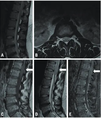
Since the patient’s laboratory and clinical findings were not specific to HSP, a repeat MRI study with contrast me-dium was performed despite the initial MRI showing no abnormality. Brain MRI showed no structural abnormalities. However, spinal MRI performed in August 2018 revealed a newly developed fusiform swelling at levels T12-L1 of the spinal cord with minimal homogeneous enhancement (Fig. 1A-C). The radiological findings led to astrocytoma as the suspected primary diagnosis. Laminectomy and tumor re-moval were performed. A grayish purple mass was observed during the surgery, with no definite dissection plane evident between the spinal cord and the mass (Fig. 1D, E).
The tumor was analyzed using hematoxylin-eosin staining, immunohistochemistry, and next-generation sequencing (NGS). The tumor showed positivity for placental alkaline phosphatase and CD117, and 95% positivity for Ki-67. NGS revealed loss of the KIT gene, with 25% of the germinoma being altered. Primary intramedullary spinal cord germino-Fig. 1. Clinical and radiological features of the patient. Weakness was
evident in the lower extremities. Magnetic resonance imaging (MRI) performed in December 2014 showed no abnormality in T2-weighted sagittal (A) and axial (B) images. Follow-up spinal MRI revealed fusiform swelling at levels T12-L1 (arrows) in sagittal unenhanced T1-weighted (C), T2-weighted (D), and gadolinium-enhanced T1-weighted (E) MRI imag-es, with minimal homogeneous enhancement.
A
C
B
D E
Fig. 2. Representative images of the surgical specimen. (A) Large epithelioid cells with abundant clear cytoplasm, round nuclei, and prominent nucle-oli were evident (hematoxylin-eosin, ×400). (B, C) Ancillary immunostaining revealed strong membrane positivity for placental alkaline phosphatase (B) and CD117 (c-kit; C) in the tumor cells (×400). (D) A grayish purple mass was found intraoperatively. There was no definite dissection plane between the spinal cord and the mass.
111
http://www.e-acn.org https://doi.org/10.14253/acn.2020.22.2.109
Haelim Kim, et al. Rare case of germinoma
ma was confirmed as the final diagnosis (Fig. 2).
The patient underwent chemotherapy with a combina-tion of etoposide, ifosfamide, mesna, and cisplatin. Radio-therapy was applied at 36 Gy in 20 fractions, and follow-up spinal MRI revealed shrinkage of the tumor. However, signifi-cant neurological deficits remained.
DISCUSSION
Primary intramedullary germinoma is rare and usually in-volves the lower thoracic part of the spinal cord, as in the present patient.2 A GCT looks similar to an astrocytoma in
MRI, which can be hypointense or isointense in T1-weighted images, hyperintense in T2-weighted images, and mod-erately enhanced in gadolinium-enhanced images. The initial imaging typically reveals a mass,2 but in rare cases it
can only show spinal cord atrophy, which is an early sign of intramedullary germinoma.3 These features make a
radiolog-ical diagnosis difficult, and hence biopsy or resection with pathohistological examination is necessary.4
HSP is often misdiagnosed since it may present with var-ious symptoms and the presence of gene mutation might not be verified. Therefore, clinicians must check for other treatable diseases before diagnosing HSP. An intracranial germinoma is a radiosensitive tumor whose cure rate is more than 90% with radiotherapy alone,5 which is also the
case for intramedullary germinoma.2 The response to
treat-ment is highly favorable, but an early diagnosis of germino-ma is critical for a good prognosis. It is particularly important to note that early imaging findings might not be apparent, as was the case in the present patient. Thus, if the diagnosis
is not clear, MRI follow-up is necessary. Although rare, germi-noma should be considered in the differential diagnosis of young adult patients with a tumor in the thoracic or thora-columbar spine.
Acknowledgements
We appreciate our patient for participating in this study. Conflict of Interest
The authors have no potential conflicts of interest to dis-close.
REFERENCES
1. Nikitović M, Grujičić D, Skender Gazibara M, Stanić D, Bokun J, Sa-rić M. Intramedullary spinal cord germinoma: a case report and review of literature. World Neurosurg 2016;95:392-398.
2. Loya JJ, Jung H, Temmins C, Cho N, Singh H. Primary spinal germ cell tumors: a case analysis and review of treatment paradigms. Case Rep Med 2013;2013:798358.
3. Madhukar M, Maller VG, Choudhary AK, Iantosca MR, Specht CS, Dias MS. Primary intramedullary spinal cord germinoma. J Neu-rosurg Pediatr 2013;11:605-609.
4. Aoyama T, Hida K, Ishii N, Seki T, Ikeda J, Iwasaki Y. Intramedullary spinal cord germinoma--2 case reports. Surg Neurol 2007;67:177-183.
5. Joo JH, Park JH, Ra YS, Im HJ, Koh KN, Khang SK, et al. Treatment outcome of radiation therapy for intracranial germinoma: adap-tive radiation field in relation to response to chemotherapy. Anti-cancer Res 2014;34:5715-5721.