43 www.i-mri.org
Intracystic Hemorrhage of an Arachnoid
Cyst: a Case with Prediagnostic Imaging
of an Intact Cyst
INTRODUCTION
Arachnoid cysts are benign lesions that are formed between the inner and outer layers of the arachnoid, and they account for 1% of all intracranial space-occupying lesions. Usually, arachnoid cysts are asymptomatic. However, they can be symptomatic in case of complication, such as intracystic hemorrhage or acute cyst expansion (1). There are several reported cases of arachnoid cysts with intracystic hemorrhage. However, cases with imaging study prior to a hemorrhagic event are very rare. We present a case of an incidentally diagnosed arachnoid cyst that developed intracystic hemorrhage combined with ipsilateral subdural hemorrhage (SDH) in a young male.
CASE REPORT
A 34-year-old man presented with intermittent, pulsatile headache for one week. The headache spontaneously resolved after a duration of 10-20 minutes, and there was a 1 hour interval between events. There was no other history of major trauma. The patient had headed a ball while playing football one month ago, but he did not develop any symptoms.
He had a previous brain CT scan about 8 months ago for health care work-up. It showed incidental cerebrospinal space (CSF) space widening in the left middle cranial fossa, suggesting an arachnoid cyst, Galassi type II (Fig. 1).
He had no other previous medical or neurological history. He was not taking any anticoagulant or antiplatelet agents. Initially, no abnormality was found on physical examination, neurological examination, or laboratory study.
This is an Open Access article distributed under the terms of the Creative Commons Attribution Non-Commercial License (http://creativecommons.org/licenses/ by-nc/4.0/) which permits unrestricted non-commercial use, distribution, and reproduction in any medium, provided the original work is properly cited. Received: November 8, 2020 Revised: November 29, 2020 Accepted: December 15, 2020 Correspondence to: Dongsoo Yoo, M.D. Department of Radiology, Dankook University Hospital, 201, Manghyang-ro, Dongnam-gu, Cheonan-si, Chungcheongnam-do 31116, Korea. Tel. +82-41-550-6907 Fax. +82-41-552-9674 E-mail: [email protected]
Copyright © 2021 Korean Society of Magnetic Resonance in Medicine (KSMRM)
iMRI 2021;25:43-46 https://doi.org/10.13104/imri.2021.25.1.43
Case Report
Arachnoid cysts are benign lesions that are formed between the inner and outer layers of the arachnoid, accounting for 1% of all intracranial space occupying lesions. Usually, arachnoid cysts are asymptomatic. It can be symptomatic in case of complication such as intracystic hemorrhage or acute cyst expansion. We present a case of incidentally prediagnosed arachnoid cyst which undergone intracystic hemorrhage combined with ipsilateral SDH in a young male.
Keywords: Arachnoid cyst; Intracystic hemorrage; Subdural hemorrhage
pISSN 2384-1095 eISSN 2384-1109
Donghyeon Kim, Dongsoo Yoo
www.i-mri.org 44
Intracystic Hemorrhage of an Arachnoid Cyst | Donghyeon Kim, et al.
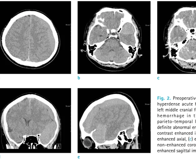
Brain CT was performed at the time of event considering the possibility of intracranial hemorrhage. CT scans showed a well-circumscribed, hyperdense, extraaxial lesion in the
left fronto-parieto-temporal lobe including the previously noted widened CSF space in the left middle cranial fossa, suggesting acute hematoma. A post-enhancement image showed no definite abnormal enhancement (Fig. 2).
He underwent trephination of the left parietal bone, without cisternostomy under the impression of acute subdural hemorrhage and intracystic hemorrhage of the arachnoid cyst. After opening the dura, the dark oil-colored, liquefied hematoma was evacuated.
The follow-up MRI 11 days after the operation showed fluid collection with a fluid-fluid level in the arachnoid cyst, probably a resolving status of intracystic hemorrhage (Fig. 3). He was discharged after 12 days of the operation without any medical or neurological complications.
On follow-up CT scan after 40 days and one year from discharge, the cyst was still evident with CSF density without any significant change in its size compared to that on preoperative CT (Fig. 4).
Fig. 1. Brain CT without contrast was performed for health care work-up. The axial image shows a Galassi type II left temporal arachnoid cyst.
Fig. 2. Preoperative brain CT shows hyperdense acute hematoma in the left middle cranial fossa and subdural hemorrhage in the left fronto-parieto-temporal lobe. There is no definite abnormal enhancement in the contrast enhanced image. (a, b) Non-enhanced axial, (c) enhanced axial, (d) non-enhanced coronal, and (e) non-enhanced sagittal images.
a b c
45 www.i-mri.org
https://doi.org/10.13104/imri.2021.25.1.43
Fig. 3. Follow-up brain MR after 11 days from the operation shows a fluid-fluid level in the arachnoid cyst, suggesting resolving state of intracystic hemorrhage in the left cranial fossa. There is a thin linear septa-like structure with T1 low, T2 high signal intensity, and enhancement crossing the hemorrhagic cystic lesion. It would be an interface between the arachnoid cyst and subdural hematoma or a possible postoperative finding. It is not visible on the previous CT image probably due to a limitation of the modality. (a) T1-weighted, (b) T2-weighted, (c) T2-FLAIR, (d) T2 fast-field-echo, and (e) T1-weighted contrast-enhanced images.
a b c
d e
Fig. 4. Follow-up CT image after 40 days (a), and one year (b). Arachnoid cyst with resolved intracystic hemorrhage is noted in the left cranial fossa with CSF density.
www.i-mri.org 46
Intracystic Hemorrhage of an Arachnoid Cyst | Donghyeon Kim, et al.
DISCUSSION
Arachnoid cysts are benign lesions that are formed between the inner and outer layers of the arachnoid, and they account for 1% of all intracranial space-occupying lesions. Their origin can be explained by a meningeal maldevelopment theory: the temporal and frontal arachnoid coverings fail to merge when the sylvian fissure is formed, thereby creating a noncommunicating fluid compartment entirely surrounded by arachnoid membranes (1).
They are found in the middle cranial fossa in 50-65% of cases, with a predilection for the left side and male gender. Computed tomography and MR imaging have increased the detection of incidental asymptomatic arachnoid cysts as in our case. Although generally asymptomatic, they can very rarely become symptomatic because of complication, such as acute cyst expansion or intracystic or subdural hemorrhage following head trauma (1, 2).
According to Wester and Helland (3), chronic SDH was found in 4.6% of the total 241 patients; all of them harboring a temporal arachnoid cyst. For temporal cysts, the hematoma frequency was 6.5%. Hematomas occurred equally frequently in individuals with small, middle-sized, and large temporal cysts.
We presented a rare case of incidentally diagnosed arachnoid cyst that developed an intracystic hemorrhagic change. There are several reported cases of arachnoid cysts with intracystic hemorrhage (2, 4-7), and other very rare cases with an imaging study prior to the hemorrhagic event (8, 9).
It is well known that intracranial hemorrhage can occur more easily with mild trauma or even without trauma in patients with an arachnoid cyst. In our case, it can be controversial whether heading a ball one month ago contributed to hemorrhage due to minor trauma. The postoperative follow-up CT scan demonstrated complete resolution of hemorrhage only after trephination.
Therefore, we suggest that in case of a previously
diagnosed arachnoid cyst, it is important to be aware of the possibility of intracranial hemorrhage even without trauma or with suspicious mild head trauma.
REFERENCES
1. Wester K. Peculiarities of intracranial arachnoid cysts: location, sidedness, and sex distribution in 126 consecutive patients. Neurosurgery 1999;45:775-779
2. Patel AP, Oliverio PJ, Kurtom KH, Roberti F. Spontaneous subdural hematoma and intracystic hemorrhage in an arachnoid cyst. Radiol Case Rep 2009;4:298
3. Wester K, Helland CA. How often do chronic extra-cerebral haematomas occur in patients with intracranial arachnoid cysts? J Neurol Neurosurg Psychiatry 2008;79:72-75 4. Kim SJ, Baek HJ, Moon JI, et al. Nontraumatic intracystic
hemorrhage of arachnoid cyst: CT and MR findings. Investig Magn Reson Imaging 2016;20:120-122
5. Holanda Ferreira TS, Malveira LRC, Gomes Neto A, Spontaneous subdural and intracavitary hemorrhage of temporal arachnoid cyst in an adult patient. Interdiscip Neurosurg 2021;23:100830
6. Johnson R, Amine A, Farhat H. Spontaneous acute subdural hematoma associated with arachnoid cyst and intra-cystic hemorrhage. Cureus 2018;10:e3383
7. Kaszuba MC, Tan LA, Moftakhar R, Kasliwal MK. Nontraumatic subdural hematoma and intracystic hemorrhage associated with a middle fossa arachnoid cyst. Asian J Neurosurg 2018;13:116-118
8. Furtado LMF, Costa Val Filho JA, Ferreira RI, Mariano I. Intracranial arachnoid cyst rupture after mild TBI in children: have we underestimated this risk? BMJ Case Rep 2019;12:e228790
9. Adin ME, Yildiz MS, Deniz MA, Behzadi AH, Mata-Mbemba D. Arachnoid cysts with spontaneous intracystic hemorrhage and associated subdural hematoma: report of management and follow-up of 2 cases. Radiol Case Rep 2018;13:516-521