저작자표시-비영리-변경금지 2.0 대한민국 이용자는 아래의 조건을 따르는 경우에 한하여 자유롭게 l 이 저작물을 복제, 배포, 전송, 전시, 공연 및 방송할 수 있습니다. 다음과 같은 조건을 따라야 합니다: l 귀하는, 이 저작물의 재이용이나 배포의 경우, 이 저작물에 적용된 이용허락조건 을 명확하게 나타내어야 합니다. l 저작권자로부터 별도의 허가를 받으면 이러한 조건들은 적용되지 않습니다. 저작권법에 따른 이용자의 권리는 위의 내용에 의하여 영향을 받지 않습니다. 이것은 이용허락규약(Legal Code)을 이해하기 쉽게 요약한 것입니다. Disclaimer 저작자표시. 귀하는 원저작자를 표시하여야 합니다. 비영리. 귀하는 이 저작물을 영리 목적으로 이용할 수 없습니다. 변경금지. 귀하는 이 저작물을 개작, 변형 또는 가공할 수 없습니다.
I
ABSTRACT
Skin is exposed to harmful environmental such as air pollution which including
various types of particulate matters (PMs). These atmospheric PMs have harmful
effect on human through increase of the reactive oxygen species (ROS). It has been
reported that ROS induced skin aging via generation of matrix metalloproteinases
(MMPs) which causes skin aging through degradation of collagen. This study
investigated the effect of fermented fish oil (FFO), which derived from mackerel, in
PM
2.5(particulate with a dimeter of < 2.5 µm)-induced skin aging in human
keratinocyte. FFO inhibited PM
2.5-induced intracellular ROS and MMPs including
MMP-1, MMP-2, and MMP-9. In addition, FFO significantly abrogated the
intracellular Ca
2+level in PM
2.5-treated cells. Furthermore, FFO blocks PM
2.5-induced MAPKs/AP-1 pathway. In conclusion, FFO has anti-aging effect on PM
2.5-induced skin aging on human keratinocyte.
Keyword: Particulate matters 2.5, Matrix metalloproteinases, Fermented fish oil,
II
CONTENTS
ABSTRACT………..Ⅰ
CONTENTS………..Ⅱ
LIST OF FIGURES………...………Ⅳ
1. Introduction………..……….……….1
2. Materials and methods………..……….…….3
2-1.
Cell culture and treatment2-2. Detection of intracellular ROS
2-3. Detection of β-galactosidase activity
2-4. MMP-1 activity
2-5. Reverse transcription –PCR (RT-PCR)
2-6. Western blot analysis
2-7. Measurement of Ca2+ level
2-8. Chromatin immunoprecipitation (ChIP) assay
III
3. Results……….………7
3-1. Effect of FFO on PM2.5-induced intracellular ROS 3-2. PM2.5-induced keratinocyte senescence 3-3. Effect of FFO on PM2.5-induced MMP-1 activation and MMPs expression 3-4. Effect of FFO on PM2.5-induced MAPKs and intracellular Ca2+ level 3-5. Effect of FFO on PM2.5-induced transcription factor activator protein 1 (AP-1) expression 4. Discussion
……….………15
5. References..
……….………...…...17
6. Abstract in Korean………...20
IV
LIST OF FIGURES
Figure 1. Scavenging effect of FFO on PM2.5-induced intracellular ROS
Figure 2. PM2.5-induced keratinocyte senescence
Figure 3. Effect of FFO on PM2.5-induced MMP-1 activation and MMPs expression
Figure 4.Effect of FFO on PM2.5-induced MAPKs and intracellular Ca 2+
level
Figure 5. Effect of FFO on PM2.5-induced transcription factor activator protein 1 (AP-1)
expression
1
1. Introduction
Since reactive oxygen species (ROS) have unpaired electrons and unstable bounds, it is able to lead cellular damage (de Jager et al. 2017; Ryu et al. 2018) and to regulate transcription factors such as activator protein 1 (AP-1), and nuclear factor kB (NF-kB) (Ranneh et al. 2017). The cellular ROS can be accumulated by exogenous sources like air pollutions (Ranneh et al. 2017).
Skin is the largest organ in body and acts as the first defense barrier against harmful stimuli such as ultraviolet (UV) and air pollution including particulate matters (PMs). PMs can be classified as ultrafine PM (particulate with a dimeter of <0.1 µm, PM0.1), fine PM
(particulate with a dimeter of <2.5 µm, PM2.5), and coarse PM (particulate with a dimeter of
<10 µm, PM10) which depending on the particle size (Kim et al. 2015). PMs lead to the
development of various skin diseases such as skin aging, alopecia and skin cancer through inducing oxidative stress (Kim et al. 2016). In addition, PMs induced oxidative stress via production of ROS and increase matrix metalloproteinases (MMPs) (Kim et al. 2016; Seok et al. 2018). MMPs including MMP-1, MMP-2, and MMP-9 caused skin aging through the degradation of collagen (Chaiprasongsuk et al. 2017; Kim et al. 2017).
ROS generations have been reported to affect skin aging by increasing the expression of MMP-1 in keratinocyte (Leiros et al. 2017). Several studies were reported that UVB-induced ROS caused skin photoaging via generation of MMP-1 in human keratinocytes and dermal fibroblasts (Kim et al. 2018; Xuan et al. 2017). Therefore, it is important to find an effective antioxidant to prevent skin aging.
Oxidative stress stimulates mitogen-activated protein kinases (MAPKs) signaling pathway, which affect the regulation of transcription factor AP-1 activity (Pittayapruek et al. 2016).
2
Activation (phosphorylation) c-Jun and c-Fos can be comprised of homodimer or heterodimer to bind to AP-1 binding sites in the promoter region of target genes to promote gene transcription (Lu et al. 2016) such as MMPs transcription (Kim et al. 2017).
Previous study was reported that fermented fish oil (FFO) has antioxidant effects and protective effect against UVB-induced oxidative damage (Park et al. 2018). But the effect of FFO in PM2.5-induced skin aging is poorly understood. Therefore, this study demonstrates
3
2. Materials and methods
2-1. Cell culture and treatment
The HaCaT human keratinocyte (CLS Cell Lines Service GmbH, Eppelheim, Germany) were cultured in DMEM medium (Gibco, Life Technologies Co., Grand Island, NY, USA) supplemented with 10% fetal bovine serum and antibiotics (100 units/mL penicillin, 100 μg/mL streptomycin and 0.25 µg/mL amphotericin B) (Gibco, Life Technologies Co.) at 37°C in an incubator with a humidified atmosphere of 5% CO2. Cell were treated to 50
μg/mL of diesel particulate matter NIST 1650b (PM2.5) (Sigma-Aldrich Chemical Company,
St. Louis, MO, USA) and 20 μg/mL of FFO. Preparation of PM2.5 was described previous
study (Piao et al. 2018) and preparation of FFO was described previous study (Park et al. 2018).
2-2. Detection of intracellular ROS
To detect intracellular ROS in HaCaT cells, cells were seeded in plates at a density of 1.0×10⁵ cells/well, cultured for 16 h, and treated 20 μg/mL of FFO, 50 μg/mL of PM2.5. After
30 min later, 50 μM 2',7'-dichlorodihydrofluorescein diacetate (DCF-DA, Molecular Proves, Eugene, OR, USA) solution was added. DCF fluorescence was measured using a BD LSRFortessa flow cytometry (PerkinElmer, Waltham, MA, USA) and images were collected by using a FV1200 laser scanning confocal microscope (Olympus, Tokyo, Japan).
2-3. Detection of β-galactosidase activity
To detect the cell senescence, cells were seeded in plates at a density of 1.0×10⁵ cells/ml. After 16 h of incubation period at 37C, cells were treated 20 μg/mL of FFO, 50 μg/mL of PM2.5. After 24 h later, 2 μM SPiDER-βGal solution (Dojindo Molecular Technologies, Inc.,
4
medium containing DAPI to label nuclei. SPiDER-βGal fluorescence was measured by using a BD LSRFortessa flow cytometry and images were collected using a FV1200 laser scanning confocal microscope (Olympus).
2-4. MMP-1 activity
The MMP-1 activity was measured by using the Fluorokine® E human active MMP-1 fluorescent assay kit (R&D Systems Inc., Minneapolis, MN, USA). HaCaT cells were seeded on a 60 mm culture dish at 1.0×10⁵ cells/mL. After 16 h of incubation period at 37C, cells were treated with 20 μg/mL of FFO and after 1 h, cell were treated with 50 μg/mL of PM2.5.
Then MMP-1 activity was assessed according to the manufacture’s instruction. Fluorescence was measured by using Spectra Max i3x microplate reader (Molecular devices, San Jose, CA, USA).
2-5. Reverse transcription –PCR (RT-PCR)
Cells were seeded at 1.5×10⁵ cells/mL and after 16 h later cell were treated with 20 μg/mL of FFO, 50 μg/mL of PM2.5. After 24 h, we isolated total RNA from cells using the
easy-BLUE™ total RNA extraction kit (iNtRON Biotechnology Inc., Seongnamsi, Korea). And then the cDNA was amplified by using reverse transcription reaction buffer, primers, dNTPs, and Taq DNA polymerase in a final volume of 20 μL. The amplified products were mixed with blue/orange 6X loading dye, resolved by electrophoresis on a 1% agarose gel which stained with RedSafe™ nucleic acid staining solution (iNtRON Biotechnology Inc., Seongnamsi, Korea), and photographed under UV light using Image Quant™ TL analysis software (Amersham Biosciences, Uppsala, Sweden). The PCR conditions were as followers: initial denaturation at 94°C for 5 min and then followed by 30 cycles of 94°C for 30 s, 55°C for 30 s, and 72°C for 1 min. The primers were used in this study: human MMP-1, forward (5'-GGAGGAAATCTTGCTCAT-3′) and reverse (5′-CTCAGAAAGAGCAGCATC-3′);
5
human GAPDH, forward TCAAGTGGGGCGATGCTGGC-3′) and reverse (5′-TGCCAGCCCCAGCGTCAAAG-3′).
2-6. Western blot analysis
The protein lysates (30 μg per lane) were electrophoresed on 12% SDS-polyacrylamide gels. Then transferred to a nitrocellulose membrane which was incubated with the primary antibodies and incubated with HRP-conjugated secondary antibodies (Invitrogen, Carlsbad, CA, USA). Next, membranes were exposed to a Western blotting detection kit (GE Healthcare Life Sciences, Little Chalfont, UK) for to detect the protein bands and then exposed to X-ray film. Primary antibodies were used in this study: MMP-1 (Cusabio Tecnology LLC., Houston, TX, USA), MMP-2 (Abcam, Cambridge, UK), MMP-9 (Abcam), phospho-c-Jun (Cell Signaling Technology, Danvers, MA, USA), c-Fos (Cell Signaling Technology), phospho-SEK (Cell Signaling Technology), phospho-MEK (Cell signaling Technology), ERK (Santa Cruz Biotechnology, Santa Cruz, CA, USA), phospho-JNK (Cell Signaling Technology), Actin (Sigma-Aldrich Chemical Company).
2-7. Measurement of Ca2+ level
To detect Ca2+ level, cells were seeded in plates at a density of 1.0×10⁵ cells/well, cultured for 16 h, and treated 20 μg/mL of FFO, 50 μg/mL of PM2.5. After 24 h later, 5 μM
Fluo-4-AM (Molocular Probes, Eugene, OR, USA) solution was added. Fluo-4-Fluo-4-AM fluorescence was measured by using a BD LSRFortessa flow cytometry and images were collected by using a FV1200 laser scanning confocal microscope (Olympus).
2-8. Chromatin immunoprecipitation (ChIP) assay
6
(Cell Signaling Technology). HaCaT cells were seeded at 1.5×10⁵ cells/mL and after 16 h, cells were treated with 20 μg/mL of FFO, 50 μg/mL of PM2.5. All processes were performed
according to the instructions. The following antibody and primers were used in this study: c-Jun antibody (Invitrogen), MMP-1 gene promoter (-67 to +94 of the MMP-1 gene sequence
from the transcription starting site, Bionics) were designed as sense
5′-CCTCTTGCTGCTCCAATATC-3′ and antisense 5′-TCTGCTAGGAGTCACCATTTC-3′. The PCR products were separated on 1% agarose gel, DNA bands were photographed under UV light using Image Quant™ TL analysis software (Amersham Biosciences).
2-9. Statistical analysis
All data were performed in triplicate and all values are expressed as the mean ± standard error of the means. This study used Tukey’s test analysis to determine the statistical significance of differences between means. p < 0.05 were considered statistically significant.
7
3. Results
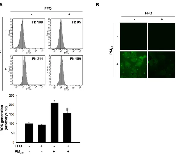
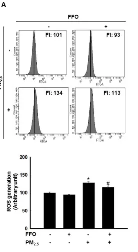
3-1. Effect of FFO on PM2.5-induced intracellular ROS
Because ROS has been reported to affect skin aging via generation of MMPs (Leiros et al. 2017), this study measured generation of intracellular ROS by DCF-DA fluorescence dye. As shown by flow cytometry data, PM2.5-treated cell induced ROS and pretreatment of FFO
reduced PM2.5-indeuced ROS in HaCaT cell and which was confirmed by using confocal
microscopy (Figure 1A and B).
Figure 1. Scavenging effect of FFO on PM2.5-induced intracellular ROS. Intracellular ROS
8
< 0.05, #p < 0.05 compared to untreated cells and PM2.5-treated cells, respectively.
3-2. PM2.5-induced keratinocyte senescence
Next, this study measured β-galactosidase activity for to detect of HaCaT cell senescence using flow cytometry and confocal microscopy after SPiDER-βGal staining. PM2.5-treated
cells increased β-galactosidase activity in the cytosol and FFO-treated cells decreased PM2.5
-induced β-galactosidase activity (Figure 2A and B).
Figure 2. PM2.5-induced keratinocyte senescence. (A) The β-galactosidase activity was
9
0.05, #p < 0.05 compared to untreated cells and PM2.5-treated cells, respectively.
3-3. Effect of FFO on PM2.5-induced MMP-1 activation and MMPs expression
Treatment of PM2.5 significantly increased the activation of MMP-1 in after 6, 12, 24 h
(Figure 3A) and FFO pretreatment decreased the PM2.5-induced activation of MMP-1
(Figure 3B). Expression of MMP-1 mRNA and protein levels are also increased in PM2.5
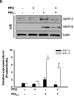
-treatment cells and decreased in FFO-pre-treatment cells (Figure 3C and D). Since MMP-2 and MMP-9 are also reported to be involved in skin aging through degradation of collage, I analyzed the expression of MMP-2 and MMP-9 protein levels. MMP-2 and MMP-9 protein level also increased by PM2.5 treatment and decreased by FFO pretreatment (Figure 3E).
11
Figure 3. Effect of FFO on PM2.5-induced MMP-1 activation and MMPs expression. (A)
The MMP-1 activity of PM2.5-treated cells at the indicated times and (B) The MMP-1
activity of FFO and PM2.5 treated cells was determined using the human active MMP-1
fluorescent assay kit. *p < 0.05, #p < 0.05 compared to untreated cells and PM2.5-treated cells,
respectively. (C) Expression level of MMP-1 was analyzed by western blot. Actin was used to loading control. *p < 0.05, compared to untreated cells. (D) The mRNA level of MMP-1 and protein level of MMP-1 were analyzed by RT-PCR and western blot, respectively. GAPDH and actin were used to loading control. *p < 0.05, #p < 0.05 compared to untreated cells and PM2.5-treated cells, respectively. (E) Expression level of MMP-2 and MMP-9
were analyzed by western blot. Actin was used to loading control. *p < 0.05, #p < 0.05 compared to untreated cells and PM2.5-treated cells, respectively.
3-4. Effect of FFO on PM2.5-induced MAPKs and intracellular Ca 2+
level
MAPKs, which enhance expression of MMP-1, are activated by an increase in the intracellular Ca2+ level (Liu et al. 2010). As shown western blot data, PM2.5 treatment
12
decreased. Furthermore, PM2.5 treatment induced the activation (phosphorylation) MAPK
kinase (MEK) 1/2 and SAPK/ERK kinase (SEK) 1 (Figure 4A). In addition, PM2.5
significantly increased intracellular Ca2+ level and FFO decreased PM2.5-induced Ca 2+
level (Figure 4B and C).
14
Figure 4. Effect of FFO on PM2.5-induced MAPKs and intracellular Ca 2+
level. (A) Expression level of p-JNK, p-ERK, p-MEK, and p-SEK by western blot analysis. Actin was used to loading control. *p < 0.05, #p < 0.05 compared to untreated cells and PM2.5-treated
cells, respectively. (B) Intracellular Ca2+ level was detected by flow cytometry and (C) confocal microscopy after Flou-4-AM staining. *p < 0.05, #p < 0.05 compared to untreated cells and PM2.5-treated cells, respectively.
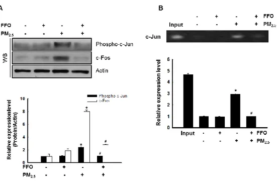
3-5. Effect of FFO on PM2.5-induced transcription factor activator protein 1 (AP-1) expression
The nuclear transcription factor AP-1 regulated by MAPKs, which increase MMP expression (Kim et al. 2018). Activation of MAPKs results in the heterodimeratization of c-Jun/c-Fos and the formation of the AP-1 complex (Kim et al. 2017). As shown Figure 4, PM2.5 treatment increased MAPKs and intracellular Ca
2+
level. Next, I determine c-Fos, phopho-c-Jun level using by western blot analysis. FFO significantly decreased PM2.5
-indeced phopho-c-Jun and c-Fos level (Figure 5A). In addition, FFO reduced the PM2.5
15
Figure 5. Effect of FFO on PM2.5-induced transcription factor activator protein 1 (AP-1)
expression. (A) Expression level of phospho-c-Jun and c-Fos by western blot analysis. Actin was used to loading control. *p < 0.05, #p < 0.05 compared to untreated cells and PM2.5
-treated cells, respectively. (B) AP-1 binding to the MMP-1 promoter was assessed by ChIP assay. *p < 0.05, #p < 0.05 compared to untreated cells and PM2.5-treated cells, respectively.
16
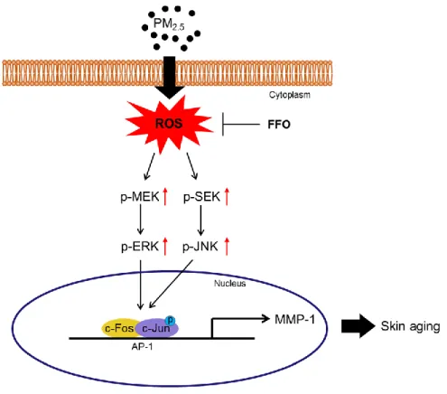
Figure 6. Schematic diagram of effect of FFO on PM2.5-induced skin aging. Expose of PM2.5
increased intracellular ROS and induced skin aging in HaCaT cells. The mackerel derived FFO has anti-aging effect against PM2.5-induced skin aging through reduced intracellular
17
4. Discussion
Skin is the largest organ in body which acts as the first defense barrier against harmful stimuli. Therefore, skin is always exposed harmful environment including PMs. In several studies, PM2.5 has been reported to have harmful effects such as inflammatory skin diseases,
skin aging, and damage of respiratory system through generation of intracellular ROS (Kim et al. 2016; Romani et al. 2018; Xing et al. 2016).
The accumulation of ROS has been reported that induced skin aging through the expression of MMPs such as MMP-1, MMP-2, and MMP-9 (Subedi et al. 2017). Therefore, it is important to find an effective antioxidant to prevent skin aging.
Previous study demonstrated that mackerel derived FFO has directly ROS scavenging effect and protective effect on UVB-induced oxidative damage (Park et al. 2018). The present study focused on the effect of FFO which derived from mackerel against PM2.5
-induced skin aging.
First, this study measured generation of intracellular ROS after PM2.5 treatment. Induction
of intracellular ROS by PM2.5 treatment significantly reduced pretreatment of FFO (Figure
1A and B). Since the generation of ROS cause skin aging, this study detected keratinocyte senescence using β-galactosidase (Yoshimoto et al. 2018). PM2.5 treatment increased
β-galactosidase activity and FFO significantly reduced PM2.5-induced β-galactosidase activity
(Figure 2A and B). Next, because of MMPs caused skin aging through degradation of collagens (Chaiprasongsuk et al. 2017; Kim et al. 2017), this study detected activity of MMP-1 and expression level of MMP-1, MMP-2, and MMP-9. FFO has effect on PM2.5
-induced MMPs (Figure 3). These results indicate that PM2.5 induced intracellular ROS and
18
scavenging ROS.
ROS activates the MAPK signaling pathway and activation of MAPKs induced various transcription factors such as AP-1 and NF-κB (Pittayapruek et al. 2016; Sun et al. 2017). As a result of translocation of the activated AP-1, a heterodimer composed of c-Jun and c-Fos, MMPs were synthesized (Hwang et al. 2011; Kim et al. 2013). As shown Figure 4A, ERK and JNK activated by PM2.5 treatment and reduced by FFO pretreatment. MEK and SEK, the
upstream of ERK and JNK respectively, also increased by PM2.5 treatment and decreased by
FFO pretreatment (Figure 4A). In addition, intracellular Ca2+ level, regulating MAPKs, was significantly increased in PM2.5-treated cells. However, FFO was decreased the PM2.5
-induced Ca2+ level (Figure 4B and C). Furthermore, phospho-c-Jun and c-Fos levels were increased in PM2.5-treated cells and decreased in FFO-pretreatment cells (Figure 5A). PM2.5
-induced AP-1 binding to the MMP-1 promoter also reduced by FFO-pretreatment cells (Figure 5B). These results demonstrate that FFO can block PM2.5-induced MAPKs/AP-1
pathway in human keratinocyte.
In conclusion, mackerel derived FFO has anti-aging effect against PM2.5-induced skin
19
5. References
Chaiprasongsuk, A.; Lohakul, J.; Soontrapa, K.; Sampattavanich, S.; Akarasereenont, P.; Panich, U. Activation of Nrf2 reduces UVA-mediated MMP-1 upregulation via MAPK/AP-1 signaling cascades: the photoprotective effects of sulforaphane and hispidulin. J Pharmacol Exp Ther. 2017, 360, 388-398.
de Jager, T.L.; Cockrell, A.E.; Du Plessis, S.S. Ultraviolet Light Induced Generation of Reactive Oxygen Species. Adv Exp Med Biol. 2017, 996, 15-23.
Hwang, Y.P.; Oh, K.N.; Yun, H.J.; Jeong, H.G. The flavonoids apigenin and luteolin suppress ultraviolet A-induced matrix metalloproteinase-1 expression via MAPKs and AP-1-dependent signaling in HaCaT cells. J Dermatol Sci. 2011, 61, 23-31.
Kim, J.; Kim, M.B.; Yun, J.G.; Hwang, J.K. Protective effects of standardized siegesbeckia glabrescens extract and its active compound kirenol against UVB-induced photoaging through inhibition of MAPK/NF-κB pathways. J Microbiol Biotechnol. 2017, 27,242-250.
Kim, J.M.; Kim, S.Y.; Noh, E.M.; Song, H.K.; Lee, G.S.; Kwon, K.B.; Lee, Y.R. Reversine inhibits MMP-1 and MMP-3 expressions by suppressing of ROS/MAPK/AP-1 activation in UV-stimulated human keratinocytes and dermal fibroblasts. Exp Dermatol. 2018, 27, 298-301.
Kim, K.E.; Cho, D.; Park, H.J. Air pollution and skin diseases: Adverse effects of airborne particulate matter on various skin diseases. Life Sci. 2016, 152, 126-134.
Kim, K.H.; Kabir, E.; Kabir S. A review on the human health impact of airborne particulate matter. Environ Int. 2015, 74, 136-143.
Kim, M.S.; Oh, G.H.; Kim, M.J.; Hwang, J.K. Fucosterol inhibits matrix metalloproteinase expression and promotes type-1 procollagen production in UVB-induced HaCaT cells.
20
Photochem Photobiol. 2013, 89, 911-918.
Leirós, G.J.; Kusinsky, A.G.; Balañá, M.E.; Hagelin, K. Triolein reduces MMP-1 upregulation in dermal fibroblasts generated by ROS production in UVB-irradiated keratinocytes. J Dermatol Sci. 2017, 85, 124-130.
Liu, W.H.; Chang, L.S. Caffeine induces matrix metalloproteinase-2 (MMP-2) and MMP-9 down-regulation in human leukemia U937 cells via Ca2+/ROS-mediated suppression of ERK/c-fos pathway and activation of p38 MAPK/c-jun pathway. J Cell Physiol. 2010, 224, 775–785.
Lu, J.; Guo, J.H.; Tu, X.L.; Zhang, C.; Zhao, M.; Zhang, Q.W.; Gao, F.H. Tiron inhibits UVB-induced AP-1 binding sites transcriptional activation on MMP-1 and MMP-3 promoters by MAPK signaling pathway in human dermal fibroblasts. PLoS One. 2016, 11, e0159998.
Park, J. E.; Hyun, Y. J.; Piao, M. J.; Kang, K. A.; Ryu, Y. S.; Shilnikova, K.; Zhen, A.X.; Ahn, M.J.; Ahn, Y.S.; Koh, Y.S.; Kang, H.K.; Hyun, J.W. Mackerel-derived fermented fish oil protects skin against UVB-induced cellular damage by inhibiting oxidative stress. J Funct Foods. 2018, 46, 147-158.
Piao, M.J.; Ahn, M.J.; Kang, K.A.; Ryu, Y.S.; Hyun, Y.J.; Shilnikova, K.; Zhen, A.X.; Jeong, J.W.; Choi, Y.H.; Kang, H.K.; Koh, Y.S.; Hyun, J.W. Particulate matter 2.5 damages skin cells by inducing oxidative stress, subcellular organelle dysfunction, and apoptosis. Arch Toxicol. 2018, 92, 2077-2091.
Pittayapruek, P.; Meephansan, J.; Prapapan, O.; Komine, M.; Ohtsuki, M. Role of matrix metalloproteinases in photoaging and photocarcinogenesis. Int J Mol Sci. 2016, 17, E868.
Ranneh, Y.; Ali, F.; Akim, A. M.; Hamid, H. A.; Khazaai, H.; Fadel, A. Crosstalk between reactive oxygen species and pro-inflammatory markers in developing various chronic
21
diseases: a review. Appl Biol Chem. 2017, 60, 327-338.
Romani, A.; Cervellati, C.; Muresan, X.M.; Belmonte, G.; Pecorelli, A.; Cervellati, F.; Benedusi, M.; Evelson, P.; Valacchi, G. Keratinocytes oxidative damage mechanisms related to airbone particle matter exposure. Mech Ageing Dev. 2018, 172, 86-95.
Ryu, O.; Park, B.K.; Bang, M.; Cho, K.S.; Lee, S.H.; Gonzales, E.L.T.; Yang, S.M.; Kim, S.; Eun, P.H.; Lee, J.Y.; Kim, K.B.; Shin, C.Y.; Kwon, K.J. Effects of several cosmetic preservatives on ROS-dependent apoptosis of rat neural progenitor cells. Biomol Ther (Seoul). 2018, 26, 608-615.
Seok, J.K.; Lee, J.W.; Kim, Y.M.; Boo, Y.C. Punicalagin and (-)-Epigallocatechin-3-gallate rescue cell viability and attenuate inflammatory responses of human epidermal keratinocytes exposed to airborne particulate matter PM10. Skin Pharmacol Physiol. 2018, 31, 134-143.
Subedi, L.; Lee, T.H.; Wahedi, H.M.; Baek, S.H.; Kim, S.Y. Resveratrol-Enriched Rice Attenuates UVB-ROS-Induced Skin Aging via Downregulation of Inflammatory Cascades. Oxid Med Cell Longev. 2017, 2017, 8379539.
Sun, Z.; Park, S.Y.; Hwang, E.; Zhang, M.; Seo, S.A.; Lin, P.; Yi, T.H. Thymus vulgaris alleviates UVB irradiation induced skin damage via inhibition of MAPK/AP-1 and activation of Nrf2-ARE antioxidant system. J Cell Mol Med. 2017, 21, 336-348.
Xing, Y.F.; Xu, Y.H.; Shi, M.H.; Lian, Y.X. The impact of PM2.5 on the human respiratory system. J Thorac Dis. 2016, 8, 69-74.
Xuan, S.H.; Park, Y.M.; Ha, J.H.; Jeong, Y.J.; Park, S.N. The effect of dehydroglyasperin C on UVB-mediated MMPs expression in human HaCaT cells. Pharmacol Rep. 2017, 69, 1224-1231.
22
repetitive UVA irradiation: induction of characteristic markers of senescence and its prevention by PAPLAL with potent catalase activity. Photochem Photobiol. 2018, 94, 438-444.
23
6. Abstract in Korean
피부는 미세먼지(particulate matters, PMs)를 비롯한 대기오염과 같은 유해한 환경에 항상 노출된다. 이러한 미세먼지는 활성 산소 종(reactive oxygen species, ROS)의 증가를 통해 인체에 해로운 영향을 미친다. 세포 내 ROS는 콜라겐을 분해하는 단백질 분해효소(matrix metalloproteinases)인 MMP-1을 생성할 수 있고 그로 인해 피부 노화를 유발 할 수 있다고 알려져 있다. 이 연구는 고등어에서 추출한 발효 어유(FFO)가 인간각질세포에서 초미세먼지(PM2.5)로 유도되는 피부노화에 미치는 영향을 조사하였다. PM2.5를 처리한 세포에서 ROS증가와 노화인자가 증가하는 것을 확인하였고 FFO 전처리 그룹에서 PM2.5로부터 유도되는 ROS 증가, 노화인자 증가가 감소되는 것을 확인 할 수 있었다. 또한, 콜라겐분해를 통해 피부노화를 일으킨다고 알려져 있는 MMP-1의 활성도와 mRNA, 단백질 발현 수준 역시 PM2.5 처리로 증가되었지만 FFO전처리로 감소되는 것을 볼 수 있었다. 또한 FFO는 MMP-1의 발현을 조절하는데 영향을 주는 세포 내 Ca2+ 수준과 MAPKs/AP-1 경로를 차단 하는 것을 보여준다. 따라서 이러한 결과는 고등어로부터 유래된 FFO는 PM2.5로 유도되는 피부노화에 있어서 세포 내 ROS를 소거함으로써 피부노화에 효과를 가진다는 것을 시사한다.
24
7. Acknowledgement
긴 방황 끝에 다시 돌아온 저를 응원해 주시고 학업에 열중 할 수 있도록 항상 격려를 아끼지 않으시고 열정과 책임감을 가지고 지도해 주신 현진원 교수님과 강희경 교수님에게 감사 드립니다. 덕분에 석사과정 2년이라는 시간 동안 학업적인 면뿐만 아니라 다른 부분에서도 많이 배울 수 있었고 제 스스로 발전 할 수 있는 시간을 가질 수 있었습니다. 다시 한번 감사의 말씀 드립니다. 그리고 바쁘신 와중에도 불구하고 저의 학위 논문 심사에 참여해주시고 아낌없이 조언해 주신 박덕배 교수님께도 감사의 말씀 드립니다. 수업시간에 부족한 저에게 하나라도 더 일깨워 주시기 위해 꼼꼼하고 날카로운 질문과 조언을 아끼지 않았던 조문제 교수님, 유은숙 교수님, 고영상 교수님께도 감사 드립니다. 그리고 가장 중요한 생화학 교실원들에게도 감사함을 전합니다. 항상 변함없이 같은 자리에서 저를 이끌어주고 많이 조언해 주었던 박미경 박사님, 지금은 러시아로 잠시 떠나갔지만 유창한 한국어로 즐거움을 주었던 크리스티나 (Kristina Shilnikova), 돌아와서 박사과정 무사히 마무리 짓길 바랄게요. 같이 입학하여 수업도 같이 듣고, 힘들 때 서로 조언하고 격려하며 추억이 가장 많은 진오현(Ao Xuan Zhen), 비록 나는 학위과정이 끝나서 먼저 떠나지만 너는 똑똑하고 성실하니깐 남은 기간도 무사히 해낼꺼라고 믿어. For the last 2 years, I will never forget all the memories with you. And I hope you will always be happy with your BF. Fighting! 가장 바쁘고 정신 없을 때 들어와서 고생해 준 연구조교 송혜원 선생님과 행정조교 박은희 선생님도 수고 하셨습니다.25
마지막으로 항상 저를 믿고 제 옆에서 묵묵히 응원을 보내준 친구들과 가족 모두에게 감사의 말씀 드립니다.
이 모든 분들께 계속해서 성장하고 발전하는 모습으로 보답하겠습니다. 감사합니다.