2008;22:226-231 □
원 저
□226
책임저자:김유선, 서울시 서대문구 신촌동 134번지 연세대학교 의과대학 외과학교실, 120-752 Tel: 02-2228-2115, Fax: 02-313-8289 E-mail: [email protected] 접수일 : 2008년 9월 30일, 게재승인일 : 2008년 11월 10일신장 이식 후 림프증식질환의 임상양상: 단일기관 25년간의 경험
연세대학교 의과대학 외과학교실
1및 장기이식연구소
2이정준
1ㆍ주동진
1ㆍ김수진
1ㆍ허규하
1ㆍ주만기
1ㆍ김명수
1,2ㆍ김순일
1,2ㆍ김유선
1,2Posttransplant Lymphoproliferative Disorders in
Kidney Transplant Patients: Report from a
Single-center over 25 Years
Jung Jun Lee, M.D.
1, Dong Jin Joo, M.D.
1, Soo Jin Kim,
M.D.
1, Kyu Ha Huh, M.D.
1, Man Ki Ju, M.D.
1, Myoung Soo
Kim, M.D.
1,2, Soon Il Kim, M.D.
1,2and Yu Seun Kim,
M.D.
1,2Department
of
Surgery
1and
Research
Institute
for
Transplantation
2,
Yonsei University College of Medicine, Seoul, Korea
Background:
Posttranplant
lymphoproliferative
disorder
(PTLD)
is a fatal complication of organ transplantation and standard
treatment is either ineffective or too toxic to tolerate. This
study aims to evaluate the characteristics of PTLD patients
retrospectively. Methods: We enrolled 2,630 kidney
recipi-ents who underwent transplantation from April 1979 to June
2007. And we retrospectively reviewed clinical manifestations
of PTLD. Results: Among one hundred ninety
post-trans-plant malignancies from 2,630 renal recipients, 11 PTLD
were diagnosed during 195.3±11.5 months (0∼388 months)
of mean follow up duration. PTLD predominantly occurred
in male (Male : Female=10 : 1) and mean age of PTLD
patients at the time of PTLD diagnosis was 51±15 year (18∼
71 year). Mean time interval to PTLD diagnosis were
126.6±74.8 months (6∼240 months). In aspect of WHO
classification, there were no early lesion, 1 polymorphic
PTLD (9.1%), 10 monomorphic PTLD (90.9%) and no other
types. In aspect of involved organ, GI tract was involved in
1 case, lung in 2 cases, bone in 2 cases, spleen in 2 cases,
neck node in 2 cases, liver in 1 case, and multiple organs
in 1 case. Conclusions: Our findings showed that the
preva-lence of PTLD was 0.46%, which was less than reports from
Western countries. We also found that the late onset PTLD
was more than early onset one, which was another
differ-ence from previous reports. (J Korean Soc Transplant
2008;22:226-231)
Key Words: Posttransplant lymphoproliferative disease (PTLD),
Posttransplant malignancy, Lymphoma
중심 단어: 이식후 림프증식질환, 이식후 악성종양,
림프종
서 론
1968년 장기이식 환자에서 면역억제제를 감량하는 것에 의하여 퇴행되는 가성 림프종(pseudolymphomas)이라고 불 려졌던 lymphoid tumor가 처음 알려 졌다.(1) 현재 장기이식 후 림프증식 질환(posttranplant lymphoproliferative disorder, PTLD)은 장기 이식 후 발생하는 잘 알려진 합병증 중 하나 로 이식 장기의 종류나 환자의 나이, 면역 억제의 정도에 따라 차이가 있으나, 일반적으로 이식환자의 1∼20% 발생 한다고 보고되고 있다.(2,3) 본 연구에서는 단일기관에서 발 생한 PTLD 환자의 특징을 분석함으로써 PTLD의 위험요소 와 예후, 치료방법에 대하여 고찰하고자 한다.대상 및 방법
1979년 4월부터 2007년 6월까지 연세대학교 세브란스병 원 장기이식센터에서 신장이식을 시행 받은 총 2,630명을 대상으로 후향적 방법으로 PTLD 발생 여부를 조사하였다. 대상 환자의 이식 후 임상적 경과와 처치를 기본적으로 조 사하였으며, 이식 후 PTLD가 발생한 환자에 대하여서는 발 병 시기, 진단 방법, 침범 장기, 진단 당시의 이식신 상태, PTLD 치료 방법의 종류와 치료 결과 등을 조사하였다. 대상 환자들의 면역억제요법은 1984년 이전에는 아자시 오프린(azathioprine)과 부신피질호르몬(prednisolone)을 사용 하였으며, 이후에는 사이크로스포린(cyclosporine A)과 부신 피질호르몬으로 이중요법 혹은 아자시오프린이나 mycophenolicTable 1. Clinical manifestations of posttransplant lymphoproliferative disorder Patient Sex/ age Interval to PTLD diagnosis (months) Acute rejection episode Immuno-suppressants at PTLD diagnosis EBV serology at diagnosis (IgM)/(IgG) Immuno-suppressants reduction (mg/day) Antiviral agent Chemo-therapy Radio- therapy Graft status Patient survival 1 2* 3 4 5 6 7* 8 9 10* 11 M/40 M/30 M/37 M/60 M/50 M/29 M/59 M/33 F/57 M/33 M/18 198 240 187 80 142 81 147 145 6 157 10 0 0 0 0 0 1 1 0 0 0 1 Aza+PL CsA+PL CsA+PL CsA+PL CsA CsA+PL+Aza CsA+Aza CsA+PL CsA+PL+Aza CsA+PL Tac+PL+MMF (−)/(−) (−)/(+) (+)/(−) (+)/(−) (−)/(+) (−)/(+) (+)/(−) NA NA (−)/(+) (+)/(+) Aza 100 → 75 CsA 200 → 50 CsA 50 → 25 CsA 200 → 100 No reduction CsA 200 → 100 Aza 25 → 0 CsA 200 → 125 Aza 25 → 0 CsA 225 → 125 CsA 125 → 50 Aza 25 → 0 CsA 125 → 50 Tac 10 → 4 MMF 1000 → 0 Acyclovir Acyclovir − Acyclovir − Acyclovir − − Acyclovir Acyclovir Acyclovir − RCHOP − CHOP VPD − Melphalan+ dexamethasone − BACOP RCHOP − − − − − + − + − − + − Survival Survival Failure Failure Survival Failure Survival Failure Failure Survival Failure Death Live Death Death Death Death Live Live Death Live Death
Abbreviations: CsA, cyclosporin A; PL, prednisolone; Aza, azathioprine; Tac, tacrolimus; MMF, mycophenolate mofetil; NA, data not available; CHOP, cyclophosphamide+adriamycin+vincristine+prednisone; BACOP, bleomycin+adriamycin+cyclophosphamide+oncovin+ dexamethasone; VPD, VP-16+isofamide+methotrexate; RCHOP, rituximab+CHOP.
*Complete remission cases.
acid를 추가한 삼중요법을 사용하였다. 1998년부터는 타크 로리무스(tacrolimus)가 도입되어 사용되었고, 1999년부터 는 부분적으로 인터루킨-2 수용체 차단제(IL-2 receptor blocker)인 daclizumab이나 basiliximab을 면역유도요법으로 사용하였다. 이식 후 면역억제제는 싸이클로스포린의 경우 혈중농도를 150∼200 ng/ml에 맞추어 조절하였으며 타크로 리무스의 경우 5∼10 ng/ml에 맞추어 조절하였다. 급성거부반응 치료로는 스테로이드 강타요법(steroid pulse therapy, methylprednisolone 500 mg/day×4 times for 5 days)을 기본적으로 시행하였으며 이에 반응하지 않는 경우 항 림 프구항체(anti-lymphocyte antibody, ALG), OKT3 혹은 an-ti-thymocyte antibody (ATG)를 사용하였다. 환자사망, 이식 신의 제거 및 투석으로의 전환 등을 이식신 소실로 간주하 였다. PTLD 진단은 생검을 통해 조직학적으로 진단된 경우 로만 정의하였고, 조직학적 소견은 세계보건기구(WHO) 분류에 의해 구분하였다. 환자의 성별, 연령, 급성 거부 반응의 경력, 주 면역억제제의 종류 및 사용기간, 항 림 프구항체의 사용 경력 등을 PTLD 발생의 위험인자로 가 정하였다.
결 과
1) PTLD의 발생률 및 발병시기 총 2,630명의 환자 중 남자는 1,763명(67.3%) 여자는 867명 (32.7%)이었고, 이식 수술 후 평균 추적 검사 기간은 195.3± 11.5개월(0∼338개월)이었다. 신장 이식 후 새롭게 악성종양 을 진단 받은 경우는 177명 환자에서 총 190예(13.8%, 190/ 2,630)이었으며, 그 중 PTLD는 11예(5.8%, 11/190)로 누적발생 률은 0.42% (11/2,630)이었다. PTLD 환자들의 이식 당시의 평균 연령은 41±14 (18∼60)세였고 PTLD를 진단받을 당시의 평균 연령은 51±15 (18∼71)세였다. 그 외 환자군의 임상양상 은 이식 시행일을 기준으로 배열한 Table 1과 같다. 2) PTLD의 발병 양상 이식 수술 후 PTLD 진단받을 때까지의 기간의 평균은 126.6±74.8개월(6∼240개월)이었다. 1년 이내에 PTLD가 발 생한 경우는 2예(18.2%), 1∼10년 사이에 발생한 경우는 2 예(18.2%), 10년 이후에 발생한 경우는 7예(63.6%)였다. PTLD를 WHO에서 분류한 기준에 따라 분류했을 때 early lesion은 없었고 polymorphic PTLD가 1명(9.1%), monomorphic PTLD가 10명(90.9%), other type은 없었다. MonomorphicTable 2. Risk factor affecting to incidence of posttransplant
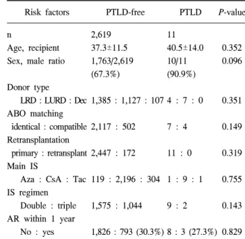
lymphoproliferative disorder; univariate analysis Risk factors PTLD-free PTLD P-value n
Age, recipient Sex, male ratio Donor type LRD : LURD : Dec ABO matching identical : compatible Retransplantation primary : retransplant Main IS
Aza : CsA : Tac IS regimen Double : triple AR within 1 year No : yes 2,619 37.3±11.5 1,763/2,619 (67.3%) 1,385 : 1,127 : 107 2,117 : 502 2,447 : 172 119 : 2,196 : 304 1,575 : 1,044 1,826 : 793 (30.3%) 11 40.5±14.0 10/11 (90.9%) 4 : 7 : 0 7 : 4 11 : 0 1 : 9 : 1 9 : 2 8 : 3 (27.3%) 0.352 0.096 0.351 0.149 0.319 0.755 0.143 0.829 Abbreviations: LRD, living related donor; LURD, living unrelated donor; Dec, deceased donor; IS, immunosuppressive agent; CsA, cyclosporine A; Aza, azathioprine; Tac, tacrolimus; AR, acute rejection.
Fig. 2. Involved organs of posttransplant lymphoproliferative
disorder.
Fig. 1. Histologic types of posttransplant lymphoproliferative
dis-order (WHO classification). PTLD, posttransplant lympho-proliferative disorder.
PTLD에는 diffuse large B cell lymphoma 7명(70%), Burkitt lymphoma 2명(20%), anaplastic plasmacytoma는 1명(10%)이 었다(Fig. 1). 11명의 환자 중 9명에서 PTLD 진단시 EBV에 대한 혈청학적 검사를 시행하였다. 혈청학적 검사를 시행 한 환자 중 1명(11.1%)은 EBV IgG와 IgM 모두 양성을 나타 내었고, 3명(33.3%)은 EBV IgM 양성 / IgG 음성, 4명(44.4%) 은 EBV IgM 음성 / IgG 양성, 1명(11.1%)은 IgM, IgG 모두 음성이었다. 침범한 장기 별로는, 위장관에서 1예(9.1%), 폐 에 2예(18.2%), 골침범이 2예(18.2%), 비장이 2예(18.2%), 경 부 림프절 침범 2예(18.2%), 간에 1예(9.1%), 다장기에서 동 시에 발견된 경우가 1예(9.1%)로 나타났다(Fig. 2). 3) PTLD의 위험인자 PTLD 발생의 위험인자 검증을 위한 일변량 분석에서, 수여 자의 성별, 연령, 급성거부반응의 경력, 주 면역억제제의 종류 및 사용기간, 급성거부반응 치료 목적으로 항 림프구항체의 사용 경력 등은 통계학적으로 유의하지 않았다(Table 2). 4) PTLD의 치료 및 예후 11명의 PTLD환자의 치료로 10예에서 면역억제제의 감 량, 7예에서 항바이러스제, 6예에서 항암화학치료, 그 중 2 예에서 Rituximab (anti CD20 monoclonal antibody)과 병용하 여 항암화학요법을 시행하였고, 방사선치료는 3예에서 시 행하였다. PTLD 환자 11명 중 7명(63.6%)이 사망하였고, 그 중 2예에서 사망 당시 이식신의 기능은 유지되었다. 11명의 환자 중 8예의 이식신 소실이 있었는데, 사망과 관련된 이 식신 소실 2예를 제외하면 6예(54.5%)에서 이식신 소실이 있었다. PTLD 진단 후 이식신 소실이 있기까지의 평균 기 간은 7.8± 10.4개월(0∼24개월)이었다. 항암화학요법을 받 은 6명중 3명(50%)은 완전관해 상태로 추적 중이며 2명은 치료 중 폐렴에 의한 패혈증으로 사망하였고 1명은 심혈관 질환으로 사망하였다. 항암화학요법과 더불어 방사선 치료 를 병행했던 환자는 3명이었으며 그 중 2명(66.7%)은 이식 신의 소실 없이 완전관해를 보였다. 항암화학요법을 시행 받지 않고 면역억제제 감량과 항바이러스 치료만 시행 받 은 5명의 환자 중 4명(80%)이 사망하였고, 사망하지 않은 1명은 이식신의 소실이 있었다(Table 1).
고 찰
장기 이식 환자에서 악성 종양의 발생위험은 정상인에 비하여 100배정도 높다는 것은 잘 알려진 사실이다.(1) PTLD 는 악성 종양의 20∼30%정도를 차지하며 피부암 다음으로 발생률이 높은 것으로 알려져 있다.(4) 이식 장기에 따라 차 이가 있지만 지금까지의 연구들에서 소장 이식 환자에서 19%, 폐 이식 환자에서 8%, 심장이식 환자에서 3%, 간이식 환자에서 1∼3%, 신장 이식 환자에서 1% 정도로 보고 되고있다.(5-8) 본 연구에서는 PTLD 발생률이 0.42%로 이전 연 구에 비하여 낮은 PTLD 발생률을 나타내고 있으며, 이식 수술 후 PTLD 진단까지의 평균 기간은 126.6±74.8개월로 다른 연구결과에서 보고된 26.3개월에서 55.6개월에 비해 발병 시기가 늦은 것으로 나타났다.(9,10) 또한 본 연구에서 는 이식 후 1년 이내에 발생한 경우가 2예(18.2%)에 불과하 여, PTLD같이 바이러스와 연관된 질환이 이식 후 1년 이내 에 가장 많이 발병한다는 결과와도 차이를 보였다.(11,12) 2005년 보건복지부에서 보고한 암 발생 통계를 보면, 일 반인구에서 발생하는 암 중에 림프종이 차지하는 비율은 2.4% (2,393/99,025)인데 반하여 본 연구에서는 신이식 후 발생한 암 중 5.8% (11/190)로 나타났다. 비호지킨 림프종의 인구 100,000명당 발생률은 8.79명으로 0.00879%인데 반하 여 본 연구에서 이식 환자에서의 PTLD 발생률은 0.42%로 이전 연구에서 보고된 PTLD 발생률 보다는 낮지만 일반 인 구에 비하여 월등히 높음을 알 수 있다.(13) 또한 본 연구에 서는 11명의 PTLD 환자 중 남자는 10명이었다. 일반 인구 에서 비호지킨 림프종의 발생은 인구 100,000명당 남자 5.21명, 여자 3.58명으로 남성에서 더 많이 발생하는 것으로 알려져 있다.(13) 이식환자에서도 이러한 비율을 따를 것으 로 기대되나 적은 수의 환자를 대상으로 한 본 연구에서 남녀 비율에 대한 통계적 유의성은 찾을 수 없었다. 그러나, 적은 수의 환자를 대상으로 한 PTLD에 대한 다른 연구들에 서도 남성에서 더 많은 PTLD 발생을 보고 하고 있 다.(14,15) EBV 감염이 PTLD의 발생의 위험성을 높인다는 것은 잘 알려진 사실이다.(16) 이식 후 투여되는 면역억제 제의 영향으로 EBV-특이 T 세포가 EBV에 감염된 B 세포 의 증식을 억제하지 못하게 되고, EBV 감염으로 인해 숙주 의 면역세포가 세포사멸의 기능을 상실하게 되어 무제한 적으로 증식이 일어나 림프증식성 질환의 형태로 발생되는 것이 PTLD의 발병기전으로 설명되고 있다.(17) 본 연구에 서도 PTLD진단 시 EBV에 대한 혈청학적 검사를 시행한 9 명의 환자 중 8명(88.9%)에서 EBV IgM 혹은 EBV IgG에 양 성 반응을 보였다. 이러한 결과는 이전의 PTLD환자의 80∼ 90%가 EBV 초감염 혹은 이전 감염의 재활성화와 밀접한 관계가 있다는 연구와 동일한 결과를 보여주는 것이 다.(18,19)
EBV의 혈청학적 검사는 EBV 바이러스 캡시드 항원(viral capsid antigen)에 대한 IgG와 IgM 항체를 측정하는 것으로 써, 이식 전 혈청학적으로 음성인 환자에서 이식 후 EBV 감염으로 인한 PTLD 발생을 감시하는 수단으로 사용될 수 있다. 그러나, EBV에 대한 혈청학적 검사는 면역억제제와 거대세포바이러스 면역글로부린 사용에 의해 영향을 받을 수 있어서 연쇄중합효소반응(PCR)을 이용한 EBV DNA 검 출이 PTLD의 예측 인자로서 더 유용하다고 보고하고 있 다.(20-25) 본 연구의 결과 PTLD의 조직학적 소견상 diffuse large B cell lymphoma가 가장 많은 형태로 나타났다. 일반적으로 PTLD를 조직학적으로 분류하면 비호지킨 림프종이 가장 많으며 그 중에서도 diffuse large B cell lymphoma가 가장 많은 분포를 차지한다. 호지킨 림프종은 일반 인구에서 발 생하는 림프종의 34%에 해당하지만 이식환자의 PTLD에서 는 2% 정도에 불과하다.(26) PTLD의 발생은 항 림프글로부린의 사용과 면역억제제에 노출 된 기간에 밀접한 연관성이 있는 것으로 알려져 있다. Cockfield 등은 이식 환자에서 PTLD의 발생은 두 개의 정점 (bimodal peak)을 나타낸다고 보고하면서 이식 초기인 1년 내에서는 강력한 면역억제 상태로 인하여 바이러스 감염에 의하여 발생하고 1년 이후에는 환자의 나이와 오랜 기간의 면역 억제 상태로 인하여 발생한다고 하였다.(27) 본 기관 에서는 서구에서 많이 사용하는 면역 유도 방법으로 항 림 프글로부린을 사용하지 않으며, 이식후 면역억제제의 유지 요법으로 주면역억제제의 혈중농도를 낮게 유지하는 것과 관련이 있을 것이다. 또한 이식 수술 후 급성 거부반응이 발생하여 구조 치료를 받은 사람들이 강력한 면역 억제 효 과로 인하여 PTLD의 발생 위험성 증가와 관련이 있을 수 있으나, 본 연구에서는 PTLD 발생군의 급성거부반응 경력 이 PTLD 비발생군과 비교하여 유의한 차이가 없어, 이에 대한 효과를 입증할 수 없었다. 본 연구에서 PTLD 발생에 미치는 위험인자 분석에서 위험인자로 간주하였던 항 림프 글로부린 사용 경력이나 주면역억제제의 종류 등은 면역억 제 정도와 비례하는 척도로, 이 또한 PTLD 발생률에 미치 는 영향은 유의하지가 않았다. 그러나 이식 후 기간이 증가 함에 따라 면역제제요법의 장기적인 유지가 PTLD 발생률 에 영향을 미치는 것으로 판단된다. EBV 감염으로 인한 PTLD 발생기전이 알려지면서 PTLD 의 일차 치료로 면역억제제의 감량이 시도되어 왔다. 그러 나 이식장기의 소실 없이 면역억제제를 얼마나 감량해야 하는지에 대한 명확한 기준은 없는 상태이다. Tsai 등의 보 고에 의하면 PTLD 환자에서 이식신의 5년 생존율은 50%정 도 기대할 수 있다고 하였다.(18) 다른 치료 방법으로 EBV 와 관련성 때문에 acyclovir 등의 항 바이러스 제제가 사용 되었으나 그 효과에 대해서는 정확히 알려진 바 없다.(19) 이상의 방법들만으로는 충분한 효과를 나타내지 못하여 악 성 림프종에서 사용되는 항암화학요법이 추가로 사용되고 있다. 항암화학요법이 PTLD에서 50%정도의 완전관해율을 가진다는 보고가 있지만,(20) 면역억제 상태인 환자들에게 강력한 항암화학요법을 사용함으로써 감염 발생으로 인한 사망률이 높아진다는 보고도 있다.(21-23) 본 연구에서도 항암화학요법을 시행 받은 6명의 환자 중 3명의 환자가 완 전관해를 보여 50%의 관해율을 보였다. 그러나 2예(33.3%) 에서 치료 중 폐렴에 의한 패혈증으로 사망하였다. 이러한 항암화학요법의 합병증 때문에 보다 합병증이 적은 rituximab 같은 항 CD20 단클론항체(anti CD20 monoclonal antibody)와
의 병용이 시행되고 있다. 최근 보고에 의하면 항암화학요 법과 항 CD20 단클론항체의 치료 효과는 비슷하나 항암화 학요법 단독 시행시의 합병증과 사망률이 의미 있게 높다 고 보고되었다.(24) 비록 본 연구의 결과가 많은 환자를 대 상으로 한 것은 아니지만 2명의 환자에서 기존의 CHOP (Cyclophosphamide, Adriamycin, Vincristine and Predisone) 병용 화학요법에 rituximab을 추가하여 2명 모두 완전관해를 보 였다. 문헌고찰에서 뿐만아니라 본 연구의 결과로 볼 때 PTLD는 항암화학요법 및 항 CD20 단클론항체, 방사선 치 료 등의 다양한 치료법을 사용하여 적극적인 치료를 시행 하는 것이 이식신의 보존과 생존에 도움이 되는 것으로 나 타났다. 현재까지 PTLD의 치료에 대한 표준 치료법은 확립 되지 않은 실정이다. 본 기관에서도 면역억제제 감량을 기 본치료법으로 하여 환자의 상태에 따라 항암화학용법 및 방사선 치료를 병행하여 사용하였으며 치료자에 따라 다른 치료법을 적용하여 왔다. 최근에는 rituximab을 병용한 항암 화학요법을 적극적으로 시행하고 있으며 여러 보고들에서 도 적극적인 항암화학요법이 PTLD 치료에 도움이 된다고 보고하고 있다.(35-37)
결 론
PTLD는 장기 이식 후 발생하는 잘 알려진 합병증 중 하 나이다. 장기 이식 후 면역억제 상태와 EBV 감염에 의하여 발생한다고 알려져 있다. 혈연간 생체 이식이 많은 비중을 차지하는 국내의 실정에 따라 PTLD 발생이 서구의 결과에 비하여 적고 이식 후 초기 발병보다 후기 발병이 많은 것으 로 나타났다. PTLD 발생시 높은 사망률 및 이식신의 소실 을 보인다. 따라서 면역억제 기간이 길어지는 장기 생존환 자에 대해서도 PTLD의 발생에 대하여 지속적인 관찰이 필 요하며 PTLD 발생시에는 항암화학요법과 방사선 치료를 병행하는 적극적인 치료가 필요하겠다.감사의 글
본 논문은 2008년도 연세대학교 의과대학 장기이식연구 소의 연구비 지원으로 이루어졌음.REFERENCES
1) Murray JE, Wilson RE, Tilney NL, Merrill JP, Cooper WC, Birtch AG, et al. Five years’ experience in renal trans-plantation with immunosuppressive drugs. Survival, function, complications, and the role of lymphocyte depletion by thora-cic duct fistula. Ann Surg 1968;168:416-35.
2) Opelz G, Dohler B. Lymphomas after solid organ trans-plantation: a collaborative transplant study report. Am J
Transplant 2004;4:222-30.
3) Muti G, Cantoni S, Oreste P, Klersy C, Gini G, Rossi V, et al: Post-transplants lymphoproliferative disorders: improved out-come after clinico-pathologically tailored treatment. Haemato-logica 2002;87:67-77.
4) Penn I. The price of immunotherapy. Curr Probl Sug 1981;18: 681-751.
5) Finn L, Reyes J, Bueno J, Yunis E. EpsteBarr virus in-fections in children after transplantation of the small intestine. Am J Surg Pathol 1998;22:299-309.
6) Mihalov ML, Gattuso P, Abraham K, Holmes EW, Reddy V. Incidence of posttransplant malignancy among 674 solid-organ- transplant recipients at a single center. Clin Transplant 1996; 10:248-55.
7) Hanto DW, Frizzera G, Gajl-Peczalska KJ, Simmons RL. Epstein-Barr virus immunodeficiency and B cell lymphoproli-feration. Transplantation 1985;39:461-72.
8) Boubenider S, Hiesse C, Goupe C, Kriaa F, Merchand S, Charpentier B. Incidence and consequences of post-trans-plantation lymphoproliferative disorders. J Nephrol 1997;10: 136-45.
9) Melchor J L, Cancino J, Gracida C. Lymphoproliferative dis-orders following kidney transplantation. Transplant Proc 2002; 34:2537-8.
10) Gerstenkorn C, Jackson G, Di Franco D, Thomusch O, Talbot D. Outcome of PTLD in renal and liver allograft recipients. Transplant Proc 2001;33:2469-72.
11) Shroff R, Rees L. The posttransplant lymphoproliferative dis-order-a literature review. Pediatr Nephrol 2004;19:369-77. 12) Cao S, Cox K, Esquivel CO, Berquist W, Concepcion W, Ojogho
O, et al. Posttransplant lymphoproliferative disorders and gastro-intestinal manifestations of Epstein-Barr virus infection in chil-dren following liver transplantation. Transplantation 1998;66: 851-6.
13) 보건복지부. 암 발생 통계 1999-2001. 2005:97-8.
14) Timuragaoglu A, Ugur-Bilgin A, Colak D, Tuncer M, Golbasi I, Hazar V, et al. Posttransplant lymphoproliferative disorders in transplant recipients. Transplant Proc 2006;38:641-5. 15) Wasson S, Zafar MN, Best J, Reddy HK. Post-transplantation
lymphoproliferative disorder in heart and kidney transplant pa-tients: a single-center experience. J Cardiovasc Pharmacol Ther 2006;11:77-83.
16) Armitage JM, Kormos RL, Stuart RS, Fricker FJ, Griffith BP, Nalesnik M, et al. Posttransplant lymphoproliferative disease in thoracic organ transplant patients: Ten years of cyclo-sporine-based immunosuppression. J Heart Lung Transplant 1991;10:877-86.
17) Young L, Alfieri C, Hennessy K, Evans H, O'Hara C, Anderson KC. Expression of Epstein-Barr virus transformation- associated genes in tissues of patients with EBV lymphoproli-ferative disease. N Engl J Med 1989;321:1080-5.
maneu-vers in the development of PTLD. Am J Transplant 2007;7: 271-7.
19) Hanto DW, Frizzera G, Purtilo DT, Sakamoto K, Sullivan JL, Saemundsen AK. Clinical spectrum of lymphoproliferative dis-orders in renal transplant recipients and evidence for the role of Epstein-Barr virus. Cancer Res 1981;41:4253-61. 20) Davis JE, Sherritt MA, Bharadwaj M, Morrison LE, Elliott
SL, Kear LM, et al. Determining virological, serological and immunological parameters of EBV infection in the develop-ment of PTLD. Int Immunol 2004;16:983-9.
21) Green M, Cacciarelli TV, Mazariegos GV, Sigurdsson L, Qu L, Rowe DT, et al. Serial measurement of Epstein-Barr viral load in peripheral blood in pediatric liver transplant recipients during treatment for posttransplant lymphoproliferative disease. Transplantation 1998;66:1641-4.
22) Cockfield SM, Preiksaitis JK, Jewell LD, Parfrey NA. Post-transplant lymphoproliferative disorder in renal allograft recipients. Clinical experience and risk factor analysis in a sin-gle center. Transplantation 1993;56:88-96.
23) Green M, Bueno J, Rowe D, Mazariegos G, Qu L, Abu- Almagd K, et al. Predictive negative value of persistent low Epstein-Barr virus viral load after intestinal transplantation in children. Transplantation 2000;70:593-6.
24) Smets F, Latinne D, Bazin H, Reding R, Otte JB, Buts JP, et al. Ratio between Epstein-Barr viral load and anti-Epstein- Barr virus specific T-cell response as a predictive marker of posttransplant lymphoproliferative disease. Transplantation 2002;73:1603-10.
25) Toyoda M, Moudgil A, Warady BA, Puliyanda DP, Jordan SC. Clinical significance of peripheral blood Epstein-Barr viral load monitoring using polymerase chain reaction in renal transplant recipients. Pediatr Transplant 2008;12:778-84. 26) Penn I. Malignant lymphomas in organ transplant recipients.
Transplant Proc 1981;13:736-8.
27) Cockfield SM. Identifying the patient at risk for post-trans-plant lymphoproliferative disorder. Transpl Infect Dis 2001;3: 70-8.
28) Tsai DE, Hardy CL, Tomaszewski JE, Kotloff RM, Oltoff
KM, Somer BG, et al. Reduction in immunosuppression as ini-tial therapy for posttransplant lymphoproliferative disorder: analysis of prognostic variables and long-term follow-up of 42 adult patients. Transplantation 2001;71:1076-88.
29) Humar A, Hebert D, Davies HD, Humar A, Stephens D, O'Doherty B, et al. A randomized trial of ganciclovir versus ganciclovir plus immune globulin for prophylaxis against Epstein-Barr virus related posttransplant lymphoproliferative disorder. Transplantation 2006;81:856-61.
30) Choquet S, Trappe R, Leblond V, Jäger U, Davi F, Oertel S. CHOP-21 for the treatment of post-transplant lymphoprolifer-ative disorders (PTLD) following solid organ transplantation. Haematologica 2007;92:273-4.
31) Dotti G, Fiocchi R, Motta T, Mammana C, Gotti E, Riva S, et al. Lymphoma occuring late after solid-organ transplantation: influence of treatment on the clinical outcome. Transplantation 2002;74:1095-102.
32) Mamzer-Bruneel MF, Lome C, Morelon E, Levy V, Bourquelot P, Jacobs F, et al. Durable remission after aggressive chemo-therapy for very late post-kidney transplant proliferation: a report of 16 cases observed in a single center. J Clin Oncol 2000;18: 3622-32.
33) Swinnen LJ, Mullen GM, Carr TJ, Costanzo MR, Fisher RI. Aggressive treatment for postcardiac transplant lymphoprolife-ration. Blood 1995;86:3333-40.
34) Elstrom RL, Andreadis C, Aqui NA, Ahya VN, Bloom RD, Brozena SC, et al. Treatment of PTLD with rituximab or chemotherapy. Am J Transplant 2006;6:569-76.
35) 윤설희, 김수정, 이혜원, 황도유, 김진석, 정준원 등. R-CHOP 요법으로 치료한 장기이식 후 림프증식 질환 2예. 대한혈액 종양학회지 2008;43:106-12.
36) Smets F, Vajro P, Cornu G, Reding R, Otte JB, Sokal E. Indications and results of chemotherapy in children with post-transplant lymphoproliferative disease after liver post-transplantation. Transplantation 2000;69:982-4.
37) Everly MJ, Bloom RD, Tsai DE, Trofe J. Posttransplant lym-phoproliferative disorder. Ann Pharmacother 2007;41:1850-8.