Fig 1. Diagram illustrating the chemotherapy protocol utilized in this study.---12
Fig. 2. Case 1. Axial contrast-enhanced magnetic resonance images(MRI) showing medulloblstoma in 4th ventricle and cerebellar hemisphere.---16
Fig. 3. Case 3. MRI showing medulloblastoma in the 4th ventricle.---17
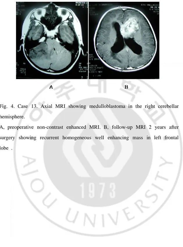
Fig. 4. Case 13. Axial MRI showing medulloblastoma in the right cerebellar hemisphere.---18
Table 1. Summary of clinical characteristics of patients---8 Table 2. Chang staging system for posterior fossa medulloblastoma---9 Table 3. Distribution of Chang tumor stage. ---10
Bailey Cushing
primitive neuroectodermal tumor
prone position Selector
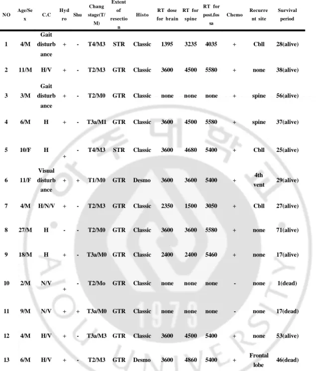
Table 1. Summary of clinical characteristics of patients
C.C, chief complaint; Hydro, hydrocephalus; Shu, shunt; STR, subtotal resection; GTR, grossly total resection; Histo, histological type; RT, radiotherapy; Chemo, chemotherapy; H, headache; V, vomiting; N, nausea; Vent, ventricle ; Desmo, desmoplastic.
NO Age/Se x C.C Hyd ro Shu Chang stage(T/ M) Extent of resectio n Histo RT dose for brain RT for spine RT for post.fos sa Chemo Recurre nt site Survival period 1 4/M Gait disturb ance + - T4/M3 STR Classic 1395 3235 4035 + Cbll 28(alive)
2 11/M H/V + - T2/M3 GTR Classic 3600 4500 5580 + none 38(alive)
3 3/M
Gait disturb
ance
+ - T2/M0 GTR Classic none none none + spine 56(alive)
4 6/M H + - T3a/M1 GTR Classic 3600 4500 5580 + spine 37(alive)
5 10/F H + - T4/M3 STR Classic 3600 4680 5400 + Cbll 25(alive) 6 11/F Visual disturb ance + + T1/M0 GTR Desmo 3600 3600 5400 + 4th vent 29(alive) 7 4/M H/N/V + - T2/M3 GTR Classic 2350 1500 3050 + Cbll 27(alive)
8 27/M H - - T2/M0 GTR Classic 3600 3600 5580 + none 71(alive)
9 18/M H + - T3a/M0 GTR Classic 2400 2400 5460 + none 17(alive)
10 2/M N/V
+ - T2/Mo GTR Classic none none none - none 1(dead)
11 9/M N/V + + T3a/M0 GTR Classic none none none - none 17(dead)
12 4/M H/V + - T3a/M3 GTR Classic 3600 4500 5400 + none 53(alive)
13 6/M H/V + - T2/M3 GTR Desmo 3600 4860 5400 + Frontal
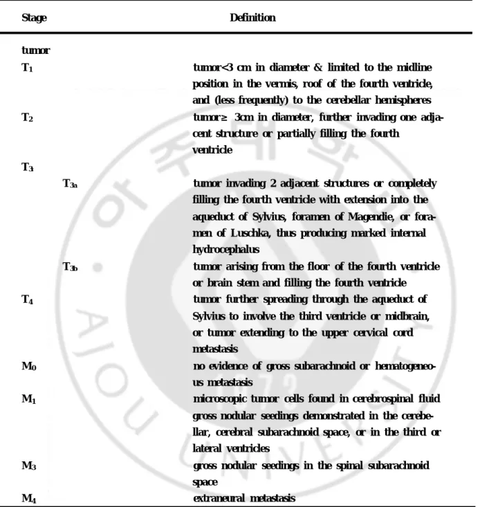
Table 2. Chang staging system for posterior fossa medulloblastoma
Stage Definition
tumor
T1 tumor<3 cm in diameter & limited to the midline position in the vermis, roof of the fourth ventricle, and (less frequently) to the cerebellar hemispheres T2 tumor≥ 3cm in diameter, further invading one
cent structure or partially filling the fourth ventricle
T3:
T3a tumor invading 2 adjacent structures or completely filling the fourth ventricle with extension into the aqueduct of Sylvius, foramen of Magendie, or men of Luschka, thus producing marked internal hydrocephalus
T3b tumor arising from the floor of the fourth ventricle or brain stem and filling the fourth ventricle
T4 tumor further spreading through the aqueduct of Sylvius to involve the third ventricle or midbrain, or tumor extending to the upper cervical cord metastasis
M0 no evidence of gross subarachnoid or us metastasis
M1 microscopic tumor cells found in cerebrospinal fluid gross nodular seedings demonstrated in the llar, cerebral subarachnoid space, or in the third or lateral ventricles
M3 gross nodular seedings in the spinal subarachnoid space
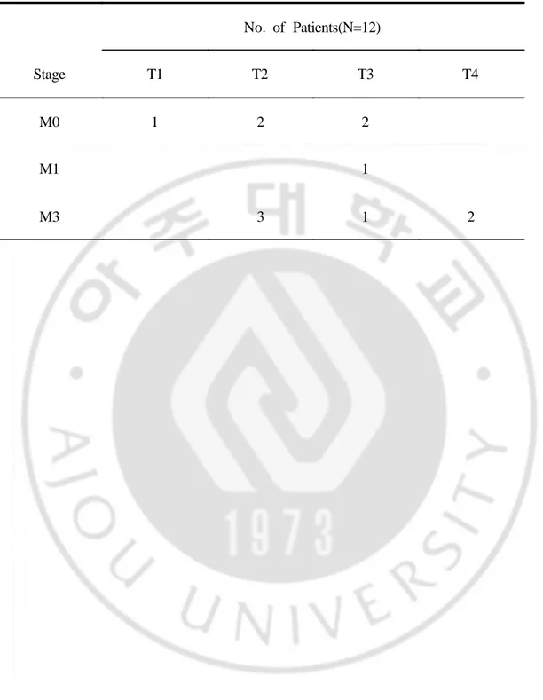
Table 3. Distribution of Chang tumor stage. No. of Patients(N=12) Stage T1 T2 T3 T4 M0 1 2 2 M1 1 M3 3 1 2
booster
vincristine, CCNU(1-(2-chloroethyl)-3-cyclohexyl-1-nitrourea), cisplatin
CCNU Cisplatin
Vincristine
Fig
GE high advanced CT MR Sigma
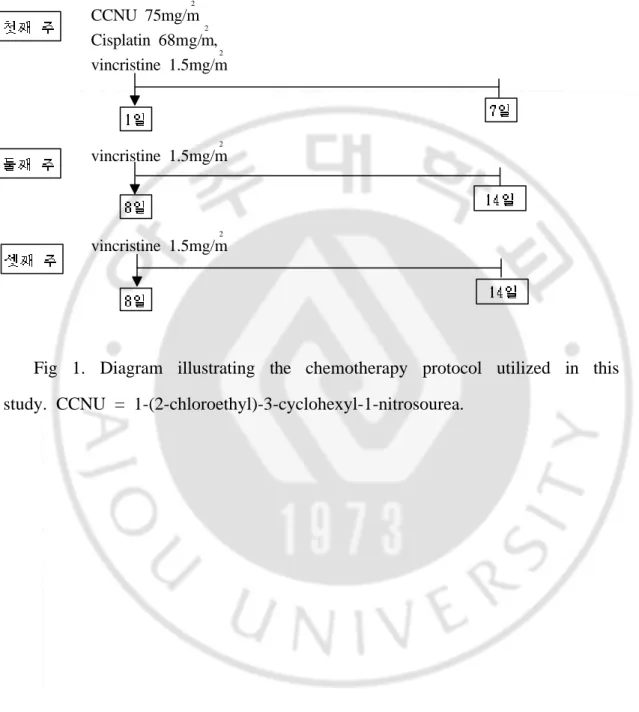
CCNU 75mg/m 2 Cisplatin 68mg/m 2 , vincristine 1.5mg/m 2 vincristine 1.5mg/m 2 vincristine 1.5mg/m 2
Fig 1. Diagram illustrating the chemotherapy protocol utilized in this study. CCNU = 1-(2-chloroethyl)-3-cyclohexyl-1-nitrosourea.
complete resection
Table 1, Case
desmoplastic variance) classic
external ventricular drainage)
ventricular peritoneal shunt
Chang
posterier fossa Fig Fig
Fig
MV linear accelerator(Mevatron, Siemen radiotherapy
CLINA 2100C/D,Variau Mevatron,
Siemens cerebellar hemisphere
vermis
stereotactic radiosurgery
CCNU, cisplatin, VCR cisplatin,
A B C
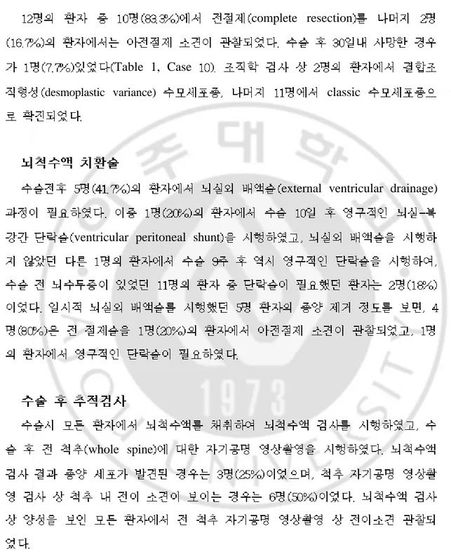
Fig. 2. Case 1. Axial contrast-enhanced magnetic resonance images(MRI) showing medulloblstoma in 4th ventricle and cerebellar hemisphere.
A, preoperative MRI. B, postoperative MRI showing the recurrent small tumor in the 4th ventricle 2 years after tumor removal. C, MRI showing tumor regression 6 months after radiosurgery.
A B C
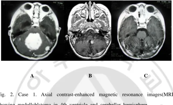
Fig. 3. Case 3. MRI showing medulloblastoma in the 4th ventricle.
A, preoperative contrast enhanced MRI. B, postoperative contrast enhanced MRI showing no recurrence in the brain 3 years after tumor removal. C, spinalMRI showing recurrent mass in lumbar area 4 years after tumor removal.
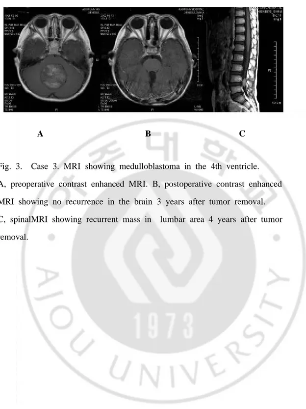
Fig. 4. Case 13. Axial MRI showing medulloblastoma in the right cerebellar hemisphere.
A, preoperative non-contrast enhanced MRI. B, follow-up MRI 2 years after surgery showing recurrent homogeneous well enhancing mass in left frontal lobe .
A B
Fig. 5. Case 7. Axial contrast-enhanced MRI.
A, preoperative MRI showing homogeneous enhancing mass in the 4th. ventricle. B, postoperative MRI showing a peanut sized recurrent tumor in the right cerebellar hemisphere.
Sutton Albright vincristine CCNU vincristin CCNU vincristine cisplatin craniospinal irradiation
CSF diversion
1. Albright AL, Wisoff Jm, Zelter PM, Boyett JM, Rorke LB, Stanley P: Effects of medulloblastoma resection on outcome in children: A report from the Children's Cancer Group. Neurosurg 38: 265-271, 1996
2. Annie WC, Nancy JT, Peter MB, David NL, Matthew PF, Marek A, Paul C, Jay SL: Adult medulloblastoma: Prognostic factors and patterns of relapse. Neurosurg 47: 623-632, 2000
3. Bailey P, Cushing H: Medulloblastoma cerebelli: A common type of midcerebellar glioma of childhood. Arch Neurol Neurosurg Psychiatry 4: 192-224, 1925
4. Berry MP, Jenkin RD, Keen CW et al: Radiation therapy for medulloblastoma: A 21-year review. J Neurosurg 55: 43-51, 1981
5. Bloom HJG, Bessell EM: Medulloblastoma in adults: A review of 47 patients treated between 1952 and 1981. Int J Radiat Oncol Biol Phys 18: 763-772, 1990
6. Carrie C, Lasset C, Alapetite C et al: Multivariate analysis of prognostic factors in adult patients with medulloblastoma. Cancer 74: 2362-2360, 1994
7. Duffner PK, Cohen ME, Mayers MH: Brain tumors in children younger than two years ago (abstract No. 62). Ann Neurol 14: 375, 1996
8. Evans AC, Jenkin RD, Sposta R et al: The treatment of medulloblastoma: Results of a prospective randomized trial of radiation therapy with and without CCNU, vincristine and prednisone. J Neurosurg 72: 572-582, 1990
9. Frost PJ, Laperpirre NJ, Wong CS, et al: Medulloblastoma in adults. Int J Radiat Oncol Biol Phys 32: 951-957, 1995
10. Garton GR, Schomberg PJ, Scheithauer BW et al: Medulloblstom l; -prognostic factors and outcome of treatment: review of the Mayo clinic experience. Mayo Clin Proc 64:1077-1086, 1990
11. Halperin EC, Friedman HS: Is there a correlation between duration of presenting symptoms and stage of medulloblastoma at the time of diagnosis? Cancer 78: 874-880, 1996
12. Harry TW, Hendrikus GK, Meic HS, Kenneth WR, Edward HK: Current therapy and new perspectives in the treatment of medulloblastoma. Pediatr Neurol 18: 103-115, 1998
13. Hughes EN, Shillito J, Sallan SE et al: Medulloblastoma at the Joint Center for Radiation Therapy between 1986 and 1984. Cancer 62: 1992-1998, 1988
14. Karoly MD, Adrian TH, Richard DH, Willian FI, Kim P, Angie MW: Medulloblastoma: Is the 5-year survival rate improving: J Neurosurg 86: 13-21, 1997
15. Packer RJ: Childhood medulloblastoma: Progress and future challenges. Brain & Development 21: 75-81, 1999
16. Paterson F, Farr R: Cerebellar meduloblastoma: Treatment by irradiation of the whole central nervous system. Acta Radiol 39: 323-336, 1953
17. Packer RJ, Sutton LN, D'Angio G, Evans AE, Schut L: Management of children with primitive neuroectodermal tumors of the posterior fossa/medulloblastoma. Pediatr Neurosci 12: 272-282, 1986
et al: Outcome for children with medulloblastoma treated with radiation and cisplatin, CCNU and vincristine chemotherapy. J Neurosurg 81: 690-698, 1994
19. Rorke LB, Gilles FH, Davis RL, Becker LE: Revision of the World Health Organization classification of brain tumors for childhood brain tumors. Cancer 56: 1869-1886, 1985
20. Rorke LB: The cerebellar medulloblastoma and its relationship to primitive neuroectodermal tumors. J Neuropathol Exp Neurol 42: 1-15, 1983
21. Russell DS, Rubinstein LJ: Pathology of tumors of the nervous system. Medulloblastoma (including cerebellar neuroblastoma). Baltimore: Williams & Wilkins, 1989, pp251-279
22. Skolyszewski J, Glinski B: Results of postoperative irradiation of medulloblastoma in adults. Int J Radiat Oncol Biol Phys 16: 479-482, 1989
23. Sutton LN, Phillips PC, Molloy PT: Surgical management of medulloblastoma. J Neurooncol 29: 9-21, 1996
24. Tait DM, Thorton-Jones H, Bloom HJ, Lemerle J, Morris-Jones P: Adjuvant chemotherapy for medulloblastoma: The first multi-centre control trial of the International Society of Paediatric Oncology (SIOP I). Eur J Cancer 26: 464-469, 1990
25. Tobias JS, Hayward RD: Brain and spinal cord tumors in children, in Thomas DGT(ed): Neuro-oncology Primary Malignant Brain Tumors. London: Edward Arnold, 1990, pp164-190
26. Ufuk A, Omer U, Meric S, Sedat T, Ahmet O: Medullobalstoma in adults: Treatment results and prognostic factors. Int J Radiat Oncol Biol
Phys 54(3): 855-860, 2002
27. Youmans: Neurological Surgery, Fourth edition. W.B. Saunders company, 1996, pp 2570-2592
ABSTRACT
-Clinical study of factors affecting the outcome of the patients with medulloblastoma.
Lee Sung Un
Department of Medical Sciences The Graduate School, Ajou University
(Supervised by Professor Kyung Gi Cho)
Purpose : The goal of this study is to investigate clinical outcome according to the treatment modality and to define the prognostic factors in the patients with medulloblastoma.
Materials and methods : Between 1996 and 2002, the clinical and radiological data of 12 patients (10 male, 2 female) were retrospectively reviewed. The mean age was 9.5 years (range, 3-27 years) and mean follow-up duration was 38.8 months (range, 7-71 months). Preoperative neuroimaging revealed hydrocephalus in 11 patients and spinal seeding of the tumor in 7 patients. Among 12 patients, 5 patients were classified to low risk group and 7 were classified to high risk group. The treatment protocol was surgical resection followed by radiation therapy and/or additional chemotherapy according to the status of patient and the tumor staging. Complete surgical resection was obtained in 10 patients (83.3 %) and incomplete resection in 2 (16.7 %). Ten out of 12 patients received craniospinal irradiation. The mean
radiation dose was 31 Gy for whole brain (range, 13.95 - 36 Gy), 54.7 Gy for the posterior fossa (range, 30.5 - 55.8 Gy), and 37 Gy for the spinal axis (range, 15 - 48 Gy). Eleven patients underwent adjuvant chemotherapy and the regimen consisted of 1-(2-chloroethyl)-3-cyclohexyl-1-nitrosourea(CCNU), vincristine, and cisplatin.
Results : Seven of 12 patients (58.3 %) developed a tumor recurrence and the median time to recurrence was 20.8 months (range, 2-36 months). Among 7 cases, local recurrence was developed in the posterior fossa in 5 cases, and distant recurrence through cerebrospinal fluid seeding in 2 cases. The incidence of recurrence was higher in the cases with pre-existing leptomeningeal seeding of the tumor and underwent radiation therapy less than 50 Gy to the posterior fossa. The estimated 2-year survival rate was 81.8 %. There was no significant difference in survival time between low risk group and high risk group. Permanent shunt operation was required in two of 11 patients who had a preoperative hydrocephalus. The survival time of 2 patients underwent a permanent shunt surgery was shorter compared to those not.
Conclusion : According to our results, we suggest that the radiation dose to the posterior fossa is important factor to prevent tumor recurrence and surgical resection followed by both radiation therapy and chemotherapy can improve the survival time of the patient of high risk group.