In vivo와 in vitro에서 DicA 단백질의 온도 의존적 DNA 결합
이연호1 ・ 윤상훈2 ・ 임헌만1*1충남대학교 생명시스템과학대학 생물과학과, 2㈜알테오젠
Temperature-dependent DNA binding of DicA protein
in vivo and in
vitro
Yonho Lee1 , Sang Hoon Yun2 , and Heon M. Lim1*
1
Department of Biological Sciences, College of Biological Sciences and Biotechnology, Chungnam National University, Daejeon 34134, Republic of Korea
2
Alteogen, Daejeon 34054, Republic of Korea
(Received February 27, 2019; Revised June 15, 2019; Accepted June 26, 2019)
*For correspondence. E-mail: hmlim@cnu.ac.kr; Tel.: +82-42-821-6276; Fax: +82-42-822-9690
In Escherichia coli, DicA protein is involved in cell division control. DicA protein is known to bind DNA better at 25°C than at 37°C. However, the molecular cause of the temperature dependent binding is not clear. In this study, we investigated how DicA binds DNA and why its DNA binding activity depends on temperature. An unique in vivo DNA binding assay developed in this laboratory showed that unlike the homologous proteins such as RovA or SlyA, DicA uses its N-terminal domain for DNA binding. The in vivo DNA binding assay of DicA also demonstrated that the temperature-dependent DNA binding activity does not come from Cnu or H-NS that is known to bind DNA better at 25°C than at 37°C. Electrophoretic Mobility Shift Assay (EMSA), when performed with purified DicA protein, did not show temperature-dependent DicA binding activity. However when EMSA was performed with crude protein from WT E. coli cells, temperature-dependent DicA binding activity was observed, suggesting that there is a factor(s) that confers temperature DNA binding activity of DicA in vivo.
Keywords: Escherichia coli, DicA, DNA binding, filamentous growth, temperature-dependent DNA binding activity
Phage에서 유래한 DNA는 대장균 genome의 20%를 차지 하지만 그 기능은 잘 알려지지 않았다(Casjens, 2003). 대장균 에는 총 166 kb 크기인 9가지 종류의 prophage로부터 유래된 DNA element들이 있으며 그들 중 하나인 dic (Division Control) 으로 명명된 Qin prophage로부터 유래한 유전자들은 대장균 의 DNA 복제가 끝나는 곳에 위치하고 있다(Wang et al., 2010). 이 유전자들의 산물 중 하나인 DicB 단백질은 MinC와 결합하여 FtsZ의 Z-ring 형성을 방해한다(de Boer et al., 1990). Cell division inhibitor인 DicB는 1985년 dicA의 연구로부터 시작되었으며, 관련된 유전자들은 genomic DNA에서 서로 가 까운 곳에 위치하고 있다(Bejar and Bouche, 1985). dicA의 연 구에서 온도 의존적으로 대장균이 세포분열을 하지 못하는 돌 연변체로부터 dicA1 변이 유전자를 찾게 되었고, DicA 단백질 의 108번째 아미노산인 류신이 페닐알라닌으로 치환된 돌연 변이 단백질임을 genetic mapping을 통해 확인하였다(Bejar and Bouche, 1985). 또한 dicB 유전자는 DicA에 의해 발현이 억제된다고 보고되었다(Bejar and Bouche, 1985). dicC 유전 자 역시 DicA에 의하여 발현이 억제 되는 것으로 보고되었다 (Bejar et al., 1986). 온도 의존적인 dicA1 돌연변이는 온도에 따른 DicA1 단백질의 DNA의 결합의 문제로 25°C에서는 dimer를 형성하여 dicB 유전자의 발현을 억제하고 37°C에서 는 dimer를 형성하지 못해 DicB의 과발현에 의한 filamentous
(A)
(B)
Fig. 1. A schematic of dic genes and DNA sequence of the promoter region of the dicA and dicC genes (Yun et al., 2012). A portion of the Qin prophage in the E. coli genome is shown with genes involved in cell division inhibition. Boxes indicate the location and relative size of the genes and arrows indicate promoter. The dicF gene produces RNA only (A). DNA sequence of the promoter region of the dicA and dicC genes is shown. The DicA-binding site (Oc) and additional putative DicA binding site (Om, Oa) were boxed with dashed line. And single nucleotide change in Oc-mutant is shown obove the arrows. A putative -10 and -35 promoter sequence of the dicC gene is underlined. The transcription initiation site of the dicC gene is indicated as +1 for dicC. The promoter sequence for the dicA gene is not obvious with DNA sequence information only (B).
성장을 야기 한다고 보고되어 있다(Bejar et al., 1988). 또 다른 세포분열 억제자인 dicF는 dicA와 dicB 유전자의 연구에서 발 견 되었으며, 단백질을 합성하는 유전자가 아닌 non-coding RNA로서 작용하는 것으로 확인되었다(Bouche and Bouche, 1989). small RNA인 dicF는 ftsZ의 mRNA에 결합하여 단백질 합성을 방해하고 있다(Tetart and Bouche, 1992). 이렇듯 dicF, dicB 두 유전자는 Qin phage로부터 유래한 대장균의 분열 억 제 유전자이며, 대장균이 정상적인 분열을 할 때에는 DicA가 두 유전자의 발현을 억제하고 있는 것이다.
DicA 단백질은 Oc (Operator of dicC)로 명명한 20 bp로 이루 어진 염기서열과 결합한다(Fig. 1B). dicA와 dicC 유전자 사이 에는 83 bp의 DNA 염기서열이 존재하며 이 PdicAC (Promoter region of dicA and dicC gene) 안에서 dicC의 전사 시작점을 기 준으로 -8에서부터 +12 위치에 걸친 20 bp의 Oc가 자리잡고 있다(Fig. 1B). Oc에 DicA 단백질이 결합하면 dicC의 유전자 발현은 억제되며 dicA의 발현이 이루어진다. 이렇게 두 유전 자의 발현은 dicA 또는 dicC 한쪽으로만 진행될 수 있다고 보 고되었다(Yun et al., 2012).
Cnu 단백질은 H-NS와 복합체를 이루어 대장균의 DNA 복 제가 시작하는 OriC 복제기점에서 16개 염기서열로 이루어진 cnb (Cnu binding site)로 명명한 위치에 특이적으로 결합함이 보고 되어있다(Kim et al., 2005). Cnu 단백질은 혼자만으로는 DNA에 결합할 수 없으며 H-NS와 복합체를 형성하여 DNA와 결합한다. 본 실험실에서 발견된 CnuK9E 돌연변이 단백질은 Cnu 단백질의 9번째 라이신이 글루탐산으로 치환된 돌연변 이로, H-NS와 복합체를 이루어 37°C에서 DicA의 Oc 결합을 억제한다. 흥미로운 사실은 이러한 CnuK9E 돌연변이체가
dicA의 발현 억제를 통해 대장균의 filamentous growth를 야기 한다는 사실이다(Yun et al., 2012).
본 연구를 통해 in vivo와 in vitro에서 DicA 단백질의 Oc 에서의 DNA 결합을 분석하여 보았다. 실험 결과, DicA 단백 질은 N-말단 도메인을 통하여 Oc에 결합하며, H-NS, 또는 Cnu/H-NS 복합체에 상관없이 또 다른 in vivo factor에 의하여 온도 의존적 DNA 결합양상을 보인다.
재료 및 방법
사용균주와 배양실험에 사용한 균주는 MG1655와 HB101으로부터 λ-Red recombinase를 이용한 유전자 제거 방법(Datsenko and Wanner, 2000)을 이용하여 dicF, dicB, dicA, cnu, hns를 제거한 균주를 사용하였으며 그 내용은 Table 1에 표시하였다. LB 배지는 D.W. 1 L 당 10 g Bacto trypton, 5 g Bacto yeast extract, 10 g NaCl을 넣어 121°C에서 20분 동안 멸균하여 사용하였다. 항 생제는 Ampicillin (Amp)은 100 μg/ml, Kanamycin (Kan)은 50 μg/ml, Chloroamphenicol (Cm)은 15 μg/ml, Streptomycin (Str)은 100 μg/ml 농도로 사용하였다. 플라스미드 실험에 사용한 플라스미드는 Table 2와 같다. DicA 돌연변이 제작 DicA 단백질의 아미노산 서열을 바꾸기 위하여 megaprimer
Table 1. E. coli strains used in this study
Strain Genotype Reference or source
MG1655 Wild type, F- λ- ilvG- rfb-50 rph-1 CGSC#: 6300
MG1655 cnu Wild type, F- λ- ilvG- rfb-50 rph-1 Δcnu Kim et al. (2005)
MG1655 hns Wild type, F- λ- ilvG- rfb-50 rph-1 Δhns Kim et al. (2005)
MG1655 dicA Wild type, F- λ- ilvG- rfb-50 rph-1 ΔdicF ΔdicB ΔdicA In this study
HB101 F-, thi-1, hsdS20 (rB–, mB–), supE44, recA13, ara-14, leuB6, proA2, lacY1, galK2, rpsL20 (strr), xyl-5, mtl-1 CGSC#: 12554
HL100 HB101 ΔdicF ΔdicB Yun et al. (2012)
HL100 cnu HB101 Δcnu In this study
HL100 hns HB101 Δhns Yun et al. (2012)
HL100 dicA HB101 ΔdicF ΔdicB ΔdicA Yun et al. (2012)
BL21 (DE3) F- ompT gal dcm lon hsdSB(rB–mB–) λ(DE3 [lacI lacUV5-T7p07 ind1 sam7 nin5]) [malB+]K-12(λS) Novagen
Table 2. Plasmid DNA used in this study
Name Plasmid information Usage Reference or source
pHL 355 tac promoter, rrnB T1T2 terminator, lacI Yun et al. (2012)
pHL 1153 pHL355 [EcoRI & HindIII] + PdicAC dicA pDicA Yun et al. (2012)
pHL 1396 pHL355 [EcoRI & HindIII] + PdicAC dicA-Q39E pDicA-Q39E In this study
pHL 1398 pHL355 [EcoRI & HindIII] + PdicAC dicA-R42E pDicA-R42E In this study
pHL 204 Contains the rpsL gene Lee et al. (1998)
pHL 1105 pHL204 [EcoRI & EcoRV] + Oc In vivo DNA binding assay Yun et al. (2012)
pHL 1292 pCC1BAC [NotI] + dicC ~ ydfD PdicAC Yun et al. (2012)
pET22b-dicA pET22b [BamHI & HindIII] + dicA DicA protein purification Yun et al. (2012)
pET22b-dicA1 pET22b [BamHI & HindIII] + dicA1 L108F DicA1 protein purification In this study
PCR 기법을 이용하였다(Ke and Madison, 1997). 1차 PCR 반 응은 pHL1153 (Yun et al., 2012) 10 ng, EcoRV-5'dicA 0.5 μM (5'-TTTGATATCTTAAAACACCTTTATTG-3'), dicAQ39E (5'-GTTCCCATTCTGATACAG-3') 또는 dicAR42E (5'-CT ATCACCTTCTTCCCATTG-3') 0.5 μM, 1 Unit phusion DNA polymerase (Thermo), 200 μM dNTP, 1X PCR buffer를 반응 액에 첨가하여, 98°C 10초, 55°C 10초, 72°C 10초 반응을 30회 반복하여 증폭하였다. 2차 PCR 반응은 pHL1153 10 ng, 1차 PCR 산물 0.5 μM, EcoRI-3'dicA 0.5 μM (5'-AAAGAATTCT TATCTTTTATTTGTC-3'), 1 Unit phusion DNA polymerase (Thermo), 200 μM dNTP, 1X PCR buffer를 반응액에 첨가하 여, 98°C 10초, 58°C 10초, 72°C 10초 반응을 30회 반복하여 수행하였다. 2차 PCR 산물을 pHL355 (Yun et al., 2012) 플라 스미드에 EcoRV, EcoRI 제한효소를 이용해 삽입하여 pDicA- Q39E, pDicA-R42E를 제작하였다.
Growth ratio 측정을 통한 in vivo DNA binding assay
Growth ratio (GR) 값은 특정 DNA 서열에 대한 단백질의
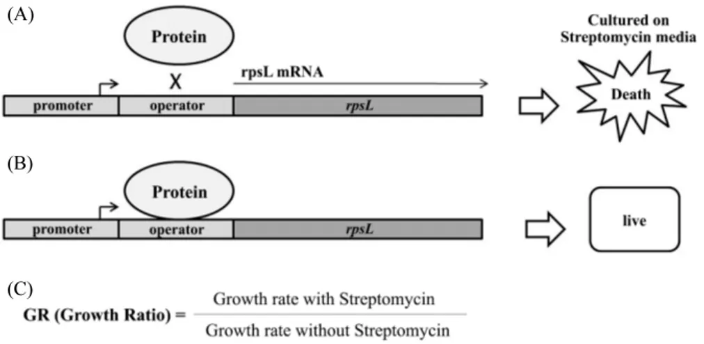
결합 정도를 수치화 한 값으로 rpsL 유전자(Lee et al., 1998)의 발현에 따른 streptomycin 배지에서의 성장 정도를 측정한 것 이다. 이러한 in vivo 방법론은 streptomycin에 저항성이 있는 rpsL20 돌연변이 유전자를 가진 HB101 균주에서 rpsL 유전자 가 발현하게 되면 streptomycin 배지에서 단백질의 번역에 문제 를 유발하여 대장균의 성장이 억제되는 원리를 이용한 것으로, 대장균의 성장 속도 측정하여 growth rate ratio (Streptomycin 이 첨가된 액체배지에서의 성장 기울기 / streptomycin이 없는 액체배지에서의 성장 기울기) GR 값을 결정한다(Fig. 3C). 이 GR 수치가 1에 가까울수록 단백질이 DNA와 더 강하게 결합 한다. Oc의 20 bp와 rpsL 유전자를 가지고 있는 플라스미드 (pHL1105)를 cnu, hns, dicA 유전자가 제거된 HL100 균주에 도입하고, 이 대장균을 3 ml의 Kan 액체배지에 접종하여 16시 간동안 배양 후 새로운 Kan 액체배지와 Kan/Str 액체배지에 OD600가 0.05가 되도록 희석시킨 뒤 25°C 또는 37°C에서 배양 하면서 1시간마다 OD600를 측정하였다.
DicA와 DicA1 단백질 정제
DicA와 DicA1 (L108F) 단백질 정제는 dicA 또는 dicA1 유전자를 BamHI, HindIII 제한효소를 이용하여 pET22b(+) (Novagen)에 클로닝한 플라스미드 DNA를 BL21(DE3) 균주 에 넣어 사용하였다. 균주는 OD600이 0.6일때 0.1 mM IPTG를
첨가한 후 18°C 250 rpm 조건으로 16시간 배양하였다. 배양된 세포는 Lysis Buffer (8 M urea, 100 mM NaH2PO4, 100 mM
Tris-HCl pH 8.0) 50 ml로 resuspend 시킨 후 sonication하여 세 포벽을 부수었다. 4°C, 6000G, 15분간 원심분리한 Cell lysate 에서 상층액을 분리하고, 0.22 μm filter에 여과후 Ni-NTA affinity chromatography column (Qiagen)에 여과시켰다. 그 다음 washing buffer (8 M urea, 100 mM NaH2PO4, 100 mM Tris-
HCl; pH 6.3) 10 ml로 3회 반복하여 washing하였다. 이후 elution buffer (8 M urea, 100 mM NaH2PO4, 100 mM Tris-
HCl; pH 4.5) 500 μl씩 8회에 나누어 용출시킨 단백질을 SDS- PAGE gel에 로딩하여 DicA 또는 DicA1 단백질이 포함된 분획을 확인하였다. 확인된 단백질 분획 500 μl를 12KD dialysis bag에 넣고 refolding buffer (4 M urea, 100 mM NaH2PO4, 100 mM Tris-HCl, 0.5% NP-40, pH 8.0) 500 ml 안에서 24시간 동안 회 전시킨다. 이후 urea 농도가 2 M, 1 M, 0 M로 감소하는 refolding 버퍼 500 ml에 각각 24시간씩 회전시킨후, 침전을 분리한 상층액을 모아 15% SDS-PAGE 로 전기영동한다.
Western blot analysis
15% SDS-PAGE 로 전기영동 한 gel을 Transfer buffer (20 mM Tris, 150 mM glycine, 20% Methanol)로 전처리한 Nitrocellulose blotting membrane (Amersham)에 전기영동을 통해 이동시킨다. Membrane은 Anti-HIS antibody (Millipore)로 1차, HRP-2차 antibody (Santa Cruz)로 처리한 후, ECL 반응액(Amersham) 반응후 X-ray에 감광하여 DicA-6Xhis 또는 DicA1-6Xhis 단 백질을 확인하였다.
EMSA (Electrophoretic mobility shift assay)
Oc를 포함하는 PdicAC 89 base pair (PdicA89)의 DNA는 pHL1292 10 ng (Table 2), 89_F (5'-CATTTAAAACACCTTT ATTGTTAG-3') 0.5 μM, 89_B (5'-CATAGTTAGCTAA TGCTAAATCGT-3') 0.5 μM primer, 1 Unit phusion DNA polymerase (Thermo), 200 μM dNTP, 1X PCR buffer를 반응 액에 첨가하여, 98°C 20초, 58°C 30초, 72°C 30초 반응을 35회 반복하여 증폭하였다. Oc-mutant 포함하는 PdicAC 89 base pair의 DNA는 Oc*-up 0.5 μM (5'- TTGTTAGTCATAACTC
ACAA-3')을 이용한 megaprimer PCR 기법을 이용하였다. 증 폭한 DNA를 10 nM 로 희석하여 1X T4 polynucleotide kinase (PNK) buffer, PNK (TaKaRa) 5 Unit, 2 μl [r-32P]ATP (6000Ci/ mmol) (PerkinElmer)으로 37°C에서 30분, 65°C에서 10분 반 응 시킨 후 G-50 column (GE Healthcare)으로 용출시켰다. 방사 선동위원소로 표지된 DNA 0.4 nM, 1X EMSA buffer (40 mM HEPES; pH 8.0, 8 mM MgCl2, 60 mM potassium glutamate, 5
mM DTT, 0.05% NP-40), salmon sperm DNA (250 ng)와 정제 된 단백질 또는 Crude 단백질과 정해진 온도에서 10분간 반응 시킨 뒤 EMSA loading buffer (50% Glycerol, 2X EMSA buffer, 0.5% Bromophenol blue, 0.5% Xylene cyanol)와 섞어 10% polyacrylamide gel에 1X TBE를 이용하여 100 V의 전압 으로 전기영동한다. 전기영동이 끝난 gel을 Whatman 3 M paper에 붙여 떼어낸 후 비닐과 스카치테이프로 밀봉후 X-ray 필름에 감광하여 EMSA 패턴을 확인하였다.
결과 및 고찰
DicA 단백질은 N-말단의 DNA 결합 도메인으로 Oc에 결합 한다.
DicA 단백질의 아미노산서열 분석 결과, N-말단의 70개 의 아미노산 서열이 Salmonella phage P22의 C2 repressor 단 백질의 N-말단과 59%의 일치함을 보이며, DicA의 C-말단의 65개 아미노산은 Yersinia의 RovA, Salmonella의 SlyA 단백 질의 N-말단의 아미노산 서열과 각각 29%, 27% 일치한다 (Fig. 2A). 이렇듯 DicA 단백질은 두개의 DNA 결합 도메인과 homology를 보이는데, DicA의 N-말단과 homology를 보이는 C2 단백질은 DNA 결합 도메인인 alpha-helix 3 (α3)가 9T- Operator로 알려진 20개의 DNA 염기, 그중 TTAA 서열과 상 호작용하고 있다(Watkins et al., 2008). 이 TTAA 서열은 Oc의 TAA와 염기서열과 그 위치가 유사하며, 이 염기서열과 상호 작용하는 α3의 아미노산 서열은 DicA와 70% 일치한다. 반면 RovA와 SlyA 단백질은 rovA1, slyA operator에서 염기와 상 호작용하는 alpha-helix 4 (α4)의 아미노산 서열이 DicA와 14.3% 의 낮은 일치율을 보인다(Fig. 2A). DicA의 C-말단에 α4와 같 은 구조가 보존된다고 가정하여도, in silico 분석만으로는 어 떤 아미노산이 DNA 염기와 상호작용하는지 특정할 수 없었 다(Dolan et al., 2011; Quade et al., 2012).
그리하여 C2 repressor의 결합구조 분석을 기반으로 DicA 의 N-말단 도메인에 위치하는 39번째 아미노산인 글루타민 과 42번째 알지닌이 Oc 와의 상호작용에 주요할 것으로 판단
(A)
(B) (C) (D)
Fig. 2. Structural analysis of DicA-homologous proteins for DNA interaction. Amino acid sequence alignment homologous to DicA protein. The N-terminal part of DicA lines up with the N-terminal of the C2 repressor protein from the Salmonella phage P22. The C-terminal part of DicA shares homology with the N-termini of RovA of Yersinia and SlyA of Salmonella. The identical amino acids are shown in black. The DNA-binding domain is underlined with the secondary structure labeled (A) (Yun et al., 2012). C2 repressor crystal structure (PBD: 2R1J) (B), RovA crystal structure (PBD: 4AIJ) (C) and SlyA crystal structure (PBD: 3Q5F) (D) with its interacting DNA sequence comparing with Oc. The protein structure of dimer was represented by a solid line structure and a meshed form. Alpha-helix interacting with DNA was indicated with label. DNA binding sequences are given below the structure and nucleotides contacting with alpha-helix structures are indicated in boldface.
(A)
(B)
(C)
Fig. 3. A principle of the In vivo DNA binding assay. The pHL1105 plasmid system should be introduced to HL100 strain which has rpsL20 mutant that streptomycin resistant. If the target protein could not bind to operator, rpsL is expressed resulting in streptomycin sensitive phenotype (A). If the target protein binds to operator, rpsL is repressed, resulting in streptomycin resistance phenotype (B). GR (Growth Ratio) is the binding parameter that indicates binding activity of target protein. If GR value close to 1, means protein binding to operator well. If GR value close to 0, means protein doesn’t bind to operator (C).
하여 두 위치의 아미노산을 글루탐산으로 치환한 돌연변이 DicA를 제작하였다. 글루타민 또는 알지닌을 글루탐산으로 치환하였을 때, 아미노산 한 개의 치환으로 인한 단백질 구조 의 변화는 최소화 하면서 아미노산 변화에 의한 DicA의 Oc 결 합에 변화가 생길 것으로 기대하였고, 그 결과는 in vivo DNA
binding assay를 통하여 알아보았다(Fig. 3).
DicA 단백질의 in vivo DNA binding assay는 pHL1105 플 라스미드의 rpsL 유전자 앞에 Oc를 위치시켜, DicA 단백질이 Oc에 결합하여 streptomycin에 감수성이 있는 rpsL 유전자의 발현을 억제하는 정도를 측정한다. Streptomycin이 존재하는
(A) (B) (C) (D)
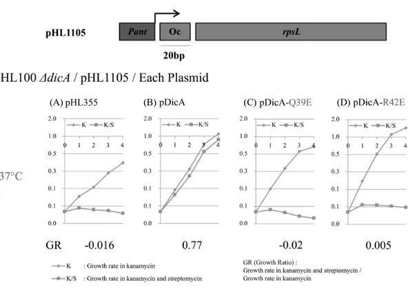
Fig. 4. In vivo DNA binding activity of DicA and DicA-Q39E, DicA-R42E on Oc. In vivo DNA-binding activity of DicA and its mutant DicA-Q39E, DicA-R42E to Oc-20 bp was measured by the growth rate of the host cell. The growth rate at 37°C of the strains HL100ΔdicA/pHL1105/pHL355 (A), HL100 ΔdicA/pHL1105/pDicA (B), HL100ΔdicA/pHL1105/pDicA-Q39E (C), and HL100ΔdicA/pHL1105/pDicA-R42E (D) was measured in LB medium (circle) and in LB containing streptomycin (rectangle). Growth rate was determined from the slope of the fitted curve of the bacterial growth measured by OD600. The regression coefficient for the fitting indicated that all the fittings are statistically significant.
배지와 streptomycin이 존재하지 않는 배지에서 대장균이 자 라는 성장속도를 측정하고 GR (Growth ratio) 수치로 나타내 면, GR 수치가 1에 가까울수록 DicA가 Oc에 잘 결합한다고 해석할 수 있다(Yun et al., 2012). dicA 유전자가 제거된 균주(HL100ΔdicA)에 pHL1105와 pDicA를 도입하여 GR 수치를 측정한 결과 GR값은 0.77로 나 타났고, dicA를 포함하지 않는 플라스미드인 pHL355를 도 입하였을때의 GR값은 -0.016로 나타났다. 이는 WT DicA 단 백질이 Oc에 잘 결합함을 나타낸다(Fig. 4A and B). 그러나 DicA의 N-말단 아미노산에 돌연변이가 도입된 pDicAQ39E, pDicAR42E를 dicA 유전자가 제거된 균주(HL100 ΔdicA)에 pHL1105와 도입한 결과, GR 수치가 -0.02, 0.005로 감소하였 다(Fig. 4C and D). 이 결과는 DicA의 N-말단 도메인의 39번째 글루타민, 42번째 알지닌이 Oc의 염기서열과 상호작용하는 데 중요한 아미노산이며, 이 아미노산이 글루탐산으로 치환된 돌연변이 DicA 단백질은 Oc에 결합하지 못하였음을 의미한 다. 이는 DicA가 N-말단을 이용하여 DNA에 결합하고 있음을 말해준다.
DicA의 온도 의존적인 DNA 결합은 Oc에서 Cnu, H-NS와 독립적으로 일어난다. DicA의 온도 의존적인 DNA 결합 특성을 알아보기 위해 WT 균주(HL100)와 dicA 유전자가 제거된 균주(HL100 ΔdicA)에 pHL1105를 도입하여 25°C와 37°C에서 대장균의 growth rate (GR)을 측정하였다. WT의 경우 25°C에서의 GR은 0.36으로, 37°C에서의 GR인 0.02 보다 높게 나타나 25°C에서 더 강한 결 합이 확인되며(Fig. 5A), dicA 유전자가 제거되면 GR이 25°C 에서 -0.21, 37°C에서 -0.04로 현저하게 감소하는 것으로 보아 WT에서 보인 Oc에서의 온도 의존적 결합은 dicA에 의한 것임 을 확인할 수 있었다(Fig. 5B).
Cnu와 H-NS가 DicA의 온도의존적인 Oc 결합에 영향을 미 치는지를 알아보기 위하여 cnu (HL100 Δcnu)와 hns (HL100 Δhns) 유전자가 제거된 대장균에서 DicA의 in vivo Oc binding assay를 수행하였다. H-NS는 37°C 보다 25°C에서 DNA에 더 잘 결합한다고 알려져 있다(White-Ziegler et al., 1998; Azam and Ishihama, 1999). Cnu와 H-NS가 DicA의 온도의존적인 Oc 결합에 영향을 준다면, 온도에 따른 GR에 변화가 생길 것 으로 예상하였으나 cnu 제거된 균주의 25°C와 37°C에서의
(A) (B) (C) (D)
Fig. 5. In vivo DNA binding activity to Oc. In vivo DNA-binding activity to Oc-20 bp was measured by the GR (growth rate) of the host cell. The GR at 25°C and 37°C of the strains HL100/pHL1105 (A), HL100ΔdicA/pHL1105 (B), HL100Δcnu/pHL1105 (C), and HL100Δhns/pHL1105 (D) was measured in LB medium (circle) and in LB containing streptomycin (rectangle). Growth rate was determined from the slope of the fitted curve of the bacterial growth measured by OD600.
GR 수치는 각각 0.36 과 0.01로(Fig. 5C), hns 유전자가 제거된 균주의 GR은 0.35와 0.02로(Fig. 5D) 여전히 온도 의존적인 DNA 결합을 보였다. 이 결과는 in vivo에서 Oc 20 bp에서의 DicA 결합은 자체적으로 온도 의존적 결합을 하고 있으며, DicA의 이 DNA 결합은 H-NS, 또는 Cnu-H-NS 복합체와 상 관없이 일어나고 있음을 말해준다(Fig. 5A and B).
DicA의 온도 의존적 DNA 결합은 아직 알려지지 않은 in vivo factor에 의해 일어난다
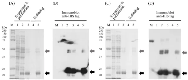
DicA와 DicA1 단백질의 온도의존적 DNA 결합을 in vitro 에서 EMSA (Electrophoretic Mobility Shift Assay)를 통하 여 확인하였다. 대장균에서 DicA와 DicA1의 발현은 대부분 in-soluble 형태로 나타나므로, 발현된 DicA와 DicA1을 8 M urea로 변성 시킨 상태에서 정제 후, 재접힘(refolding) 시키는 방법으로 정제하였다(Fig. 6A and C). 정제후 재접힘이 일어 난 DicA와 DicA1을 확인하기 위하여 anti-HIS 항체를 이용한 western blot을 수행하였다(Fig. 6B and D). 그 결과 재접힌 된
상태의 DicA와 DicA1 단백질이 존재함을 확인하였다(Fig. 6B and D lane 4).
정제된 DicA의 DNA 결합은 EMSA 방법을 통하여 알아보 았다. DicA의 농도 변화에 의해 substrate DNA가 up-shift 된 pattern을 분석하여, substrate DNA에 절반의 DicA가 결합하 는 농도(Half binding concentration of DicA)를 DNA 결합 친 화도인 Kd (dissociation constant) 값으로 정의하였다. DicA가 DNA에 결합했을 때, ‘DicA-major’가 증가하며, 상대적으로 substrate DNA의 양은 줄어드는 것을 확인할 수 있다(Fig. 7). 앞서 수행한 in vivo DNA binding assay에서 측정된 DicA의 DNA 결합 활성이 37°C 보다 25°C에서 크게 나왔던 것과 다르 게(Fig. 5A), 정제된 DicA의 EMSA 실험결과, 25°C, 37°C의 온도에서 Kd 수치는 0.102 nM, 0.100 nM로 DNA 결합활성에 서 큰 차이를 보이지 않았다(Fig. 7A). 이러한 결과는 in vitro에 서 정제된 DicA의 Oc 결합이 온도 의존적이지 않다는 것을 말 해 주고 있다. 반면 정제된 DicA1 돌연변이 단백질은 온도에 따른 DNA 결합활성을 보인다(Fig. 7C). DicA 단백질은 N-말
(A) (B) (C) (D)
Fig. 6. 15% SDS-PAGE and Immunoblot analysis of DicA-6Xhis protein (A, B) and DicA1-6Xhis mutant protein (C, D). Expression, purification of DicA-6Xhis and DicA1-6Xhis protein and their refolding products. Arrows indicate a 6Xhis fused DicA or DicA1 proteins. Monomer indicated with black arrow, Dimer indicated with grey arrow (A). Lanes: M, protein molecular weight markers (Bio-Rad); 1, Total Cell lysate of before IPTG induction; 2, Total Cell lysate of after IPTG induction; 3, purified protein; 4, soluble fraction separated from refolding; 5, precipitated protein from the refolding.
Over expressed DicA and DicA1 proteins have severe insoluble property, purification procedures were performed under denaturation condition using 8 M urea. Coomassie blue staining did not detect DicA or DicA1 protein in the soluble fraction (lane 4 in A, C). However, we were able to detect DicA and DicA1 protein in the soluble fraction when we used the anti-His antibody (lane 4 in B, D).
(A) (B) (C)
Fig. 7. In vitro DNA binding activity to PdicA89 of purified DicA (A), crude extract of E. coli (B), purified DicA1 (C). 0.4 nM of substrate DNA, PdicA89 and purified protein or Crude protein were reacted for 10 min at the specified temperature, 25°C or 37°C. The reaction product was electrophoresed on a 10% polyacrylamide gel. Half binding concentration is shown in the table.
단의 DNA 결합 Domain을 이용하여 Oc에 결합하는데, DicA1 의 경우 C-말단에 위치한 108번째 류신이 페닐알라닌으로 치 환된 돌연변이다(Fig. 2A). dicA1 돌연변이는 25°C에서는 정 상적인 분열을 하지만 37°C에서 dicB 유전자를 억제하지 못 해 대장균의 filamentous growth를 야기한다고 알려져 있다 (Bejar et al., 1988). DicA와 같은 방법으로 정제한 DicA1 단백 질을 이용하여 수행한 EMSA 실험 결과, DicA1의 Kd 수치는 25°C에서 49.51 nM, 37°C에서 27.726 nM로 DicA 단백질보 다 25°C일 때 약 27배, 37°C일 때 50배 가량 DNA 결합활성이 떨어졌고, 특히 37°C의 경우에는 DicA 단백질처럼 DNA와 복 합체를 이루지 못하고 ‘smeared-DicA1 band’를 형성하였다
(Fig. 7C). Substrate DNA가 감소하는 것으로 보아 DicA1 돌연 변이의 경우 DNA 결합 도메인이 DNA에 결합하지만, dimer 를 이루어 안정적인 단백질/DNA 복합체를 형성하지 못해 band를 형성하지 못한 결과로 생각된다.
온도 의존적 in vitro EMSA 실험은 추가적으로 Crude protein 을 이용해서도 분석하였다. Crude protein은 37°C에서 16시간 배양한 대장균을 특정 단백질의 정제과정 없이 sonication으 로 분쇄한 후 원심분리한 상층액을 total protein 정량 이후 사 용하였다. Crude protein을 25°C와 37°C에서 10분간 substrate DNA 결합시킨 후 EMSA를 수행하였다. 그 결과, Crude protein 의 EMSA에서도 정제된 DicA로 수행하였던 EMSA와 같은
(A) (B)
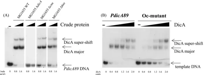
Fig. 8. In vitro DNA binding to PdicAC89 of crude extract of E. coli (A) and purified DicA binding assay with different DNA templates, PdicA89 or Oc-mutant (B). Crude protein was exracted from MG1655 and each dicA, cnu, hns gene deleted strain (A). Purified DicA and different template DNA, PdicA89 or Oc-mutant (Fig. 1B) which single nucleotide change has introduced (B). 0.4 nM of substrate DNA and proteins were reacted for 10 min at 25°C. The reaction product was electrophoresed on a 10% polyacrylamide gel.
‘DicA-band’를 확인할 수 있었다(Fig. 7B). 또한 Crude EMSA 의 Kd 수치는 25°C에서 0.293 μg/μl, 37°C에서는 1.658 μg/μl 으로 25°C에서 37°C 보다 5.7배 높은 Kd 수치를 나타냈다 (Fig. 7B). 이는 crude protein에 DicA 단백질 이외의 다른 인자 들도 존재하므로, in vivo 실험결과와 흡사하게 DicA가 25°C 에서 더 강한 DNA 결합을 할 수 있도록 하거나, 또는 37°C에 서 DNA 결합을 약하게 하는 DicA 이외의 ‘in vivo factor’가 관 여하고 있다는 사실을 보여준다.
DicA는 in vitro에서 Oc에서의 결합을 매개로 복합적인 DNA 결합을 한다.
DicA의 DNA 결합에 Cnu, H-NS의 관여 여부를 알아보기 위하여 dicA, cnu 그리고 hns 유전자가 각각 제거된 MG1655 대장균으로부터 추출한 Crude protein을 이용하여 EMSA를 수행하였다. cnu 와 hns 유전자가 제거된 대장균에서는 WT과 같이 ‘dicA major’와 ‘dicA super-shift’ band를 형성하였으나, dicA가 제거된 대장균의 경우 어떠한 단백질/DNA 복합체를 형성하지 못하였다(Fig. 8A). 이는 ‘dicA major’와 ‘dicA super- shift’ band가 Cnu, H-NS와 상관없이 DicA 단백질에 의해 형 성됨을 말해준다.
PdicA89에서 DicA의 DNA 결합 양상을 분석하기 위하여 Oc-mutation (Fig. 1B)이 도입된 PdicA89에서 EMSA를 정제 된 DicA 단백질을 이용하여 수행하였다. Oc-mutant는 선행된 in vivo 연구에서 DicA의 DNA 결합이 일어나지 않는다고 보 고된 단일염기치환 돌연변이다(Yun et al., 2012). 실험 결과
PdicA89에 Oc-mutant가 도입되면 DicA의 Kd 수치가 0.402 nM에서 2.187 nM로 DNA 결합 활성이 5.4배 감소하였다(Fig. 8B). 또한 두드러지게 ‘DicA major’ band의 형성이 지연되며, DicA 단백질 농도가 1.2 nM에서 2.0 nM로 높아지는 구간에서 ‘DicA major’ band가 점차적으로 감소하며 ‘DicA super-shift’ 가 증가하는 PdicA89의 결과와 대조적으로, 약한 ‘DicA major’ band를 유지하다 ‘DicA super- shift’ band를 형성하는 양상을 보인다(Fig. 8B). PdicA89에는 Oc 이외에 Om, Oa로 알려진 operator 염기서열이 존재한다(Bejar et al., 1988)(Fig. 1B). 각 각의 Om과 Oa 염기서열 20 bp를 이용한 in vivo DNA binding assay에서는 DicA가 결합하지 않는 것으로 나오지만(Yun et al., 2012), Oc와 Om, Oa가 연속적으로 존재하는 PdicA89에 서는 Oc에서의 DicA 결합을 매개로 Om 또는 Oa 위치에 DicA 가 추가적으로 결합하는 식의 ‘cooperative DNA binding’에 의하여 ‘DicA supershift’ band와 같은 단백질/DNA 복합체를 형성되는 것으로 생각되며 이에 대한 추가적인 연구가 필요 하다.
적 요
대장균 세포분열 조절에 관여하는 DicA 단백질은 37°C보 다 25°C에서 DNA에 더욱 잘 결합한다. 그러나 DicA 단백질 의 온도의존적 DNA 결합에 대한 분자적 원인은 명확하지 않 다. 본 연구에서는 DicA 단백질이 어떻게 DNA에 결합하며, 왜 온도 의존적 결합양상을 보이는지 알아보았다. In vivoDNA 결합 분석 결과 RovA나 SlyA와 같은 DicA의 상동성 단 백질과는 달리 DicA는 N 말단에 있는 DNA 결합 도메인을 이 용하여 20개의 염기쌍으로 이루어진 dicC 조절자 유전자(Oc) 에 결합함을 보여주었다. 또한 in vivo 실험에서 DicA는 37°C 보다 25°C에서 DNA에 더 잘 결합하는 것으로 알려진 Cnu 또 는 H-NS의 영향을 받지 않고 자체적으로 Oc에서의 온도 의존 적 DNA 결합을 보인다. 하지만 정제된 DicA 단백질을 이용한 in vitro binding 실험에서는 온도 의존적 DNA 결합이 관찰되 지 않았다. Crude 단백질을 이용한 실험에서 DicA 단백질의 온도 의존적 DNA 결합이 관찰되는 것으로 보아 DicA의 온도 의존적 DNA (Oc) 결합은 crude 단백질내에 존재하는 아직 알 려지지 않은 in vivo factor에 의해 일어난다.
감사의 말
이 논문은 2015년 충남대학교 학술연구지원사업(2015- 1420-01)에 의하여 연구되었음.
References
Azam TA and Ishihama A. 1999. Twelve species of the nucleoid- associated protein from Escherichia coli. Sequence recognition specificity and DNA binding affinity. J. Biol. Chem. 274, 33105– 33113.
Bejar S and Bouche JP. 1985. A new dispensable genetic locus of the terminus region involved in control of cell division in Escherichia coli. Mol. Gen. Genet. 201, 146–150.
Bejar S, Bouché F, and Bouché JP. 1988. Cell division inhibition gene dicB is regulated by a locus similar to lambdoid bacteriophage immunity loci. Mol. Gen. Genet. 212, 11–19.
Bejar S, Cam K, and Bouche JP. 1986. Control of cell division in Escherichia coli. DNA sequence of dicA and of a second gene complementing mutation dicA1, dicC. Nucleic Acids Res. 14, 6821–6833.
Bouche F and Bouche JP. 1989. Genetic evidence that DicF, a second division inhibitor encoded by the Escherichia coli dicB operon, is probably RNA. Mol. Microbiol. 3, 991–994.
Casjens S. 2003. Prophages and bacterial genomics: what have we learned so far? Mol. Microbiol. 49, 277–300.
Datsenko KA and Wanner BL. 2000. One-step inactivation of chro-mosomal genes in Escherichia coli K-12 using PCR products. Proc. Natl. Acad. Sci. USA 97, 6640–6645.
de Boer PA, Crossley RE, and Rothfield LI. 1990. Central role for the Escherichia coli minC gene product in two different cell division- inhibition systems. Proc. Natl. Acad. Sci. USA 87, 1129–1133. Dolan KT, Duguid EM, and He C. 2011. Crystal structures of SlyA
protein, a master virulence regulator of Salmonella, in free and DNA-bound states. J. Biol. Chem. 286, 22178–22185.
Ke SH and Madison EL. 1997. Rapid and efficient site-directed mutagenesis by single-tube ‘megaprimer’ PCR method. Nucleic Acids Res. 25, 3371–3372.
Kim MS, Bae SH, Yun SH, Lee HJ, Ji SC, Lee JH, Srivastava P, Lee SH, Chae H, Lee Y, et al. 2005. Cnu, a novel oriC-binding protein of Escherichia coli. J. Bacteriol. 187, 6998–7008. Lee SY, Lee HJ, Lee H, Kim S, Cho EH, and Lim HM. 1998. In vivo
assay of protein-protein interactions in Hin-mediated DNA inversion. J. Bacteriol. 180, 5954–5960.
Quade N, Mendonca C, Herbst K, Heroven AK, Ritter C, Heinz DW, and Dersch P. 2012. Structural basis for intrinsic thermosensing by the master virulence regulator RovA of Yersinia. J. Biol. Chem. 287, 35796–35803.
Tetart F and Bouche JP. 1992. Regulation of the expression of the cell-cycle gene ftsZ by DicF antisense RNA. Division does not require a fixed number of FtsZ molecules. Mol. Microbiol. 6, 615–620.
Wang X, Kim Y, Ma Q, Hong SH, Pokusaeva K, Sturino JM, and Wood TK. 2010. Cryptic prophages help bacteria cope with adverse environments. Nat. Commun. 1, 147.
Watkins D, Hsiao C, Woods KK, Koudelka GB, and Williams LD. 2008. P22 c2 repressor-operator complex: mechanisms of direct and indirect readout. Biochemistry 47, 2325–2338.
White-Ziegler CA, Angus Hill ML, Braaten BA, van der Woude MW, and Low DA. 1998. Thermoregulation of Escherichia coli pap transcription: H-NS is a temperature-dependent DNA methylation blocking factor. Mol. Microbiol. 28, 1121–1137.
Yun SH, Ji SC, Jeon HJ, Wang X, Kim SW, Bak G, Lee Y, and Lim HM. 2012. The CnuK9E H-NS complex antagonizes DNA binding of DicA and leads to temperature-dependent filamentous growth in E. coli. PLoS One 7, e45236.


