Abstract
BACKGROUND: Biological iron redox transformation alters iron minerals, which may act as effective adsorbents for arsenate [As(V)] in the environments. In the viewpoint of alleviating arsenate, microbial Fe(III) reduction was sought under high concentration of As(V). In this study, Fe(III)-reducing bacteria were isolated from the wild plant rhizosphere soils collected at abandoned mine areas, which showed tolerance to high concentration of As(V), in pursuit of potential agents for As(V) bioremediation.
METHODS AND RESULTS: Bacterial isolation was per-formed by a series of enrichment, transfer, and dilutions. Among the isolated strains, two strains (JSAR-1 and JSAR-3) with abilities of tolerance to 10 mM As(V) and Fe(III) reduction were selected. Phylogenetic analysis us-ing 16S rRNA genesequences indicated the closest mem-bers of Pseudomonas stutzeri DSM 5190 and Paenibacillus selenii W126, respectively for JSAR-1 and JSAR-3. Ferric and ferrous iron concentrations were measured by ferrozine assay, and arsenic concentration was analyzed by ICP-AES, suggesting inability of As(V) reduction whereas ability of Fe(III) reduction.
CONCLUSION: Fe(III)-reducing bacteria isolated from the enrichments with arsenate and ferric iron were found to be resistant to a high concentration of As(III) at 10 mM. We
suppose that those kinds of microorganisms may suggest good application potentials for As(V) bioremediation, since the bacteria can transform Fe while surviving under As- contaminated environments. The isolated Fe(III)-reducing bacterial strains could contribute to transformations of iron minerals which may act as effective adsorbents for arsenate, and therefore contribute to As(V) immobilization
Key words: Arsenic-Resistant, Microbial iron reduction, Paenibacillus, Pseudomonas
Introduction
Arsenic (As) contamination is widespread due to many kinds of anthropogenic activities and natural sources as well, and arsenic in polluted environments can adversely affect the health of millions of people, such as in Bangladesh and West Bengal [1]. Arsenic is stable in several oxidation states, but mostly found as arsenate [As(V) as H2AsO4- and HAsO42-] and arsenite [As(III) as H3AsO30 and H2AsO3-] in aqueous environ-ments [2]. Arsenate is known to be strongly adsorbed to the surface of iron minerals, such as ferrihydrite and hematite [3, 4]. There have been many studies demonstrating biologicalformation of iron minerals by many types of microorganisms, such as Fe(III)-reducing bacteria and Fe(II)-oxidizing bacteria [5, 6]. In nature, in addition to abiotic iron minerals, those biogenic iron minerals could play as adsorbents for arsenate *Corresponding author: Ji-Hoon Lee
Phone: +82-63-270-2546; Fax: +82-63-270-2550; E-mail: [email protected]
Korean J Environ Agric. 2021;40(1):67-72. English Online ISSN: 2233-4173
Published online 2021 March 31. https://doi.org/10.5338/KJEA.2021.40.1.8 Print ISSN: 1225-3537
Reduction of Dissolved Fe(III) by As(V)-tolerant
Bacteria Isolated from Rhizosphere Soil
Anamika Khanal, Yoonjin Song, Ahyeon Cho, Ji-Hoon Lee
*Department of Bioenvironmental Chemistry, College of Agriculture & Life Sciences, Jeonbuk National University, Jeonju 54896, Korea
Received: 15 March 2021/ Revised: 22 March 2021/ Accepted: 25 March 2021 Copyright ⓒ 2021 The Korean Society of Environmental Agriculture
This is an Open-Access article distributed under the terms of the Creative Commons Attribution Non-Commercial License (http://creativecommons.org/licenses/by-nc/3.0) which permits unrestricted non-commercial use, distribution, and reproduction in any medium, provided the original work is properly cited.
ORCID Ji-Hoon Lee
https://orcid.org/0000-0001-8916-149X
67
and also ferrous iron [Fe(II)] in some iron minerals may have potentials to reduce arsenate as well.
Under a decreasing redox potential condition, ferric iron tends to be reduced to Fe(II) and Fe(III) reduction occurs relatively readily than As(V) reduction. So, Fe(II)-containing iron minerals might act as adsorbents for As(V) in some reducing conditions. Those interactions between solid phase Fe and dissolved As might occur under microaerobic to anoxic conditions such as sediments, flooded soils, and aquifers. Those environments might be niches for Fe(III)-reducing bacteria, which can use solid phase Fe as well as dissolved Fe as electron acceptors for the anaerobic respiration. In such environments, Fe(II)- reducing bacteria may contribute to formation of Fe-minerals and thus interactions between Fe-minerals and arsenic.
Here, we have focused to find Fe(III)-reducing bacteria, which have tolerance to high concentration of As, so that suggest potentials for biotransformation of Fe-minerals and adsorption of As by those minerals in arsenic-contaminated environments. Furthermore, the bacterial strains may suggest potentials to be used for bioremediation of dissolved arsenic in microaerobic to anoxic conditions.
Materials and Methods
Sample collectionThe soil samples were collected from rhizosphere of wild plants from the abandoned mine area, Jeongseon, Gangwon-do, during the collaborative research field tour, July 2018. Sampleswere transported to the laboratory with icepacks and stored at 4ºC until uses for bacterial isolation.
Enrichment and isolation of As(V)-resistant and Fe(III)-reducing bacteria
Bacterial isolation was performed in anaerobic condition using M1 medium [7], containing 1.5 g NH4Cl, 0.1 g KCl, 0.6 g NaH2PO4, 2.52 g NaHCO3, 1.75 g NaCl, 7.15 g HEPES, and 0.05 g yeast extract per 1 liter. To each of 50 ml M1 media in 100 ml-serum bottles, one of three organic substrates of glucose, lactate, and acetate was added at the final concentrations of 10 mM, 5 mM, and 5 mM, respectively, and As(V) was added as Na2HAsO4・ 7H2O (Merck KGaA, Darmstadt, Germany) at the final concentration of 10 mM. In addition, ferric citrate was
supplemented at 2 mM as the potential terminal electron acceptor. The serum bottles were sealed with butyl rubber stoppers and aluminum seals and mixture gas of N2:CO2 (90:10) was purged for 20 min. After autoclave sterilization, one gram of the rhizosphere soilswas inoculated toeach of the serum bottles and incubated at dark at 28°C for 2 weeks. After the enrichment incubations, 1% of the culture mediawere transferred to the fresh serum bottle media consecutively 4 times. The cultures were then diluted to 10-1 to 10-7, respectively and spread onto anaerobically prepared agar plate mediamade of M1 with the same condition, followed by incubation in the anaerobic jars (Thermo Scientific, Massachusetts, USA) with anaerobic gas generating sachets (Oxoid AnaeroGen, Massachusetts, USA) at 28°C for 2 weeks. Grown colonies were transferred and maintained until single and pure colonies wereobserved. From the three different types of incubations by those organic substrates, dozens of colonies were selected and isolated for further studies. Isolated colonies were tested for the aerobic growth using different types of media, such as R2A, lysogeny broth (LB), nutrient broth, and tryptic soy broth (TSB).
DNA extraction and polymerase chain reaction Genomic DNAs of the isolated colonies were extracted from the cultures grown in R2A broth at 28°C for 2 days using Inclone genomic plus DNA preparation kit (Inclone Biotech, Yongin, South Korea). Genomic DNAs were diluted to 1-5 ng µl-1 and used for the PCR. A total of 50 µl of reaction mixture was prepared for the PCR reactions, where 1-2 µl of genomic DNAs (1-5 ng µl-1) and 1 µl of each primer (27F primer, 5′-AGA GTT TGA TCM TGG CTC AG-3′; 1492R primer, 5′-GGT TAC CTT GTT ACG ACT T-3′) at the final 0.2 µM were added. The thermal cycles were performed with an initial denaturation at 95°C for 2 min, followed by 30 cycles of denaturation at 94°C for 30 sec, annealing at 50°C for 30 sec and an extension at 72°C for 1 min 30 sec. The quality and the quantity of the purified DNAswere checked, respectively by using gel electrophoresis and fluorometer (Qubit 3.0 Fluorometer, Invitrogen, Pennsylvania, USA).
Sequencing and phylogenetic analysis
The PCR products of 16S rRNA genes were sequenced at GenoTech (Daejeon, South Korea). The
forward and reverse sequenceswere assembled using BioEdit (v. 7.2.5), and the contigs were compared with GenBank nucleotide database from the National Center for Biotechnology Information (NCBI) using the Basic Local Alignment Search Tool (BLAST) for the initial screening. Sequence alignment, manual calibration, and phylogenetic tree construction were performed in ARB 6.0.6 [8] using the SILVA database 132 Ref NR 99 [9].
Biochemical characterization of the isolated strain Biochemical testson the selected isolates were performed by using API 50 CH Kit, API API 50 CHL Medium, and API 50 CHB/E Medium (bioMerieux SA, France). The color change of the strips was determined after 24 h and 48 h, as suggested in the manufacturer’s manuals.
Ironreduction under arsenic condition
Isolated strains that showedFe(III) reduction among the tested carbon sourceswerechosenfor the following experiment. The same incubation condition as the enrichment was used: M1 media supplemented with 2 mM Fe(III) as ferric citrate, 10 mM As(V) as solidum arsenate, and the selected organic carbon at the appropriate concentrations. To inoculate the cells, the isolated bacterial cells were grown aerobically in R2A, LB, or TSB, harvested, and normalized to approximately 2x10⁸ cells ml-1.
The Fe(III) reduction was measured by ferrozine assay [10], which determines dissolved ferrous iron [Fe(II)]. The aliquots of media were withdrawn using the syringes from the serum bottles, and 1 ml of the media was mixed with 1 ml of 1N HCl, followed reaction with ferrozine (3-(2-pyridyl)-5,6-diphenyl-1,2,4- triazine-p,p’-disulfonic acid monosodium salt) (Fluka, Buchs, Switzerland) solution (1 g l−1 ferrozine in 50 mM HEPES at pH 7) to make ferrouscomplex, which exhibits maximum absorbance at 562 nm. Ferrous ethylene diammonium sulfate tetrahydrate (Fluka, Buchs, Switzerland) was used to make Fe(II) standard solutions [6].
To identify potential bacterial reduction of As(V), concentrations of As(V) and As(III) were measured by using inductively coupled plasma atomic emission spectroscopy (ICP-AES) (ICPS-7500; Shimadzu, Kyoto, Japan). To separate As species from solution, solid- phase extraction cartridge (SPE) (Supelclean LC-SAX SPE 3-ml, Supelco) was used [11]. Aliquots of the
media were filtrated through the SPE, acidified with 2 vol % HNO3, and analyzed for As(III). Arsenate that was retained in the cartridge was eluted by using 1 M HCl and analyzed [12].
Results and Discussion
Bacterial isolation and phylogenetic analysis Among dozens of isolated strains, JSAR-1 and JSAR-3 were selected for this study, based on the abilities of anaerobic Fe(III) reduction in the presence of 10 mM As(V) with 10 mM glucose or 10 mM sucrose as carbon source and electron donor. Both the strains also showed aerobic growth on R2A, LB, nutrient broth, and TSB, thereby representing the strains as facultative anaerobes.
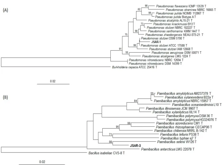
16S rRNA gene sequences from both the strains were compared with GenBank database, indicating that strain JSAR-1 was closest to Pseudomonas stutzeriW1 (KT380544) at 98.50% similarity, and strain JSAR-3 was closest to Paenibacillus anaericanus LMG 23878(AM745262) at 98.63% similarity. The 16S rRNA gene sequences of JSAR-1 and JSAR-3 were deposited to the GenBank with the accession numbers of MK305828 and MK305830, respectively. Neighbor- joining phylogenetic trees were constructed using those sequences with SILVA database in ARB, indicating the closest members of Pseudomonas stutzeri DSM 5190 and Paenibacillus selenii W126, respectively for JSAR-1 and JSAR-3 (Fig. 1).
Biochemical characterization of the isolated strains
Substrates that were utilized by the bacteria were examined by API kits. Strain JSAR-1 showed to utilize D-xylose, D-galactose, D-glucose, D-fructose, D-mannose, D-mannitol, N-acetylglucosamine, D-cellobiose, D- maltose, D-lactose, D-saccharose, D-trehalose, D- raffinose, amidon, esculin ferric citrate, glycogene, gentiobiose, D-turanose, D-lyxose after 24 h and D-ribose, amygdaline, and xylitol after 48 h. Similarly, strain JSAR-3 showed to utilize D-ribose, D-xylose, D-galactose, D-glucose, D-fructose, D-mannose, D- mannitol, N-acetylglucosamine, amygdaline, arbutine, esculin ferric citrate, D-cellobiose, D-maltose, D- lactose, D-saccharose, D-trehalose, D-raffinose, amidon, glycogene, gentiobiose, D-turanose, D-lyxose after 24 h and salicine, D-melibiose, and xylitol after 48 h.
Fig. 1. Neighbor-joining phylogenetic treesof (A) JSAR-1 and (B) JSAR-3 using ARB 6.0.6. Percentages in the bootstrap test from 1,000 replicate trees are shown next to the branches. Outgroups are included.
Fe(III) reduction experiment
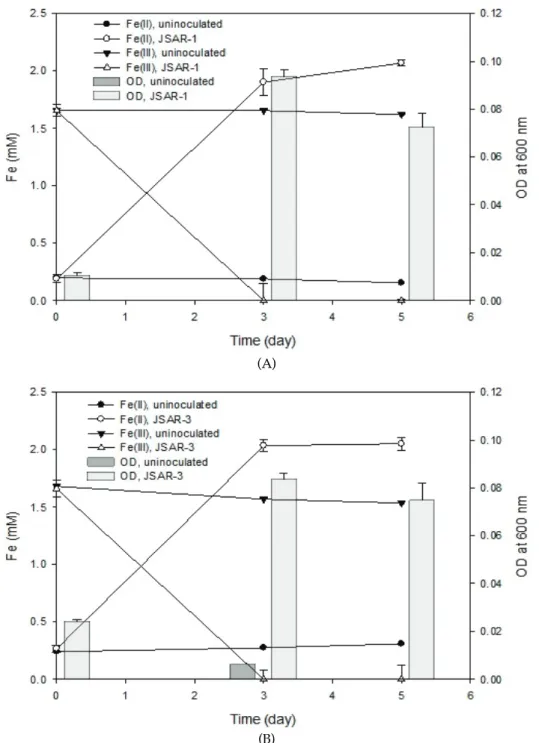
The strains were incubated anaerobically to elucidate potentials of Fe(III) reduction and As(V) reduction in the presence of both 2 mM Fe(III) and 10 mM As(V) with 10 mM glucose in M1 media. Reduction of As(V) to As(III) was not observed in the anaerobic incubations of the strains JSAR-1 and JSAR-3 by ICP-AES analysis (data not shown), although both of the strains were tolerant to dissolved arsenate upto 10 mM, which was supported by optical densities (OD) of the bacterial growths (Fig. 2).
Compared to the uninoculated control bottles, the bacterial cultures of strain JSAR-1 showed almost complete reduction of 2 mM Fe(III) (as ferric citrate) within 5 d with the use of glucose as a carbon source (Fig. 2A). During the time course bacterial growth was monitored by OD, indicating obvious anaerobic growth using Fe(III) as the sole electron acceptor. Similar type of result has been demonstrated by
JSAR-3: Fe(III) was reduced almost completely, while OD increased (Fig. 2B).
Conclusion
Fe(III)-reducing bacteria isolated from the enrichments with arsenate and ferric iron were found to be resistant to a high concentration of As(III) at 10 mM. We suppose that those kinds of microorganisms may suggest good application potentials for As(V) bioremediation, since the bacteria can transform Fe while surviving under As-contaminated environments. Microbial Fe(III) reduction could generate many types of iron minerals which may act as effective adsorbents for arsenate, and therefore contribute to As(V) immobilization. In addition, those saccharide- utilizable facultative microbes would be better option for actual applications, due to relatively easier use and maintenance.
(A)
(B)
Fig. 2. Microbial Fe(III) reduction experiments in the presence of 10 mM As(V) with (A) JSAR-1 and (B) JSAR-3. Concentrations of Fe(III) and Fe(II) shown in symbolled lines and optical density at 600 nm for bacterial growth shown in bar charts. Error bars represent standard deviation values from tree individual replicate incubations.
Note
The authors declare no conflict of interest.
Acknowledgement
This research was funded by National Research Foundation of Korea (NRF) (Grant No. 20162016R1D 1A3B01012231).
References
1. Nickson R, McArthur J, Burgess W, Ahmed KM, Ravenscroft P, Rahman M (1998) Arsenic poisoning of Bangladesh groundwater. Nature, 395: 338. https://doi.org/10.1038/26387.
2. Oremland RS, Stolz JF (2003) The ecology of arsenic. Science, 300(5621), 939-944.
3. La Force MJ, Hansel CM, Fendorf S (2000) Arsenic speciation, seasonal transformations, and co- distribution with iron in a mine waste-iInfluenced palustrine emergent wetland. Environmental Science & Technology, 34(18), 3937-3943.
https://doi.org/10.1021/es0010150.
4. Manning BA, Fendorf SE, Goldberg S (1998) Surface structures and stability of arsenic(III) on goethite: Spectroscopic evidence for inner-sphere complexes. Environmental Science & Technology, 32(16), 2383- 2388. https://doi.org/10.1021/es9802201.
5. Shelobolina E, Xu H, Konishi H, Kukkadapu R, Wu T, Blöthe M, Roden E (2012) Microbial lithotrophic oxidation of structural Fe(II) in biotite. Applied and Environmental Microbiology, 78(16), 5746-5752. https://doi.org/10.1128/AEM.01034-12.
6. Fredrickson JK, Zachara JM, Kennedy DW, Dong H, Onstott TC, Hinman NW, Li S-m (1998) Biogenic iron mineralization accompanying the dissimilatory reduction of hydrous ferric oxide by a groundwater bacterium. Geochimica et Cosmochimica Acta, 62(19-20), 3239-3257.
https://doi.org/10.1016/S0016-7037(98)00243-9. 7. Lee JH, Kennedy DW, Dohnalkova A, Moore DA,
Nachimuthu P, Reed SB, Fredrickson JK (2011) Manganese sulfide formation via concomitant
microbial manganese oxide and thiosulfate reduction. Environmental Microbiology, 13(12), 3275-3288. https://doi.org/10.1111/j.1462-2920.2011.02587.x. 8. Ludwig W, Strunk O, Westram R, Richter L, Meier
H, Yadhukumar, Arno B, Tina L, Susanne S et al. (2004) ARB: a software environment for sequence data. Nucleic Acids Research, 32(4), 1363-1371. https://doi.org/10.1093/nar/gkh293.
9. Quast C, Pruesse E, Yilmaz P, Gerken J, Schweer T, Yarza P, Jörg P, Frank OG (2013) The SILVA ribosomal RNA gene database project: improved data processing and web-based tools. Nucleic Acids Research, 41(D1), 590-596.
https://doi.org/10.1093/nar/gks1219.
10. Stookey LL (1970) Ferrozine - a new spectrophotometric reagent for iron. Analytical Chemistry, 42(7), 779-781. https://doi.org/10.1021/ac60289a016.
11. Yalçin S, Le XC (2001) Speciation of arsenic using solid phase extraction cartridges. Journal of Environmental Monitoring, 3(1), 81-85.
https://doi.org/10.1039/B007598L.
12. Jiang S, Lee JH, Kim MG, Myung NV, Fredrickson JK, Sadowsky MJ, Hur HG (2009) Biogenic Formation of As-S Nanotubes by Diverse Shewanella Strains. Applied and Environmental Microbiology, 75(21), 6896-6899. https://doi.org/10.1128/aem.00450-09.