Should an Ulnar Styloid Fracture Be Fixed Following
Volar Plate Fixation of a Distal Radial Fracture?
By Jae Kwang Kim, MD, PhD, Young-Do Koh, MD, PhD, and Nam-Hoon Do, MD
Investigation performed at the Department of Orthopedic Surgery, Ewha Medical Research Institute, School of Medicine, Ewha Womans University, Seoul, South Korea
Background: Ulnar styloid fractures often occur in association with distal radial fractures. The purpose of this study was to determine whether an associated ulnar styloid fracture following stable fixation of a distal radial fracture has any effect on wrist function or on the development of chronic distal radioulnar joint instability.
Methods: One hundred and thirty-eight consecutive patients who underwent surgical treatment of an unstable distal radial fracture were included in this study. During surgery, none of the accompanying ulnar styloid fractures were internally fixed. Patients were divided into nonfracture, nonbase fracture, and base fracture groups, on the basis of the location of the ulnar styloid fracture, and into nonfracture, minimally displaced (£2 mm), and considerably displaced (>2 mm) groups, according to the amount of ulnar styloid fracture displacement at the time of injury. Postoperative evaluation included measurement of grip strength and wrist range of motion; calculation of the modified Mayo wrist score and Disabilities of the Arm, Shoulder and Hand score; as well as testing for instability of the distal radioulnar joint at a mean of nineteen months postoperatively.
Results: Ulnar styloid fractures were present in seventy-six (55%) of the 138 patients. Forty-seven (62%) involved the nonbase portion of the ulnar styloid and twenty-nine (38%) involved the base of the ulnar styloid. Thirty-four (45%) were minimally displaced, and forty-two (55%) were considerably (>2 mm) displaced. We did not find a significant relationship between wrist functional outcomes and ulnar styloid fracture level or the amount of displacement. Chronic instability of the distal radioulnar joint occurred in two wrists (1.4%).
Conclusions: An accompanying ulnar styloid fracture in patients with stable fixation of a distal radial fracture has no apparent adverse effect on wrist function or stability of the distal radioulnar joint.
Level of Evidence: Prognostic Level II. See Instructions to Authors for a complete description of levels of evidence.
U
lnar styloid fractures are commonly associated with distal radial fractures, but their effects on functional outcome are unclear. Several clinical studies have concluded that a fracture of the ulnar styloid has no impact on anatomical, radiographic, or functional results when it ac-companies a distal radial fracture1-5; however, others have found that ulnar styloid fractures are associated with distal radioulnar joint instability, a concomitant triangular fibro-cartilage complex tear, and a decreased range of motion or decreased grip strength in affected wrists6-9
. It is important to
determine whether an accompanying styloid fracture affects distal radioulnar joint stability and outcome because styloid fracture fixation can be difficult and potentially introduces the risk of additional complications.
We believe that this uncertainty about the effect of an accompanying ulnar styloid fracture probably originates from the heterogeneity of previous study designs. Thus, we limited our study to patients with a distal radial fracture that had stable fixation with a volar locking plate. The purpose of this pro-spective study was to determine whether an accompanying A commentary by Moheb S. Moneim, MD,
is available at www.jbjs.org/commentary and as supplemental material to the online version of this article.
Disclosure: The authors did not receive any outside funding or grants in support of their research for or preparation of this work. Neither they nor a member of their immediate families received payments or other benefits or a commitment or agreement to provide such benefits from a commercial entity.
ulnar styloid fracture in patients with stable fixation of a distal radial fracture has any effect on wrist function or distal radio-ulnar joint stability. Our null hypothesis was that an un-repaired ulnar styloid fracture, regardless of fracture level or displacement, does not affect wrist function or distal radio-ulnar joint stability.
Materials and Methods
Study Population
O
ur institutional review board granted approval for this study. Inclusion criteria were an unstable distal radial fracture with one or more of the following findings: an intra-articular fracture with a step-off or gap of >1 mm, radial shortening of >5 mm, or dorsal angulation of >20°. Exclusion criteria included stable fractures treated nonoperatively, pre-existing severe illness, or a refusal to undergo surgery. Skele-tally immature patients and patients with a previous wrist injury, bilateral fractures, open fracture, or a concomitant ul-nar head or neck fracture were also excluded.Between March 2005 and March 2007, 149 consecutive patients met our inclusion criteria. The surgical procedures were performed by two surgeons (J.K.K. and Y.-D.K.). Nine of the 149 patients were excluded. One patient died, four could not be located, and four refused to return for follow-up evaluation. In addition, two patients lost initial reduction during follow-up and were excluded. This left 138 patients (eighty-five women and fifty-three men) with a mean age of forty-nine years (range, seventeen to eighty-eight years). Patients were followed for a mean of nineteen months (range, twelve to thirty-six months). Distal radial fractures were reduced and internally fixed with a 2.4-mm volar locking compression plate (Synthes, Paoli, Pennsylvania) in all study subjects. During surgery, no pro-cedure was undertaken on an accompanying ulnar styloid fracture. After fixation, laxity of the distal radioulnar joint was determined in the following manner: The distal end of the radius was grasped by the examiner, with the forearm in a neutral position, and the distal end of the ulna, grasped by the contralateral hand of the examiner, was moved in dorsal and palmar directions with respect to the radius3,10
. Intraoperative laxity of the distal radioulnar joint was determined to be present when the distal end of the ulna translated noticeably without end-point resistance relative to the contralateral, un-injured side. In thirty-two patients with intraoperative laxity of the distal radioulnar joint, a sugar tong splint was applied with the forearm in 30° of supination for four weeks. In patients in whom the distal radioulnar joint was deemed unstable, a short arm splint was applied for four weeks. Subsequently, wrist motion was allowed with intermittent protection with a short arm brace for another two weeks.
Radiographic Evaluations
Preoperative radiographs of all 138 patients were evaluated for associated ulnar styloid fractures. Ulnar styloid fracture level and displacement were determined on posteroanterior radio-graphs of the injured wrist by a single hand surgeon (J.K.K.) using a millimeter ruler (Fig. 1). Patients were assigned to a
nonfracture group, a nonbase fracture group, or a base fracture group, depending on the ulnar styloid fracture level. The nonfracture group had no accompanying ulnar styloid frac-ture. The fracture group had either a nonbase fracture located in the distal portion of the ulnar styloid or a base fracture at the styloid base or in the foveal area of the ulnar head. We also divided patients into three groups depending upon the amount of fracture fragment displacement at the time of injury: a nonfracture group, a minimally displaced (£2 mm) group, and a considerably displaced (>2 mm) group.
Plain posteroanterior and lateral radiographs of the wrist were made immediately after surgery and at the final follow-up visits. Radial inclination, radial height, volar tilt, and ulnar variance were compared with use of the immediate post-operative and final follow-up radiographs. Articular incon-gruity was measured, and ulnar styloid fracture displacements were reevaluated after internal fixation of the distal radial fractures. The healing status of ulnar styloid fractures was also evaluated.
Perioperatively, lateral radiographs of the contralateral, uninjured wrist were examined to evaluate subluxation of the distal radioulnar joint. A difference of >5 mm between the radioulnar distance of the injured and the uninjured wrist indicates subluxation of the distal radioulnar joint when a difference in the pisoscaphoid distance was £3 mm (Fig. 2)11
.
Fig. 1
Ulnar styloid fracture displacement was measured on a pos-teroanterior radiograph of the injured wrist with use of a milli-meter ruler.
Clinical Evaluations
We compared wrist function and the degree of instability of the distal radioulnar joint between groups after a minimum follow-up of one year. One author (N.-H.D.), who was un-aware of the radiographic results, examined all patients.
Wrist function was evaluated with use of grip strength, wrist range of motion, the modified Mayo wrist score12
, and the Disabilities of the Arm, Shoulder and Hand (DASH) score13
. Grip strength was measured with use of a Jamar dy-namometer (Sammons Preston, Bolingbrook, Illinois) with the elbow flexed 90° and the forearm in neutral rotation. Range of motion of the wrist and forearm (extension, flexion, supination, and pronation) was measured with use of a goni-ometer. The modified Mayo wrist score has a 100-point scale, with 100 representing normal wrist function. It comprises separate ratings for pain (25 points), range of motion (25 points), grip strength (25 points), and functional status (25 points). The DASH questionnaire consists of thirty items, twenty-one of which address the ability to perform specified activities, whereas the other nine concern symptoms. The
DASH score ranges from 0 to 100, with higher scores indi-cating greater disability.
Chronic instability of the distal radioulnar joint was assessed by physical examination3,10
. The radius was grasped by one hand of the examiner with the forearm in a neutral po-sition, and the distal end of the ulna, which was fixed by the contralateral hand of the examiner, was translated in dorsal and palmar directions with respect to the radius. Distal radio-ulnar joint instability was determined to be present when both noticeable displacement at the distal radioulnar joint relative to the contralateral, uninjured side and pain or apprehension of the distal radioulnar joint were evident.
Statistical Analysis
Analysis of variance was used to evaluate significant differences between the study groups in terms of wrist function. The Fisher exact test and the Pearson chi-square test were used to evaluate significant differences between the study groups in terms of intraoperative laxity of the distal radioulnar joint, chronic instability of the distal radioulnar joint, and frequency of ulnar styloid fracture union. The Student t test was used to compare radiographic parameters. A p value of <0.05 was considered significant throughout.
Power Analysis
A post hoc power analysis was performed with use of analysis of variance and equivalence testing as a model for our study. A sample size of twenty-six patients per group would be neces-sary to have >80% power to detect a minimum difference of 10 points in the DASH score with a standard deviation of 13. With an occurrence rate of 2% for chronic instability of the distal radioulnar joint and an acceptable difference in the risk of instability between the groups of 5%, a sample size of ninety-seven patients per group would be necessary to have >80% power to detect a difference.
Source of Funding
We received no funding for this project.
Results
Ulnar Styloid Fracture Level (Table I)
U
lnar styloid fractures were present in seventy-six (55%) of the 138 patients. Of those fractures, forty-seven (62%) involved the nonbase portion of the ulnar styloid and twenty-nine (38%) involved its base.At the time of the final follow-up, the mean flexion-extension arc (and standard deviation) was 104° ± 13° in the nonfracture group, 102° ± 15° in the nonbase fracture group, and 108° ± 14° in the base fracture group. These differences were not significant (p = 0.34). The mean supination-pronation arc was 159° ± 18° in the nonfracture group, 155° ± 24° in the nonbase fracture group, and 160° ± 21° in the base fracture group. These differences were not significant (p = 0.64). The mean grip strength was 24 ± 4 kg in the nonfracture group, 23 ± 4 kg in the nonbase fracture group, and 24 ± 6 kg in the base fracture group (p = 0.44). The mean modified
Fig. 2
The pisoscaphoid distance (P) and the radioulnar distance (S) were measured on a lateral radiograph of the wrist. The piso-scaphoid distance indicates the distance between the volar end of the pisiform and the volar end of the scaphoid, and the ra-dioulnar distance represents the distance between the dorsal cortices of the radius and ulna at the distal radioulnar joint.
Mayo wrist score was 87 ± 14 points in the nonfracture group, 85 ± 12 points in the nonbase fracture group, and 89 ± 15 points in the base fracture group (p = 0.41). The mean DASH score was 14 ± 10 points in the nonfracture group, 16 ± 11 points in the nonbase fracture group, and 13 ± 8 points in the base fracture group (p = 0.46).
Ulnar Styloid Fracture Displacement (Table II)
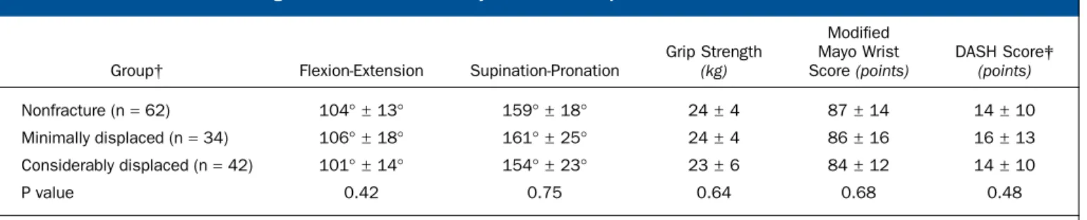
Of the seventy-six ulnar styloid fractures, thirty-four (45%) were minimally displaced and forty-two (55%) were consider-ably displaced. The mean flexion-extension arc was 106° ± 18° in the minimally displaced group and 101° ± 14° in the con-siderably displaced group (p = 0.42). The mean supination-pronation arc was 161° ± 25° in the minimally displaced group and 154° ± 23° in the considerably displaced group (p = 0.75). The mean grip strength was 24 ± 4 kg in the minimally displaced group and 23 ± 6 kg in the considerably displaced group (p = 0.64). The mean modified Mayo wrist score was 86 ± 16 points in the minimally displaced group and 84 ± 12 points in the considerably displaced group (p = 0.68). The mean DASH score was 16 ± 13 points in the minimally displaced group and 14 ± 10 points in the considerably displaced group (p = 0.48).
Intraoperative and Chronic Instability of the Distal Radioulnar Joint
Of the thirty-two patients with intraoperative laxity of the distal radioulnar joint, thirteen (41%) were in the
non-fracture group, ten (31%) were in the nonbase non-fracture group, and nine (28%) were in the base fracture group. In-traoperative laxity of the distal radioulnar joint did not cor-relate significantly with the ulnar styloid fracture level (p = 0.53). In terms of ulnar styloid fracture displacement among the thirty-two patients with intraoperative laxity of the dis-tal radioulnar joint, thirteen (41%) were in the nonfracture group, seven (21%) were in the minimally displaced group, and twelve (38%) were in the considerably displaced group. Intraoperative laxity of the distal radioulnar joint did not correlate significantly with ulnar styloid fracture displacement (p = 0.61).
Chronic instability of the distal radioulnar joint was encountered in two male patients (1.4%). One had an AO type-C3 distal radial fracture without an ulnar styloid frac-ture14
. In this patient, the postoperative volar tilt at the radius was 2°, radial inclination was 20°, and ulnar variance was 12 mm. The other patient had an AO type-C2 distal radial frac-ture with a considerably displaced ulnar styloid nonbase fracture. In this patient, the postoperative volar tilt of the ra-dius was 4°, radial inclination was 21°, and ulnar variance was 13 mm. Both patients had intraoperative laxity of the distal radioulnar joint. Although both patients had laxity of the dis-tal radioulnar joint, resulting in ulnar-sided pain during in-stability testing of the distal radioulnar joint, both refused further treatment because their symptoms were not viewed as troublesome.
TABLE I Wrist Function According to the Presence or Absence of an Ulnar Styloid Fracture*
Group Flexion-Extension Supination-Pronation
Grip Strength (kg) Modified Mayo Wrist Score (points) DASH Score† (points) Nonfracture (n= 62) 104° ± 13° 159° ± 18° 24± 4 87± 14 14± 10 Nonbase fracture (n= 47) 102° ± 15° 155° ± 24° 23± 4 85± 12 16± 11 Base fracture (n= 29) 108° ± 14° 160° ± 21° 24± 6 89± 15 13± 8 P value 0.34 0.64 0.44 0.41 0.46
*The values are given as the mean and the standard deviation. †DASH= Disabilities of the Arm, Shoulder and Hand questionnaire.
TABLE II Wrist Function According to the Amount of Ulnar Styloid Fracture Displacement*
Group† Flexion-Extension Supination-Pronation
Grip Strength (kg) Modified Mayo Wrist Score (points) DASH Score‡ (points) Nonfracture (n= 62) 104° ± 13° 159° ± 18° 24± 4 87± 14 14± 10 Minimally displaced (n= 34) 106° ± 18° 161° ± 25° 24± 4 86± 16 16± 13 Considerably displaced (n= 42) 101° ± 14° 154° ± 23° 23± 6 84± 12 14± 10 P value 0.42 0.75 0.64 0.68 0.48
*The values are given as the mean and the standard deviation. †The minimally displaced group had <2 mm of displacement of the fracture fragments, and the considerably displaced group had >2 mm of displacement. ‡DASH= Disabilities of the Arm, Shoulder and Hand questionnaire.
Radiographic Outcomes
The mean amount of displacement of the ulnar styloid fracture in the minimally displaced group was 1.2 ± 0.5 mm at the time of injury and 1.5 ± 0.7 mm immediately after fixation of the distal radial fracture. The mean displacement of the ulnar styloid fractures in the considerably displaced group was 3.8 ± 1.9 mm at the time of injury and decreased to 2.4 ± 1.4 mm immediately after distal radial fracture surgery. This was a significant change (p = 0.01). The amount of ulnar styloid fracture displacement reduced to <2 mm immediately after distal radial fracture surgery in twenty-four of the forty-two considerably displaced fractures.
With regard to the distal radial fractures immediately after surgery, the mean radial inclination was 23° (range, 17° to 25°), mean radial height was 12 mm (range, 8 to 16 mm), mean volar tilt was 9° (range, 2° to 15°), mean ulnar variance was 10.3 mm (range, 23 to 4 mm), and mean articular in-congruity was 0.2 mm (range, 0 to 2 mm). On the final follow-up radiographs, the mean radial inclination was 23° (range, 16° to 26°), mean radial height was 11 mm (range, 8 to 14 mm), mean volar tilt was 9° (range, 2° to 15°), and mean ulnar vari-ance was 10.8 mm (range, 23 to 4 mm). These four radio-graphic parameters were not significantly different in a comparison of postoperative and final follow-up measurements. No patient showed a difference of >5 mm in the radioulnar distance be-tween the injured and uninjured wrist on the lateral postop-erative radiographs.
Of the seventy-six ulnar styloid fractures, thirty-one (41%) appeared united radiographically at the time of final follow-up. Eighteen (38%) of the forty-seven ulnar styloid nonbase fractures and thirteen (45%) of the twenty-nine ulnar styloid base fractures were united. Fifteen (44%) of the thirty-four minimally displaced ulnar styloid fractures and sixteen (38%) of the forty-two considerably displaced ulnar styloid fractures were united. With the numbers studied, ulnar styloid union rates were not dependent on ulnar styloid fracture level (p = 0.74) or displacement (p = 0.77).
Discussion
D
ebate continues as to whether an ulnar styloid fracture accompanying a distal radial fracture affects wrist function and leads to chronic instability of the distal radio-ulnar joint. Concerns about instability of the distal radioradio-ulnar joint in patients with an ulnar styloid fracture originate from the anatomical feature that the radioulnar ligaments (a pri-mary stabilizer of the distal radioulnar joint) attach to the base of the ulnar styloid15,16. Thus, some authors have sug-gested that ulnar styloid fractures that have been avulsed at the styloid base and are displaced by >2 mm can lead to instability of the distal radioulnar joint7,17
. However, other authors have concluded that ulnar styloid fractures accompanying distal radial fractures do not affect wrist function or stability of the distal radioulnar joint1-5,15
. To isolate the effect of an accom-panying ulnar styloid fracture in distal radial fractures on the wrist function or distal radioulnar joint stability, it is necessary to eliminate the effect of dorsal angulation and
shortening of the distal radial fracture fragments because they are the most common causes of instability of the distal radi-oulnar joint following distal radial fractures18,19
. With the de-velopment of stable internal fixation of distal radial fractures, it may now be possible to resolve this problem. Locking plate systems have the advantage of maintaining initial reduction while preventing dorsal angulation and progressive radial shortening even in osteoporotic elderly patients19,20
. In the present study, we included forty-six patients over sixty years of age, and all except two maintained an acceptable fracture reduction after a minimum follow-up interval of one year. All of our patients had an unstable distal radial fracture treated with open reduction and internal fixation with use of a volar locking plate. Our observations suggest that, following such treatment, an unrepaired ulnar styloid fracture, regardless of fracture level or amount of displace-ment, does not affect wrist function or stability of the distal radioulnar joint. This finding is consistent with the recent report by Souer et al.21
, which suggested that an unrepaired fracture of the base of the ulnar styloid does not appear to influence function or outcome after treatment of a distal radial fracture with plate and screw fixation.
In the present study, thirty-two wrists had intraoperative laxity of the distal radioulnar joint after distal radial fixation, but no wrist showed ulnar head subluxation on the immediate postoperative radiographs. This finding suggests that intra-operative laxity of the distal radioulnar joint results from lig-amentous injury that does not disturb its congruity. In our study, the risk of intraoperative laxity of the distal radioulnar joint did not correlate with the presence of an ulnar styloid fracture, concurring with the findings of other authors3,22
. Lindau et al.3
suggested that the presence of an ulnar styloid fracture is not a good indicator of a tear of the triangular fibrocartilage complex, which is a major ligamentous stabilizer of the distal radioulnar joint. Richards et al.22
also proposed a relationship between initial displacement and the presence of a triangular fibrocartilage complex injury, but no correlation was found between ulnar styloid fractures and triangular fibro-cartilage complex injuries. The peripheral portion of the tri-angular fibrocartilage complex, which is the region usually injured in distal radial fractures, is well vascularized and has considerable healing potential if the ruptured ligaments are approximated and reasonably immobilized23
. In the present study, ulnar styloid fractures displaced by >2 mm showed less displacement after fixation of a distal radial fracture, which is consistent with the findings reported by Buterbaugh and Palmer24
. Therefore, although triangular fibrocartilage com-plex tears occur in patients with a distal radial fracture, the triangular fibrocartilage complex is likely to be well reduced after fixation of a distal radial fracture and can heal with nonoperative treatment. In the present study, four weeks of immobilization, even for patients with intraoperative laxity of the distal radioulnar joint, appears to be effective in limiting chronic instability of the distal radioulnar joint as long as an acceptable reduction of the distal radial fracture can be maintained.
Oskarsson et al.8
noted that the presence of an ulnar styloid fracture is a predictor of a poor outcome, but they treated distal radial fractures nonoperatively. Stoffelen et al.9 concluded that ulnar styloid fractures contribute to a poorer result because of their effect on the function of the distal ra-dioulnar joint, but they failed to describe the method used to treat the distal radial fractures in their series. We consider that the maintenance of an anatomical reduction of the distal radial fracture is a prerequisite for the nonoperative treatment of ulnar styloid fractures because instability of the distal radio-ulnar joint may be accentuated when there is associated dorsal angulation or shortening of the distal end of the radius.
In the present study, we, like other authors3,4,10,23,25 , relied mainly on clinical testing to assess chronic instability of the distal radioulnar joint, but this type of testing is subjective and constitutes a drawback of our study. To address this, we also performed functional and radiographic evaluations of the wrist. It has been reported that when distal radioulnar joint instability is present following distal radial fractures, wrist scores worsen and the range of motion of the injured wrist is less4,23
. For this reason, we evaluated range of motion, grip strength, and functional outcome scores. Grip strength, range of motion, and the DASH score have been found to be the most responsive outcome measurements for patients with distal radial fractures26
. Another weakness of this study is that it
is underpowered in terms of identifying the risk of chronic instability of the distal radioulnar joint among the various fracture types. However, differences in the risk of chronic in-stability of the distal radioulnar joint conferred by the ulnar styloid fracture type may be too small to justify operative treatment of the ulnar styloid fracture, provided that an ac-ceptable reduction of the distal radial fracture can be achieved and maintained.
In conclusion, we found that an unrepaired ulnar styloid fracture, regardless of fracture level or amount of displacement, does not affect wrist function or stability of the distal radioulnar joint following treatment of an unstable distal radial fracture with open reduction and internal fixation with use of a volar locking plate and well-controlled postoperative immobilization.n
Jae Kwang Kim, MD, PhD Young-Do Koh, MD, PhD Nam-Hoon Do, MD
Department of Orthopedic Surgery,
Ewha Womans University Mokdong Hospital, 911-1, Mok-6-dong, Yangcheon-gu, Seoul, 158-710, South Korea.
E-mail address for J.K. Kim: kimjk@ewha.ac.kr
References
1. af Ekenstam F, Jakobsson O, Wadin K. Repair of the triangular ligament in Colles’ fracture. No effect in a prospective randomized study. Acta Orthop Scand. 1989;60:393-6.
2. Flinkkil¨a T, Raatikainen T, H¨am¨al¨ainen M. AO and Frykman’s classifications of Colles’ fracture. No prognostic value in 652 patients evaluated after 5 years. Acta Orthop Scand. 1998;69:77-81.
3. Lindau T, Adlercreutz C, Aspenberg P. Peripheral tears of the triangular fibro-cartilage complex cause distal radioulnar joint instability after distal radial fractures. J Hand Surg Am. 2000;25:464-8.
4. Lindau T, Hagberg L, Adlercreutz C, Jonsson K, Aspenberg P. Distal radioulnar instability is an independent worsening factor in distal radial fractures. Clin Orthop Relat Res. 2000;376:229-35.
5. Tsukazaki T, Iwasaki K. Ulnar wrist pain after Colles’ fracture. 109 fractures followed for 4 years. Acta Orthop Scand. 1993;64:462-4.
6. Frykman G. Fracture of the distal radius including sequelae—shoulder-hand-finger syndrome, disturbance in the distal radio-ulnar joint and impairment of nerve function. A clinical and experimental study. Acta Orthop Scand. 1967;Suppl 108:31. 7. May MM, Lawton JN, Blazar PE. Ulnar styloid fractures associated with distal radius fractures: incidence and implications for distal radioulnar joint instability. J Hand Surg Am. 2002;27:965-71.
8. Oskarsson GV, Aaser P, Hjall A. Do we underestimate the predictive value of the ulnar styloid affection in Colles fractures? Arch Orthop Trauma Surg. 1997;116:341-4. 9. Stoffelen D, De Smet L, Broos P. The importance of the distal radioulnar joint in distal radial fractures. J Hand Surg Br. 1998;23:507-11.
10. Kim JP, Park MJ. Assessment of distal radioulnar joint instability after distal radius fracture: comparison of computed tomography and clinical examination re-sults. J Hand Surg Am. 2008;33:1486-92.
11. Nakamura R, Horii E, Imaeda T, Tsunoda K, Nakao E. Distal radioulnar joint subluxation and dislocation diagnosed by standard roentgenography. Skeletal Radiol. 1995;24:91-4.
12. Cooney WP, Linscheid RL, Dobyns JH. Triangular fibrocartilage tears. J Hand Surg Am. 1994;19:143-54.
13. Hudak PL, Amadio PC, Bombardier C. Development of an upper extremity out-come measure: the DASH (disabilities of the arm, shoulder and hand) [corrected].
The Upper Extremity Collaborative Group (UECG). Am J Ind Med. 1996;29:602-8. Erratum in: Am J Ind Med. 1996;30:372.
14. M¨uller ME, Allg¨ower M, Schneider R, Willenegger H. Manual of internal fixation. 3rd ed. New York: Springer; 1991.
15. Garcia-Elias M. Soft-tissue anatomy and relationships about the distal ulna. Hand Clin. 1998;14:165-76.
16. Lindau T. Treatment of injuries to the ulnar side of the wrist occurring with distal radial fractures. Hand Clin. 2005;21:417-25.
17. Mikic ZD. Treatment of acute injuries of the triangular fibrocartilage complex associated with distal radioulnar joint instability. J Hand Surg Am. 1995;20:319-23. 18. Faierman E, Jupiter JB. The management of acute fractures involving the distal radio-ulnar joint and distal ulna. Hand Clin. 1998;14:213-29.
19. Gehrmann SV, Windolf J, Kaufmann RA. Distal radius fracture management in elderly patients: a literature review. J Hand Surg Am. 2008;33:421-9.
20. Orbay JL, Fernandez DL. Volar fixed-angle plate fixation for unstable distal ra-dius fractures in the elderly patient. J Hand Surg Am. 2004;29:96-102. 21. Souer JS, Ring D, Matschke S, Audige L, Marent-Huber M, Jupiter JB; AOCID Prospective ORIF Distal Radius Study Group. Effect of an unrepaired fracture of the ulnar styloid base on outcome after plate-and-screw fixation of a distal radial frac-ture. J Bone Joint Surg Am. 2009;91:830-8.
22. Richards RS, Bennett JD, Roth JH, Milne K Jr. Arthroscopic diagnosis of intra-articular soft tissue injuries associated with distal radial fractures. J Hand Surg Am. 1997;22:772-6.
23. Szabo RM. Distal radioulnar instability. J Bone Joint Surg Am. 2006;88:884-94. 24. Buterbaugh GA, Palmer AK. Fractures and dislocations of the distal radioulnar joint. Hand Clin. 1988;4:361-75.
25. Ruch DS, Lumsden BC, Papadonikolakis A. Distal radius fractures: a compar-ison of tension band wiring versus ulnar outrigger external fixation for the man-agement of distal radioulnar instability. J Hand Surg Am. 2005;30:969-77. 26. MacDermid JC, Richards RS, Donner A, Bellamy N, Roth JH. Responsiveness of the Short Form-36, Disability of the Arm, Shoulder, and Hand questionnaire, patient-rated wrist evaluation, and physical impairment measurements in evaluating re-covery after a distal radius fracture. J Hand Surg Am. 2000;25:330-40.