INTRODUCTION
Hepatocellular carcinoma (HCC) constitutes one of the most common gastrointestinal malignancies worldwide, and its in-cidence continues to increase.1 The historical role of radiation therapy (RT) in HCC patients has been insignificant due to the
low radiation tolerance of the whole liver. However, radiother-apeutic technical advances using three-dimensional confor-mal RT (3D-CRT), intensity-modulated RT (IMRT) and image-guided RT (IGRT) technology have considerably contributed to the improvement of therapeutic ratios in terms of the ad-ministration of higher radiation doses to the target volumes and significant reduction in surrounding normal tissue com-plications.1-3
In locally-advanced–stage HCC, the frequency of uncon-trolled primary tumors after previous irradiation or sustained symptomatic local problems remains high. Thus, RT currently plays an important role in locally-advanced as well as early-stage, locally-confined liver tumors. The need for re-irradiation as a salvage option in HCC patients is also continuously in-creasing.
Despite the development of modern RT technology, re-irra-diation in HCC remains a challenging issue due to the relative-ly low radiation tolerance of the small and large bowels and the
Re-Irradiation of Hepatocellular Carcinoma:
Clinical Applicability of Deformable Image Registration
Dong Soo Lee*, Joong-Yeol Woo, Jun Won Kim, and Jinsil Seong
Department of Radiation Oncology, Yonsei Cancer Center, Yonsei University College of Medicine, Seoul, Korea.
Purpose: This study aimed to evaluate whether the deformable image registration (DIR) method is clinically applicable to the safe delivery of re-irradiation in hepatocellular carcinoma (HCC).
Materials and Methods: Between August 2010 and March 2012, 12 eligible HCC patients received re-irradiation using helical to-motherapy. The median total prescribed radiation doses at first irradiation and re-irradiation were 50 Gy (range, 36–60 Gy) and 50 Gy (range, 36–58.42 Gy), respectively. Most re-irradiation therapies (11 of 12) were administered to previously irradiated or marginal areas. Dose summation results were reproduced using DIR by rigid and deformable registration methods, and doses of organs-at-risk (OARs) were evaluated. Treatment outcomes were also assessed.
Results: Thirty-six dose summation indices were obtained for three OARs (bowel, duodenum, and stomach doses in each pa-tient). There was no statistical difference between the two different types of DIR methods (rigid and deformable) in terms of cal-culated ΣD (0.1 cc, 1 cc, 2 cc, and max) in each OAR. The median total mean remaining liver doses (MRLD) in rigid- and deformable-type reg-istration were not statistically different for all cohorts (p=0.248), although a large difference in MRLD was observed when there was a significant difference in spatial liver volume change between radiation intervals. One duodenal ulcer perforation developed 20 months after re-irradiation.
Conclusion: Although current dose summation algorithms and uncertainties do not warrant accurate dosimetric results, OARs-based DIR dose summation can be usefully utilized in the re-irradiation of HCC. Appropriate cohort selection, watchful interpre-tation, and selective use of DIR methods are crucial to enhance the radio-therapeutic ratio.
Key Words: Deformable image registration, hepatocellular carcinoma, radiotherapy, re-irradiation
Yonsei Med J 2016 Jan;57(1):41-49
http://dx.doi.org/10.3349/ymj.2016.57.1.41 pISSN: 0513-5796 · eISSN: 1976-2437
Received: January 8, 2015 Accepted: March 10, 2015
Corresponding author: Dr. Jinsil Seong, Department of Radiation Oncology,
Yonsei Cancer Center, Yonsei University College of Medicine, 50-1 Yonsei-ro, Seo-daemun-gu, Seoul 03722, Korea.
Tel: 82-2-2228-8111, Fax: 82-2-2227-7823, E-mail: [email protected]
*Current address: Department of Radiation Oncology, Uijeongbu St. Mary’s Hospi-tal, The Catholic University of Korea, College of Medicine, Uijeongbu-si, Gyeonggi-do, Korea.
•The authors have no financial conflicts of interest. © Copyright: Yonsei University College of Medicine 2016
This is an Open Access article distributed under the terms of the Creative Com-mons Attribution Non-Commercial License (http://creativecomCom-mons.org/ licenses/ by-nc/3.0) which permits unrestricted non-commercial use, distribution, and repro-duction in any medium, provided the original work is properly cited.
continuous target motion around the diaphragm or normal bowels.4,5 Another remaining problem for re-irradiation is the strong necessity of an accurate tool for determining radiation dose summation. Fortunately, commercially available software that applies an intensity-based free-form deformable registra-tion algorithm has been utilized at several instituregistra-tions recent-ly.6-8 Therefore, conceptually, we can perform re-irradiation more safely than before by using a more conformal treatment planning method involving a novel RT technique and by more accurately estimating cumulative radiation doses using a de-formable image registration (DIR) tool.
In the present study, we sought to evaluate the potential clini-cal applicability of the DIR method in HCC re-irradiation by analyzing the dosimetric results, clinical outcomes, and toxici-ties of re-irradiation cases. Based on the study results, we sug-gest future perspectives on re-irradiation for HCC using DIR.
MATERIALS AND METHODS
Patient populationBetween August 2010 and March 2012, 12 eligible patients re-ceived re-irradiation for HCC using helical tomotherapy (HT)-based IMRT and were included in this study. We obtained the approval of the Institutional Review Board of Severance Hospi-tal to conduct this study. The eligibility criteria of the study were as follows: re-irradiation using HT in HCC; re-irradiation delivery of full tolerable RT doses with normal tissue con-straints; re-irradiation interval of more than 5 months from the first course of RT; and adequate hepatic function and general performance status allowing toleration of the entire RT course. Patients who received additional small doses of RT with 3D-CRT or HT, which was regarded as boost RT for persistent dis-eases, within 1–3 months after the first course of irradiation were excluded from this analysis.
Radiotherapy
Simulation of 3D-CRT was routinely performed in the same manner, in the supine position. To assess the respiration-asso-ciated margins, fluoroscopic examination was regularly con-ducted before 2011. Four-dimensional computed tomography (4D-CT) was introduced after 2011 and has been commonly used to evaluate the movement of the targets using a SO-MATOM (Siemens, Berlin, Germany) CT scanner.
Prior to 2012, simulation of HT was performed using only the BodyFix system (Medical Intelligence, GmbH, Schwab-munchen, Germany) to immobilize the patients, and the up-per-abdominal area was compressed by elastic foils using low negative pressure in order to restrict body motion. Beyond that period, abdominal compressors, which directly compress the upper-abdominal areas below the xiphoid process, were uti-lized with the BodyFix system to minimize the respiration-as-sociated margins.
During HT, the simultaneous integrated boost (SIB) tech-nique, which prescribes different fractional doses in gross tu-mor volume (GTV) and clinical target volume (CTV), was commonly utilized. During re-irradiation, we were commonly confronted with problems of overdose in organs-at-risk (OARs); this was countered by routine adaptive planning, which reduced the treatment of target volumes after tumor shrinkage or reduced planning target volume (PTV) margins in close proximity to the critical organs.
Prescribed dose summation constraints for OARs were as follows: ≤50 Gy per 2 cc of small and large bowels; ≤45 Gy per 2 cc of duodenum and stomach; and mean remaining liver dose (MRLD) ≤30 Gy, with at least 700 cc of remaining liver volume. However, when we were unable to comply with those dose constraints, we endeavored to prescribe the lowest doses pos-sible to the OARs.
Target volume and critical organ assessment
OARs were slightly different according to the location of tu-mors. For example, re-irradiation was relatively safe in tumors located around the liver dome or right lobe, and the liver itself was the main dose-limiting organ. In contrast, the neighboring bowels, duodenum, or stomach were the dose-limiting OARs in tumors located in the left lobe or lower portion of the liver. The purpose of this study was to re-evaluate the dosimetric summation results in critical organs; thus, we assessed the do-simetric factors of the most important OARs for re-irradiation including the liver, bowels, duodenum, and stomach.
The dosimetric data were retrospectively retrieved from ac-tual registered treatment records. At the time of re-irradiation, we could only approximately estimate the cumulative dose of critical organ using the Pinnacle (Philips, Madison, WI, USA) RT planning system. Each contoured organ was recalled to the software, and the OARs were newly contoured if needed. The VRL was defined as the total liver volume (VTL) minus CTV. CTV was defined as the treated GTV plus a margin of 0.3–0.5 cm. Bowels were defined as the total bowels including the small and large bowels, which were shown in the planning CT. Deformable image registration method
DIR was performed using the software MIMvista version 5.2 (MIM Software, Inc., Cleveland, OH, USA), which utilizes an intensity-based, free-form deformable registration algorithm. We chose the chronological dose summation scenario for this study, as this method reflected the latest information about each anatomical structure and was considered to be more reli-able than the anti-chronological version. In the registration process, we the adjustment of important OARs was given pri-ority. Deformed prescribed doses were summed with the dos-es of the re-irradiation plan in the same manner as previously described.8 Dose summation was performed by both rigid (uniform transformation of all voxels) and deformable (de-formable transformation based on voxel similarity) registration
methods. RT dose levels were evaluated on dose-volume histo-grams. To evaluate the detailed dosimetric analyses, the cumu-lative Dmax (ΣDmax), D0.1 cc (ΣD0.1 cc), D1 cc (ΣD1 cc), and D2 cc (ΣD2 cc) in each OAR were calculated. Dx cc was defined as the RT dose re-ceiving X cc.
Evaluation of clinical outcome and toxicity
Follow-up was regularly conducted after 1 month initially and then at 3- to 6-month intervals. Complete blood counts, liver function tests, and other important blood chemistry tests were performed every 1 to 2 weeks or more frequently during treat-ment, and at 1-, 3-, and 6-month intervals thereafter.
To evaluate the treatment response, modified RECIST (mRECIST) criteria and RECIST criteria were utilized for HCC parenchymal lesions and abdominal lymph nodes (LNs), re-spectively.9 Evaluation of major treatment responses was con-ducted for in-field lesions. The timing of the response achieve-ment was recorded as the earliest period of maximal tumor response, which was evaluated by the aforementioned criteria. Change in liver function was assessed based on the Child-Pugh (C-P) classification. We hypothesized that major liver function change after RT could occur within early periods, and we evaluated the change in the C-P score for up to 6 months after each course of RT. In this study, we aimed to determine the safety of re-irradiation, and severe toxicity (≥grade 3) was evaluated. The criteria of toxicity were evaluated using the Common Toxicity Criteria for Adverse Events (CTCAE) version 4. Statistical analyses
All statistical analyses were conducted using PASW statistics 18 (SPSS Inc., Chicago, IL, USA). A non-parametric Wilcoxon rank-sum test was utilized to discriminate the difference be-tween the two different types of dose summation methods (rig-id vs. deformable). Fisher’s exact test was used to compare the distribution of variables. A p-value <0.05 was considered to be statistically significant, and all statistical analyses were based on the two-sided test.
RESULTS
Patient and treatment characteristics
The patient characteristics at the first course of RT and at re-ir-radiation are summarized in Table 1 and 2, respectively. The first course of RT was delivered by 3D-CRT (n=9) or HT (n=3). Concurrent chemotherapy (5-FU) was employed with RT in five patients, and nine patients received transarterial chemo-embolization (TACE), doxorubicin-eluting bead TACE (DC-bead TACE) or transarterial chemoinfusion (TACI) as com-bined treatment modalities. Before the first course of RT, the majority of the patients (11 of 12) had received other localized or systemic treatments. The median primary prescribed total RT dose and daily fractional dose were 50 Gy (range, 36–60 Gy)
and 1.8 Gy (range, 1.8–3 Gy), respectively. Re-irradiation was performed with a median total dose of 50 Gy (range, 36–58.42 Gy) and a median fractional dose of 2.54 Gy (range, 2.5–9 Gy). The median elapsed time from the first course of RT to re-irra-diation was 20.3 months (range, 5.3–69.4 months). Most of the re-irradiation fields were previously irradiated or were mar-ginal areas in relation to the initial course of RT.
After irradiation, three patients (patients 1, 4, and 5) re-ceived sorafenib for suspicious remnant lesions, and one pa-tient (papa-tient 2) received 5-FU-based systemic chemotherapy after extrahepatic disease progression. One patient (patient 10) received ten cycles of TACI after re-irradiation.
Therapeutic effect of RT
Treatment responses after the first irradiation and re-irradia-tion are described in Table 1 and 4, respectively. In-field tumor response [complete response (CR)/partial response (PR)/sta-ble disease (SD)] rates after the first and second courses of RT were 100% and 90.9%, respectively. One patient (patient 4) showed disease progression 3 months after re-irradiation, and we were unable to evaluate a response from one patient (pa-tient 3) due to death before response assessment.
Dose summation indices for the liver
Table 3 shows the results of liver volume and liver dose sum-mation indices.
The median VTL and the median VRL at the initial course of RT were 1531.1 cc (range, 1107.8–3041.3 cc) and 1131 cc (range, 986.3–1560.9 cc), respectively. The median VTL and the VRL at the time of re-irradiation were 1157.8 cc (range, 887.9–2078.9 cc) and 964.5 cc (range, 746.6–1335.7 cc), respectively. The me-dian ΣMRLD for rigid- and deformable-type registration were 28.3 Gy (range, 17.4–48.2 Gy) and 27.7 Gy (range, 17.2–42.7 Gy), respectively, and the results for the two different DIR methods did now show statistical difference (p=0.248, Wilcoxon rank-sum test). In patients who showed a large change in VTL (Δ-VTL) between time intervals (patients 4, 5, 8, and 11), the MRLD (Δ-MRLD) indices of the two different DIR methods also showed a large difference.
In patients 4 and 11, the calculated ΣMRLD was overestimated compared to each course of MRLD due to a large CTV at the ini-tial course and large differences in CTV between each course of RT. In this situation, the calculated ΣMRLD could be overesti-mated compared to its predicted value, and a separate assess-ment of MRLD in each course was mandatory. In patient 10, VTL and VRL in the initial course did not show a large difference as the main target was the portocaval LN.
Dose summation indices for other OARs
We assessed the usefulness of DIR methods by dosimetric cal-culation and visual review. We visually inspected whether the Dmax points were actually in the predicted areas for each OAR. The actual Dmax dose summation results (initial Dmax
+re-irradi-Table 2.
Re-Irradiation Characteristics of Patients
Pt
Re-RT field (in relation to 1st RT)
C-P score
Liver CTV (cc)
Interval (mo) (1st–2nd)
RT dose (Gy) (total/fractional)
Treatment scheme
Additional Tx after Re-RT
1 In-field/marginal A (6) 315.7 20.5 50/5 CCRT (5-FU) Sorafenib 2 In-field A (5) 64.2 25.4 50/5 RT sFP 3 In-field/marginal B (7) 13.7 12.8 36/4.5 CCRT (5-FU) followed by T ACE None 4 In-field A (6) 743.2 5.4 50/2.5 CCRT (5-FU) Sorafenib 5 In-field A (6) 179.8 22.2 45/2.5 CCRT (5-FU) RF A/sorafenib 6 In-field/marginal B (7) 99.3 15.5 52.5/3.5 RT followed by DC-bead T ACE None 7 In-field/marginal A (5) 123.4 39.8 50/2.5 RT None 8 Marginal A (5) 40.4 5.3 36/9 RT None 9 Marginal A (5) 63.2 69.4 50.8/2.54 RT followed by T ACE None 10 Out-field (liver/PVTT) A (5) 538.8 6.1 50/2.5 RT TACI 11 In-field A (5) 51 20.1 50/2.5 RT None 12 In-field (liver)/out-field (LN) A (5) 289.9 29.9 58.42/2.54 TACE followed by RT None Re-RT , re-irradiation; C-P , Child-Pugh; CTV
, clinical target volume; RT
, radiation therapy; Tx, treatment; CCRT
, concurrent chemoradiation; sFP
, systemic 5-FU/Cisplatin; T
ACE, transarterial chemoembolization; RF
A,
radiofrequency ablation; DC-bead T
ACE, doxorubicin eluting bead T
ACE; PVTT
, portal vein tumor thrombosis; T
ACI, transarterial chemoinfusion; LN, lymph node.
Table 1.
Patient Characteristics at the First Course of Radiation
Pt
Sex/age
Primary site
C-P score
Liver CTV (cc)
RT dose (Gy) (total/fractional)
Treatment scheme Treatment response C-P score change 1 M/55 Liver A (5) 509.1 45/1.8 CCRT (5-FU) PR (5 mo) 2 M/72 Liver A (5) 66.6 60/2 TACE followed by RT CR (6 mo) 3 M/50 Liver/PVTT A (5) 475 45/1.8 CCRT (5-FU) Liver -SD/PVTT -SD (3 mo) 4 M/54 Liver/PVTT A (6) 1480.4 45/1.8
TACE followed by CCRT (5-FU)
Liver -PR/PVTT -SD (3 mo) A (6) → B (7): 1 mo 5 M/52 Liver A (5) 746 50/2.5 CCRT (5-FU) PR (4 mo) A (5) → A (6): 6 mo 6 M/64 Liver/PVTT A (5) 64.1 50/2.5 TACI followed by RT Liver -SD/PVTT -SD (6 mo) 7 M/82 Liver A (5) 296.1 54/1.8 TACE followed by RT PR (4 mo) 8 F/69 Liver A (5) 113.7 54/1.8 TACE followed by RT PR (5 mo) 9 M/66 Liver A (6) 287.6 50.4/1.8 TACE followed by RT PR (6 mo) A (6) → A (5): 1 mo 10 M/61 LN A (5) 1.7 45/3 DC-bead T ACE followed by RT CR (4 mo) 11 F/56 Liver B (7) 913.9 36/1.8
TACE followed by CCRT (5-FU)
PR (4 mo) B (7) → A (6): 1 mo 12 M/69 Liver A (5) 165.5 54/2 TACE followed by RT CR (9 mo) C-P , Child-Pugh; CTV
, clinical target volume; RT
, radiation therapy; M, male; CCRT
, concurrent chemoradiation; PR, partial response; T
ACE, transarterial chemoembolization; CR, complete response; PVTT
, portal vein
tumor thrombosis; T
ACI, transarterial chemoinfusion; SD, stable disease; F
, female; DC-bead T
ACE, doxorubicin eluting bead T
ation Dmax) from each course of RT were compared to the sum-mated dose (ΣDmax) calculated by MIMvista. Among 36 avail-able indices for OARs (bowel, duodenum, and stomach doses in each patient) dose summation (Table 4), 12 indices (33.3%) were useful for utilizing DIR, and 21 (58.3%) were useless. The remaining three indices (8.3%) showed contradictory results (different results for rigid and deformable types). There was no statistical difference between the two different DIR methods (rigid and deformable types) in terms of ΣD0.1 cc, ΣD1 cc, ΣD2 cc, and ΣDmax for each OAR.
A representative illustrative case (patient 4) is shown in Fig. 1. Toxicity and evaluation of a causal relationship Liver function change as determined by C-P classification was observed in four and eight patients at the first and second courses of RT, respectively. Deterioration of liver function was found in two patients at the first course of RT (patients 4 and 5) (Table 1); however, this change was only a minor worsening of the C-P score. We also observed two patients who showed mild C-P score recovery. After re-irradiation, the majority of patients (6 of 12) showed deterioration of liver function, and severe worsening of liver function (C-P score elevation ≥3 points) was also observed in four patients (patients 3, 4, 6, and 9) (Table 5). In patients 3, 4, and 6, the causes of deaths were lung, liver abscess, and pleural effusion (patient 3), hepatore-nal syndrome and hepatic failure (patient 4), and liver function deterioration and related superimposed septic shock (patient 6). Among four patients who presented with a C-P score eleva-tion ≥3 points, only one patient (patient 4) showed a high de-formable ΣMRLD index of 42.7 Gy. A C-P score elevation ≥2 points tended to be significantly associated with the use of TACE or DC-bead TACE as a combined treatment modality at re-irradiation (p=0.061, Fisher’s exact test). The median value of deformable ΣMRLD was not significantly associated with se-vere C-P score elevation (ΣMRLD in patients with C-P score ele-vation: ≥2 vs. <2, p=0.415; ΣMRLD in patients with C-P score ele-vation: ≥3 vs. <3, p=0.465).
Patient 1 was diagnosed with gastric and duodenal ulcers 4 months before re-irradiation and was treated for Helicobacter pylori eradication. However, duodenal ulcer perforation sud-denly developed 20 months after re-irradiation and was man-aged by emergency operation. The patient’s duodenal ΣDmax and ΣD0.1 cc by deformable type DIR were 70.7 Gy and 68.4 Gy, respectively.
DISCUSSION
The present study evaluated the feasibility of using a DIR meth-od in the re-irradiation of HCC. Several publications reported the clinical utility of DIR algorithms in the head and neck re-gions.6-8 This method uses a deformation map of vector fields by connecting each voxel from the initial CT to the second
Table 3.
Assessment of Liver V
olume Change and Mean Remaining Liver Dose Using Deformable Image Registration
Pt Initial V TL (cc) Initial V RL (cc) Re-RT V TL (cc) Re-RT V RL (cc) Δ-V TL iMRLD (Gy) rM RLD (Gy) Rigid ∑M RLD (Gy) Deformable ∑M RLD (Gy) Δ-M RLD 1 1542.2 1033.1 1167.1 851.4 374.5 19.1 9.9 31.2 31.2 0.0 2 1520.0 1453.4 1137.0 1072.8 383.0 17.7 10.7 26 26.2 0.2 3 1461.3 986.3 1148.5 1134.8 312.8 10.7 24.5 24.5 24.2 0.3 4 3041.3 1560.9 2078.9 1335.7 962.4 22.0 17.5 48.2 42.7 5.5 5 1974.9 1228.9 1036.0 856.2 938.9 19.4 11.6 27.7 25.9 1.8 6 1089.1 1025 909.6 810.3 179.5 15.9 5.8 21.3 20.9 0.4 7 1311.6 1015.5 1306.8 1183.4 4.8 23.6 11 33.8 34.4 0.6 8 1107.8 994.1 829.4 789.0 278.4 21.9 9.7 28.8 30.6 1.8 9 1550.2 1262.6 1394.8 1331.6 155.4 12.2 9.7 17.4 17.2 0.2 10 1309.4 1307.7 1285.4 746.6 24.0 4.5 19.6 22.4 23.0 0.6 11 1720.2 806.3 887.9 836.9 832.3 21.0 9.6 39.3 36.6 2.7 12 1559.4 1393.9 1450.7 1160.8 108.7 13.1 21.1 30.2 29.2 1.0 VTL
, total liver volume; V
RL
, remaining liver volume; Re-RT
, re-irradiation; Δ-V
TL
, total liver volume difference between first and second courses of radiation; iM
RLD
, mean remaining liver dose at initial course; rM
RLD
,
mean remaining liver dose at re-irradiation; Δ-M
RLD
Table 4.
Organs-at-Risk (Bowel, Duodenum, Stomach) Dosimetric Analyses
Pt OARs D0.1 cc (Gy) D1 cc (Gy) D2 cc (Gy) Dmax (Gy) I/R DIR ∑ (R/D) I/R DIR ∑ (R/D) I/R DIR ∑ (R/D) I/R DIR ∑ (R/D) 1 Bowel 38.8/20.9 66.4/67 26.1/18.4 63.3/63.8 19.8/16.9 61.9/62.1 40.3/22.1 68.0/68.6 Duodenum 47.9/22 68.4/68.4 47.6/18.2 64.7/64.6 47.4/16.7 63.1/63.1 48.2/24.5 70.9/70.7 Stomach 40.4/22.1 68.6/68.5 47.6/17.4 63.8/63.9 47.5/15.3 61.6/61.7 47.4/25.5 71.9/71.7 2 Bowel 7.2/33.1 80.2/83.1 7/31.2 64.9/73.7 6.9/30.4 55.4/69.9 7.5/33.4 82.8/86.1 Duodenum 2.9/4.2 9.7/10.6 2.7/3.4 7.7/8.3 2.5/2.8 5.3/6.7 3.1/4.3 10.2/11.3 Stomach 12.3/7.8 29.3/23.2 11.9/7.4 23.6/21.8 11.7/7.2 21.8/21.1 12.5/8.1 32.1/23.9 3 Bowel 46.2/16.8 61.3/61 46.1/15.1 59.7/59.3 46/14.3 58.8/58.4 46.3/17.5 62.1/62.1 Duodenum 45.1/14.7 60.7/60.5 44.8/9.1 54.8/54.5 44.2/6.7 52.3/51.9 45.1/18.5 65.1/64.9 Stomach 45.6/17.4 62.9/61.9 45.5/14.9 59.3/56.2 45.5/13.3 56.6/52.5 45.6/18.4 64.2/63.7 4 Bowel 46.8/46.4 91.9/92 46.6/42.1 88.4/88.5 46.5/41.0 86.9/87 46.9/50.3 96.1/96.4 Duodenum 29.5/18.5 63.8/62.6 21.1/14.5 59.5/57.9 18.5/12.8 57.3/55.9 32.7/19.7 66.2/65.1 Stomach 33.8/21.4 67/67.1 32.8/17.9 62.8/62.9 32.2/16.3 60.6/60.6 36.7/22.5 69.1/69.0 5 Bowel 45.7/33.1 84/84.3 45/31.2 82.1/82.2 44.9/30.2 81.1/81.0 46.0/34.7 85.9/85.6 Duodenum 42.1/24.6 76.0/76.0 41.8/20.1 71.7/70.4 41.6/18.3 69.9/67.8 42.1/26.9 78.3/79.1 Stomach 26.1/13.6 49.9/48.8 24.5/13.0 47.2/47.7 23.4/12.7 45.6/47.0 27.1/13.9 51.2/49.4 6 Bowel 49.3/35.3 71.4/72.4 47.8/29.7 64.2/66.3 46.6/28.2 60.0/63.1 50.3/37.1 75.5/76.0 Duodenum 33.4/18.0 44.4/62.6 23.0/11.8 32.6/44.8 20.0/9.8 27.6/36.6 39.9/19.8 51.9/70.5 Stomach 24.1/19.4 38.2/40.2 21.5/15.7 32.4/34.6 20.4/13.5 29.4/31.8 25.0/20.9 41.8/43.2 7 Bowel 54.6/36.0 92.5/92.7 50.2/30.6 87.3/87.4 45.9/27.9 84.6/84.8 55.4/39.3 95.6/96.1 Duodenum 56.7/2.2 57.6/57.6 55.9/1.9 57.1/56.3 55.4/1.7 56.6/55.9 57.3/2.3 57.9/58.1 Stomach 51.7/24.3 78.4/78.7 43.2/17.0 71.4/72.0 40.2/14.6 69.1/69.6 53.4/30.9 85.5/85.9 8 Bowel 56.8/9.1 64.0/64.2 56.5/7.9 62.7/62.8 56.1/7.3 62.1/61.9 56.9/9.6 65.2/65.3 Duodenum 46.9/3.8 52.9/47.3 41.4/3.5 49.3/40.3 30.7/3.1 46.1/34.3 48.5/4.0 53.9/49.4 Stomach 35.8/9.8 53.7/50.7 23.7/9.1 49.5/42.9 20.6/8.7 45.2/38.2 41.5/10.2 54.8/53.3 9 Bowel 32.7/6.5 67.0/58.6 31.3/6.2 63.8/57.6 30.8/6.1 62.1/57.0 36.7/6.7 68.6/59.0 Duodenum 12.1/14.6 63.2/54.8 11.2/11.1 58.7/46.4 10.3/9.9 56.8/40.6 12.8/17.5 66.3/58.5 Stomach 53.0/27.7 78.8/79.4 52.5/19.1 70.2/70.8 52.4/16.1 67.2/67.6 53.1/31.7 82.8/83.2 10 Bowel 42.6/42.9 59.5/69.0 35.1/38.9 55.8/59.4 31.6/37.0 54.2/55.1 44.6/44.9 62.9/76.1 Duodenum 44.4/30.9 81.1/82.5 34.6/26.5 73.7/63.9 30.1/24.5 68.9/58.1 48.2/34.6 85.8/92.5 Stomach 23.3/31.7 61.1/82.4 20.6/26.2 49.3/54.9 19.3/23.0 43.6/45.4 24.4/34.4 67.5/94.6 11 Bowel 35.3/3.1 15.5/17.9 34.1/2.7 11.9/16.7 33.3/2.4 10.8/15.9 35.9/3.4 17.0/19.1 Duodenum 34.8/4.2 40.0/40.0 32.8/2.7 38.6/38.5 29.8/2.2 38.1/38.1 35.1/5.1 40.9/40.9 Stomach 35.6/13.5 50.4/50.3 35.3/11.3 48.2/48.0 34.9/10.3 47.2/46.9 35.8/14.8 51.6/51.6 12 Bowel 3.8/41.8 44.6/44.6 3.7/40.0 42.5/42.8 3.6/38.9 41.1/41.6 3.9/42.4 45.1/45.2 Duodenum 2.7/33.3 49.3/37.1 2.2/29.3 34.7/31.6 2.0/27.5 31.5/29.7 2.9/36.0 59.7/40.8 Stomach 5.6/34.4 35.9/39.2 5.5/31.1 33.1/34.5 5.4/29.7 32.1/32.9 5.7/36.2 38.1/42.2
OARs, organs at risk; D
x cc
, radiation dose receiving X cc; I/R, initial/re-irradiation; DIR, deformable image registration; R/D, rigid type/deformable ty
course of CT. By assigning manually-contoured structures and planned RT doses to the corresponding voxels, DIR can be used to correct the time- and spatially-dependent distribution differences.10 Clinical application of this algorithm has been evaluated, and the acceptable error rates in consistency and re-producibility have been demonstrated in head and neck areas.7 However, in the study by Brock, et al.,11 the accuracy results from one anatomical site did not translate to another site, particular-ly in the abdominal organs. A three-dimensional deformable phantom study performed by Juang, et al.12 also showed signifi-cant errors in dose mapping using commercially available de-formable registration algorithms.
Dosimetric study in the abdomen using this tool has not yet been reported. This is probably due to the unique features of uncertainty in abdominal areas, characterized by continuous movement of target volumes caused by normal bowel motion and respiration. Therefore, we could not convincingly demon-strate that our dose summation method using DIR was rational or could be used practically in these highly variable regions. However, we hypothesized that OARs-centered DIR may per-form better than not using any dose summation engines in the re-irradiation setting in terms of dose summation calculation. In addition, we were frequently confronted with challenging re-irradiation cases of HCC in the clinic and needed to exam-ine the dosimetric analyses using this tool, which was the main purpose of this pilot study. All recruited patients had been re-irradiated using HT, and commercially available DIR software was utilized to represent dose summation indices.
Re-irradiation in HCC was mainly administered as the last salvage option when the tumor was localized and there were no other possible treatment modalities. Despite re-irradiation dose limits to the in-field area, full tolerable doses were em-ployed, and the overall response rate of 90.9% was encourag-ing; moreover, PR could be achieved in three patients (27.3%) in our study. However, one duodenal perforation and four
cas-es of severe C-P score elevation developed.
When planning the treatment, we attempted to preserve the MRLD based on the critical volume model of liver.13 Liver func-tion significantly deteriorated after re-irradiafunc-tion at a relatively high frequency, whereas an evident causal dose-response was not identified. The consecutive employment of other combined treatments (TACE and DC-bead TACE) at re-irradiation was the main contributing factor for the development of severe liv-er function damage. This was probably due to the extensive liver damage caused by TACE, which could be aggravated by vascular injuries that were already formed at the first course of irradiation. One case of duodenal ulcer perforation was identi-fied; however, that patient already had a history of duodenal and gastric ulcers before re-irradiation, and ΣDmax and ΣD0.1 cc indices by deformable-type DIR were much higher than the tolerable doses. Bae, et al.14 reported the clinical importance of ulcer history in the development of severe duodenal toxicity following gastrointestinal stereotactic body radiotherapy, and the results were concordant with our experience.
In terms of intestinal OAR doses, we endeavored to adhere to normal tissue constraints in re-irradiation using stepwise adaptive planning. Although there were several patients who received significant cumulative doses in limited volumes of OARs, we did not observe clinically significant toxicities in all but one of these patients. However, the follow-up duration was limited, and additional follow-up in good performance future cohorts is needed to confirm the results.
In modern DIR calculations, different registration methods (rigid vs. deformable) did not show statistically different results in terms of dose summation for either solid (liver) or hollow (bowels, duodenum, stomach) OARs. However, when Δ-VTL was large between intervals, the Δ-MRLD between the two dif-ferent types of DIR also seemed to increase. In hollow OARs, the dose summation using DIR revealed somewhat variable results, with 58.3% being useless. Moreover, visual inspections
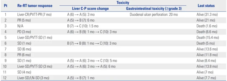
Table 5. Treatment Outcome after Re-Irradiation
Pt Re-RT tumor response Toxicity Last status
Liver C-P score change Gastrointestinal toxicity (≥grade 3)
1 Liver-CR/PVTT-PR (7 mo) A (6) → A (5): 3 mo Duodenal ulcer perforation: 20 mo Alive (31.3 mo) 2 PR (5 mo) A (5) → B (7): 6 mo Alive (21 mo) 3 N/A B (7) → C (10): 1.5 mo Death (1.6 mo) 4 PD (3 mo) A (6) → B (9): 1 mo → C (10): 3 mo Death (6.6 mo)
5 Liver-SD/PVTT-SD (1 mo) Death (15.4 mo)
6 SD (1 mo) B (7) → B (8): 1 mo → C (10): 3 mo Death (5 mo)
7 SD (6 mo) Alive (13.6 mo)
8 PR (6 mo) Alive (11.8 mo)
9 SD (1 mo) A (5) → A (6): 3 mo → C (10): 5 mo Alive (8.4 mo) 10 Liver-SD/PVTT-SD (3 mo) A (5) → A (6): 3 mo → A (5): 6 mo Alive (13.8 mo)
11 SD (4 mo) Alive (7 mo)
12 Liver-SD/LN-SD (3 mo) A (5) → B (7): 1 mo Alive (7.7 mo) Re-RT, re-irradiation; C-P, Child-Pugh; CR, complete response; PVTT, portal vein tumor thrombosis; PR, partial response; N/A, not available; PD, progressive dis-ease; SD, stable disdis-ease; LN, lymph node.
in each region were essential to confirming the summation re-sults. Thus, the modern DIR method might be more reliably utilized in solid organs, which have a limited spatial difference between intervals in terms of summated dose calculation, af-ter careful visual review.
We aimed to investigate the clinical correlation between nor-mal tissue dose summation results and high-degree clinico-functional deteriorations. Although we did not find a dosimet-ric causal relationship, subclinical mucosal changes in OARs
could be substantially high, as previously reported.15 Moreover, in cirrhotic HCC patients, the frequency of undetectable mu-cosal changes was commonly noticed, and special concern is required in this population.15,16 More intensive review involv-ing endoscopic exams will disclose the subclinical toxicities in future studies.
In conclusion, the OARs-based, DIR-based dose summation method can be utilized in re-irradiation of HCC patients after careful visual confirmation of high-risk regions. Although we
Fig. 1. Illustrative case (patient 4). Contrast-enhanced axial computed tomography before the first course of radiation (A) and at re-irradiation (B).
Recon-structed isodose lines using deformable image registration (DIR) at the first (A-1) and second course of irradiation (B-1) are shown, as well as the axial (C) and coronal (C-1) dose summation results using DIR. Narrow arrows in (A) and (B) indicate the tumor extent and wide arrows in (C) indicate potential high-risk regions (bowels) at re-irradiation.
A B C A-1 B-1 C-1
did not reach optimal and accurate dose summation results under the current algorithms and circumferential uncertain-ties, we can conclude that more favorable gastrointestinal toxic-ity profiles are expected with this modern technology. DIR may be more reliably used in solid organs with limited spatial differ-ence between intervals in comparison to the hollow organs, in terms of accurate prediction of dose summation. A more opti-mized adaptive plan using a highly conformal RT technique and appropriate use of DIR may enhance radiotherapeutic out-comes in future HCC cohorts treated with re-irradiation.
REFERENCES
1. Feng M, Ben-Josef E. Radiation therapy for hepatocellular carci-noma. Semin Radiat Oncol 2011;21:271-7.
2. Wang PM, Hsu WC, Chung NN, Chang FL, Fogliata A, Cozzi L. Radiation treatment with volumetric modulated arc therapy of hepatocellular carcinoma patients. Early clinical outcome and toxicity profile from a retrospective analysis of 138 patients. Radiat Oncol 2012;7:207.
3. Lee DS, Seong J. Radiotherapeutic options for hepatocellular car-cinoma with portal vein tumor thrombosis. Liver Cancer 2014;3: 18-30.
4. Abusaris H, Hoogeman M, Nuyttens JJ. Re-irradiation: outcome, cumulative dose and toxicity in patients retreated with stereotac-tic radiotherapy in the abdominal or pelvic region. Technol Can-cer Res Treat 2012;11:591-7.
5. Koom WS, Choi Y, Shim SJ, Cha J, Seong J, Kim NK, et al. Reirradia-tion to the pelvis for recurrent rectal cancer. J Surg Oncol 2012;105: 637-42.
6. Castadot P, Lee JA, Parraga A, Geets X, Macq B, Grégoire V. Com-parison of 12 deformable registration strategies in adaptive radia-tion therapy for the treatment of head and neck tumors. Radioth-er Oncol 2008;89:1-12.
7. Kovalchuk N, Jalisi S, Subramaniam RM, Truong MT. Deformable
registration of preoperative PET/CT with postoperative radiation therapy planning CT in head and neck cancer. Radiographics 2012; 32:1329-41.
8. Olteanu LA, Madani I, De Neve W, Vercauteren T, De Gersem W. Evaluation of deformable image coregistration in adaptive dose painting by numbers for head-and-neck cancer. Int J Radiat On-col Biol Phys 2012;83:696-703.
9. Edeline J, Boucher E, Rolland Y, Vauléon E, Pracht M, Perrin C, et al. Comparison of tumor response by Response Evaluation Crite-ria in Solid Tumors (RECIST) and modified RECIST in patients treated with sorafenib for hepatocellular carcinoma. Cancer 2012; 118:147-56.
10. Senthi S, Griffioen GH, van Sörnsen de Koste JR, Slotman BJ, Senan S. Comparing rigid and deformable dose registration for high dose thoracic re-irradiation. Radiother Oncol 2013;106:323-6. 11. Brock KK; Deformable Registration Accuracy Consortium. Re-sults of a multi-institution deformable registration accuracy study (MIDRAS). Int J Radiat Oncol Biol Phys 2010;76:583-96.
12. Juang T, Das S, Adamovics J, Benning R, Oldham M. On the need for comprehensive validation of deformable image registration, investigated with a novel 3-dimensional deformable dosimeter. Int J Radiat Oncol Biol Phys 2013;87:414-21.
13. Pan CC, Kavanagh BD, Dawson LA, Li XA, Das SK, Miften M, et al. Radiation-associated liver injury. Int J Radiat Oncol Biol Phys 2010;76(3 Suppl):S94-100.
14. Bae SH, Kim MS, Cho CK, Kang JK, Lee SY, Lee KN, et al. Predictor of severe gastroduodenal toxicity after stereotactic body radiother-apy for abdominopelvic malignancies. Int J Radiat Oncol Biol Phys 2012;84:e469-74.
15. Chon YE, Seong J, Kim BK, Cha J, Kim SU, Park JY, et al. Gastrodu-odenal complications after concurrent chemoradiation therapy in patients with hepatocellular carcinoma: endoscopic findings and risk factors. Int J Radiat Oncol Biol Phys 2011;81:1343-51. 16. Lee IJ, Kim JW, Han KH, Kim JK, Kim KS, Choi JS, et al.
Concur-rent chemoradiotherapy shows long-term survival after conver-sion from locally advanced to resectable hepatocellular carcino-ma. Yonsei Med J 2014;55:1489-97.