Adjuvant Chemoradiation Therapy in
Gallbladder Cancer
by
Seong Yeon Cho
Major in Medicine
Department of Medical Sciences
The Graduate School, Ajou University
Adjuvant Chemoradiation Therapy in
Gallbladder Cancer
by
Seong Yeon Cho
A Dissertation Submitted to The Graduate School of
Ajou University in Partial Fulfillment of the Requirements
for the Degree of Doctor of Medicine
Supervised by
Hee-Jung Wang, M.D., Ph.D.
Major in Medicine
Department of Medical Sciences
The Graduate School, Ajou University
This cert요
f모es that the d요
ssertat묘 on
0f Seong Yeon Cho
요
s approved●
SUPERV工
SORY COMMITTEE
(λ
/"- "'˘
Seong
—ι辛
竺〓 —
Byullglno Lee
느 느 —
The Graduate Schoo1,
서
ou IJn묘
versity
December, 20th, 201 1
i
- ABSTRACT –
Adjuvant Chemoradiation Therapy in Gallbladder Cancer
Gallbladder cancer is a relatively uncommon gastrointestinal malignancy. Indications
for adjuvant chemoradiation therapy after surgical resection have not yet been determined.
We aimed this study to elucidate the effectiveness of adjuvant chemoradiation therapy
according to TNM stage for gallbladder cancer. Between March 2001 and March 2009, 100
patients with gallbladder cancer underwent surgical resection. We divided the patients
according to TNM stage, and subdivided further according to whether adjuvant
chemoradiation therapy was added or not. The clinicopathologic factors, recurrence and
survival were retrospectively analyzed. Patients with gallbladder cancer at T2N0M0,
T2N1M0, T3N0M0 and T3N1M0 stages were enrolled in this study. Among the 4 stages, the
2 lymph node-negative stages (T2N0M0 and T3N0M0) did not show any gain in survival by
adding adjuvant chemoradiation therapy. Conversely, the remaining lymph node-positive
stages (T2N1M0 and T3N1M0) showed gain in disease-free survival, and the lymph
node-positive T2 stage (T2N1M0) showed gain in disease-specific survival. In patients with lymph
node-positive T2/T3 GB cancers, adjuvant chemoradiation therapy was an independent
prognostic factor for survival. Adjuvant chemoradiation therapy is recommended for lymph
node-positive T2/T3 gallbladder cancer following surgical resection.
ii
TABLE OF CONTENTS
ABSTRACT ··· ⅰ TABLE OF CONTENTS ··· ⅱ LIST OF FIGURES ··· ⅲ LIST OF TABLES ··· ⅳ ABBREVIATION ··· v . Ⅰ INTRODUCTION ··· 1 . Ⅱ MATERIALS AND METHODS ··· 2A. Materials ··· 2
B. Methods ··· 3
. Ⅲ RESULTS ··· 4
A. Comparative analysis of 18 patients with T2N0M0 stage ··· 5
B. Comparative analysis of 20 patients with T2N1M0 stage ··· 7
C. Comparative analysis of 10 patients with T3N0M0 stage ··· 10
D. Comparative analysis of 20 patients with T3N1M0 stage ··· 12
E. Prognostic factor analysis for lymph node negative T2/T3 stage ··· 14
F. Prognostic factor analysis for lymph node positive T2/T3 stage ··· 14
. Ⅳ DISCUSSION ··· 16 . Ⅴ CONCLUSION ··· 19 REFERENCES ··· 20 국문요약 ··· 24
iii
LIST OF FIGURES
Fig. 1. Survival curves according to tumor stage in 68 patients with T2N0M0,
T2N1M0, T3N0M0 or T3N1M0 stage. (A) disease-free survival (DFS).
(B) disease-specific survival (DSS). ··· 4
Fig. 2. Survival difference curves according to chemoradiation therapy.
(A) Disease-free survival (DFS) and (B) disease-specific survival (DSS) of
the 20 patients with T2N1M0 stage (NCRTx, n = 7; CRTx, n = 13).
(C) Disease-free survival (DFS) and (D) disease-specific survival (DSS) of
iv
LIST OF TABLES
Table 1. Comparative analysis of clinicopathologic factors in the patients with
2N0M0 stage. ··· 6
Table 2. Comparative analysis of clinicopathologic factors in the patients with
2N1M0 stage. ··· 8
Table 3. Comparative analysis of clinicopathologic factors in the patients with
T3N0M0 stage. ··· 11
Table 4. Comparative analysis of clinicopathologic factors in the patients with
T3N1M0 stage. ··· 13
Table 5. Significant factors for disease-free and disease-specific survival of
T2/T3N0 GB cancer by multivariate analysis. ··· 15
Table 6. Significant factors for disease-free and disease-specific survival of
v
ABBREVIATION
GB, gallbladder;
CRTx,
adjuvant
chemotherapy with or without radiation therapy; NCRTx,no adjuvant chemoradiation therapy
;DFS, disease-free survival;
DSS, disease-specific survival;
1
I. INTRODUCTION
Though it is the most common cancer of the biliary tree, gallbladder (GB) cancer is a
relatively uncommon gastrointestinal tract malignancy worldwide, and it has been known to
be a lethal malignancy (Henson et al., 1992; Furlan et al., 2008; Jemal et al., 2009).
Complete surgical resection still remains the only curative measure for GB cancer (de
Aretxabala et al., 2006; Duffy et al., 2008; Furuse, 2008). However, recurrence is common,
if it happens, it is considered to be fatal. Some auxiliary treatments, such as neoadjuvant or
adjuvant chemotherapy and radiation therapy, have been introduced to enhance survival of
patients with GB cancer. However, there is no established regimen for such measures
(Murakami et al., 2009). In addition, indications for adjuvant chemoradiation therapy have
not yet been determined.
To the best of our knowledge, there have been few published reports on TNM
stage-specific comparative analysis of the effectiveness of adjuvant chemoradiation therapy. The
aim of this study was to elucidate the effectiveness of adjuvant chemoradiation therapy
2
II. MATERIALS AND METHODS
A. Materials
Between March 2001 and March 2009, 100 patients with GB cancer underwent
surgical resection with curative intent at the National Cancer Center of Korea. Our center
policy for GB cancer has been as follows: simple cholecystectomy for T0 (carcinoma in
situ)/T1 cancer (invasion to the lamina propria or muscle layer) and extended
cholecystectomy for T2N0M0 and more advanced stages. For extended cholecystectomy, a
combination of liver resection with a more than 2-cm margin from the GB bed,
cholecystectomy, and dissection of lymph node stations 8, 12 and 13 have been performed.
Adjuvant chemotherapy with or without radiation therapy (CRTx) was administered
according to an interdisciplinary decision of a clinical oncologist and a surgeon after surgery.
If the patient’s general condition was tolerable, adjuvant concurrent chemoradiation therapy
was applied to a patient with T2N0M0, T2N1M0, T3N0M0 or T3N1M0 stage, but in more
advanced stages, only chemotherapy was applied without radiation therapy.
Among the 100 patients, 68 (68.0%) had T2N0M0, T2N1M0, T3N0M0 or T3N1M0
stage. We compared clinicopathologic factors and the survival rates between patients with
(CRTx group) and without adjuvant CRTx (group) according to tumor stage: T2N0M0 (n =
3
B. Methods
For the comparative analysis of the 2 groups in each stage, the following factors were
analyzed: age, gender, tumor marker, blood transfusion, differentiation, vascular invasion,
lymphatic invasion, perineural invasion, and resection margin. Comparisons of nominal and
continuous variables were made using the Fisher’s exact test (two-sided) and Mann-Whitney
U test, respectively. The cumulative survival rate was calculated by the Kaplan-Meier
method. For the evaluation of prognostic factors, the following factors were analyzed: age,
gender, tumor marker, blood transfusion, T-classification, differentiation, vascular invasion,
lymphatic invasion, perineural invasion, resection margin, and adjuvant chemoradiation
therapy. We performed the Cox proportional hazard regression analyses both in the
4
III. RESULTS
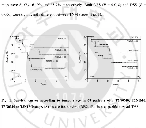
For the 68 patients, the 1-, 3- and 5-year disease-free survival (DFS) rates were 75.7%,
49.6% and 49.6%, respectively, and the 1-, 3- and 5-year disease-specific survival (DSS)
rates were 81.0%, 61.9% and 58.7%, respectively. Both DFS (P = 0.018) and DSS (P =
0.006) were significantly different between TNM stages (Fig. 1).
Fig. 1. Survival curves according to tumor stage in 68 patients with T2N0M0, T2N1M0, T3N0M0 or T3N1M0 stage. (A) disease-free survival (DFS). (B) disease-specific survival (DSS).
Among the 68 patients with T2/T3 classifications, 40 (58.8%) received adjuvant
chemotherapy (5-fluorouracil in 35 patients [87.5%]; cisplatin + capecitabine in 3 patients
[7.5%]; gemcitabine in 2 patients [5.0%]) concurrently with radiation therapy (dosage, 45 Gy
in 25 fractions for 25 days). For 5-fluorouracil-based regimen which was the most
5
on days 1, 2, 3 and on days 23, 24, 25 at a fixed dose of 500 mg/m2 during radiation therapy
period.
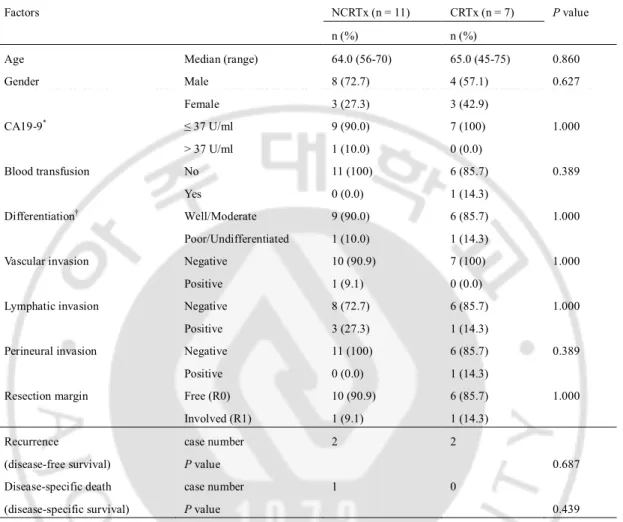
A. Comparative analysis of 18 patients with T2N0M0 stage (NCRTx, n = 11; CRTx, n = 7)
Clinicopathologic factors did not show any significant differences between the 2
groups (Table 1).
Recurrence developed in 4 of the 18 patients with T2N0M0 stage; 2 (18.2%) of the 11
patients from the NCRTx group (2 recurrences in the liver, 3 and 23 months after surgery,
respectively) and 2 (28.6%) of the 7 patients from the CRTx group (2 recurrences in
peritoneum, after 16 and 18 months after surgery, respectively), but the DFS rate showed no
statistical significance between the 2 groups (P = 0.687).
The DSS rate showed no statistical significance either because there was only 1
6
Table 1. Comparative analysis of clinicopathologic factors in the patients with T2N0M0 stage. Factors NCRTx (n = 11) CRTx (n = 7) P value
n (%) n (%)
Age Median (range) 64.0 (56-70) 65.0 (45-75) 0.860 Gender Male Female 8 (72.7) 3 (27.3) 4 (57.1) 3 (42.9) 0.627 CA19-9* ≤ 37 U/ml > 37 U/ml 9 (90.0) 1 (10.0) 7 (100) 0 (0.0) 1.000 Blood transfusion No Yes 11 (100) 0 (0.0) 6 (85.7) 1 (14.3) 0.389 Differentiation† Well/Moderate Poor/Undifferentiated 9 (90.0) 1 (10.0) 6 (85.7) 1 (14.3) 1.000
Vascular invasion Negative Positive 10 (90.9) 1 (9.1) 7 (100) 0 (0.0) 1.000
Lymphatic invasion Negative Positive 8 (72.7) 3 (27.3) 6 (85.7) 1 (14.3) 1.000
Perineural invasion Negative Positive 11 (100) 0 (0.0) 6 (85.7) 1 (14.3) 0.389
Resection margin Free (R0) Involved (R1) 10 (90.9) 1 (9.1) 6 (85.7) 1 (14.3) 1.000 Recurrence (disease-free survival) case number P value 2 2 0.687 Disease-specific death (disease-specific survival) case number P value 1 0 0.439
Abbreviation: NCRTx, no adjuvant chemoradiation therapy; CRTx, adjuvant chemoradiation therapy. * Preoperative CA19-9 level (reference, ≤ 37 U/ml) of 1 patient in the NCRTx group was not available.
† The differentiation of 1 patient in the NCRTx group was not available because initial simple cholecystectomy was performed at other hospital, followed by additional extended resection at our institute.
7
B. Comparative analysis of 20 patients with T2N1M0 stage (NCRTx, n = 7 and CRTx, n = 13)
Clinicopathologic factors did not show any significant differences between the 2
groups (Table 2).
Recurrence developed in 6 of the 20 patients with T2N1M0 stage; 3 (42.9%) of the 7
patients from the NCRTx group (1 recurrence in the peritoneum, 2 months after surgery; 2
recurrences in the lymph nodes, 5 and 9 months after surgery, respectively) and 3 (23.1%) of
the 13 patients from the CRTx group (1 recurrence in the lymph nodes, 19 months after
surgery; 2 recurrences in the peritoneum, 10 and 29 months after surgery, respectively). The
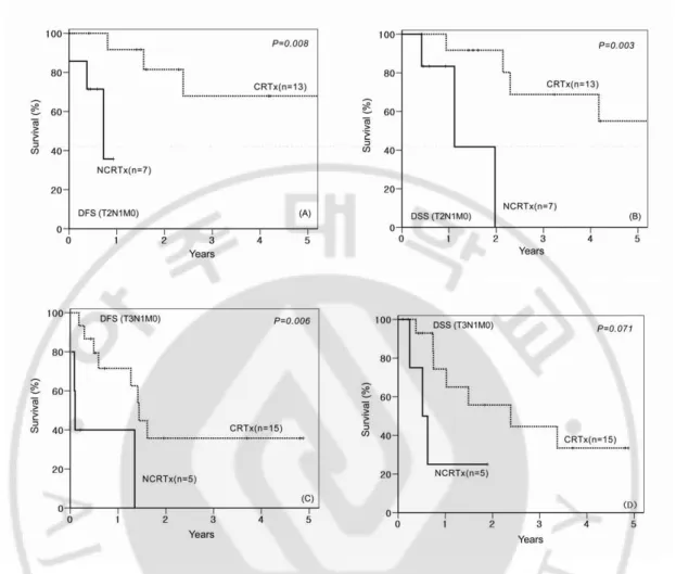
DFS rate was higher in the CRTx group than in the NCRTx group (P = 0.008) (Fig. 2A).
There were 5 disease-specific deaths; 3 (50.0%) of the 6 patients from the NCRTx
group (5, 14 and 24 months after surgery, respectively) and 2 (14.3%) of the 14 patients from
the CRTx group (12 and 26 months after surgery, respectively). The DSS rate was also
8
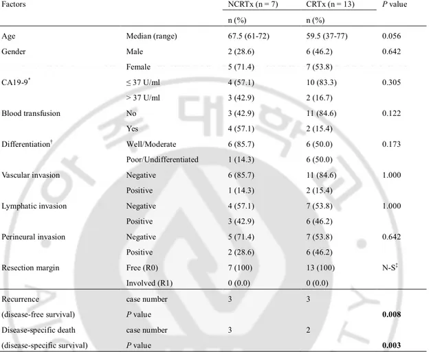
Table 2. Comparative analysis of clinicopathologic factors in the patients with T2N1M0 stage. Factors NCRTx (n = 7) CRTx (n = 13) P value
n (%) n (%)
Age Median (range) 67.5 (61-72) 59.5 (37-77) 0.056 Gender Male Female 2 (28.6) 5 (71.4) 6 (46.2) 7 (53.8) 0.642 CA19-9* ≤ 37 U/ml > 37 U/ml 4 (57.1) 3 (42.9) 10 (83.3) 2 (16.7) 0.305 Blood transfusion No Yes 3 (42.9) 4 (57.1) 11 (84.6) 2 (15.4) 0.122 Differentiation† Well/Moderate Poor/Undifferentiated 6 (85.7) 1 (14.3) 6 (50.0) 6 (50.0) 0.173
Vascular invasion Negative Positive 6 (85.7) 1 (14.3) 11 (84.6) 2 (15.4) 1.000
Lymphatic invasion Negative Positive 4 (57.1) 3 (42.9) 7 (53.8) 6 (46.2) 1.000
Perineural invasion Negative Positive 5 (71.4) 2 (28.6) 7 (53.8) 6 (46.2) 0.642
Resection margin Free (R0) Involved (R1) 7 (100) 0 (0.0) 13 (100) 0 (0.0) N-S‡ Recurrence (disease-free survival) case number P value 3 3 0.008 Disease-specific death (disease-specific survival) case number P value 3 2 0.003
Abbreviation: NCRTx, no adjuvant chemoradiation therapy; CRTx, adjuvant chemoradiation therapy. * Preoperative CA19-9 level (reference, ≤ 37 U/ml) of one patient in CRTx group was not available. † The differentiation of 1 patient in the CRTx group was not available because initial simple cholecystectomy was performed at other hospital, followed by additional extended resection at our institute.
9
Fig. 2. Survival differences according to adjuvant chemoradiation therapy. (A) Disease-free survival (DFS) and (B) disease-specific survival (DSS) of the 20 patients with T2N1M0 stage (NCRTx, n = 7; CRTx, n = 13). (C) Disease-free survival (DFS) and (D) disease-specific survival (DSS) of the 20 patients with T3N1M0 stage (NCRTx, n = 5; CRTx, n = 15).
10
C. Comparative analysis of 10 patients with T3N0M0 stage (NCRTx, n = 5 and CRTx, n = 5)
Clinicopathologic factors did not show any significant differences between the 2
groups (Table 3).
Recurrence developed in 5 of the 10 patients with T3N1M0 stage; 2 (40.0%) of the 5
patients from the NCRTx group (2 recurrences in the liver, 15 and 36 months after surgery,
respectively) and 3 (60%) of the 5 patients from the CRTx group (1 recurrence in the
peritoneum, 12 months after surgery; 2 recurrences in the liver, 5 months after surgery,
respectively), but the DFS rate showed no statistical significance between the 2 groups (P =
0.144).
There were 4 disease-specific deaths; 1 (20.0%) out of the 5 patients from the NCRTx
group (26 months after surgery) and 3 (75%) out of the 5 patients from the CRTx group (8,
10 and 28 months after surgery, respectively) but, the DSS rate showed no significant
11
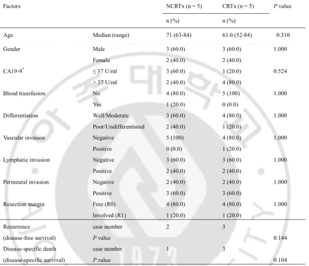
Table 3. Comparative analysis of clinicopathologic factors in the patients with T3N0M0 stage. Factors NCRTx (n = 5) CRTx (n = 5) P value
n (%) n (%)
Age Median (range) 71 (63-84) 61.0 (52-84) 0.310 Gender Male Female 3 (60.0) 2 (40.0) 3 (60.0) 2 (40.0) 1.000 CA19-9* ≤ 37 U/ml > 37 U/ml 3 (60.0) 2 (40.0) 1 (20.0) 4 (80.0) 0.524 Blood transfusion No Yes 4 (80.0) 1 (20.0) 5 (100) 0 (0.0) 1.000 Differentiation Well/Moderate Poor/Undifferentiated 3 (60.0) 2 (40.0) 4 (80.0) 1 (20.0) 1.000
Vascular invasion Negative Positive 5 (100) 0 (0.0) 4 (80.0) 1 (20.0) 1.000
Lymphatic invasion Negative Positive 3 (60.0) 2 (40.0) 3 (60.0) 2 (40.0) 1.000
Perineural invasion Negative Positive 2 (40.0) 3 (60.0) 2 (40.0) 3 (60.0) 1.000
Resection margin Free (R0) Involved (R1) 4 (80.0) 1 (20.0) 4 (80.0) 1 (20.0) 1.000 Recurrence (disease-free survival) case number P value 2 3 0.144 Disease-specific death (disease-specific survival) case number P value 1 3 0.104
Abbreviation: NCRTx, no adjuvant chemoradiation therapy; CRTx, adjuvant chemoradiation therapy. * Preoperative CA19-9 level (reference, ≤ 37 U/ml).
12
D. Comparative analysis of 20 patients with T3N1M0 stage (NCRTx, n = 5 and CRTx, n = 15)
Clinicopathologic factors did not show any significant differences between the 2
groups (Table 4).
Recurrence developed in 12 of the 20 patients with T3N1M0 stage; 4 (80.0%) of the 5
patients from the NCRTx group (2 recurrences in the liver, both 1 month after surgery,
respectively; 1 recurrence in the peritoneum, 2 months after surgery; and 1 recurrence in the
lung, 17 months after surgery) and 8 (53.3%) of the 15 patients from the CRTx group (4
recurrences in the liver, 3, 8, 16 and 18 months after surgery, respectively; 3 recurrences in
the lymph nodes, 4, 6 and 20 months after surgery, respectively; and 1 recurrence in the
peritoneum, 18 months after surgery). The DFS rate was higher in the CRTx group than in
the NCRTx group (P = 0.006) (Fig. 2C).
There were 10 disease-specific deaths; 3 (60.0%) out of the 5 patients from the
NCRTx group (3, 7 and 8 months after surgery, respectively) and 7 (46.7%) out of the 15
patients from the CRTx group (5, 9, 10, 13, 19, 29 and 41 months after surgery, respectively),
but the DSS rate showed no statistical significance between the 2 groups (P = 0.071) (Fig.
13
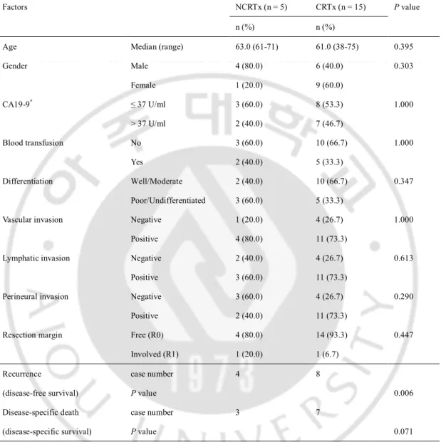
Table 4. Comparative analysis of clinicopathologic factors in the patients with T3N1M0 stage. Factors NCRTx (n = 5) CRTx (n = 15) P value
n (%) n (%)
Age Median (range) 63.0 (61-71) 61.0 (38-75) 0.395 Gender Male Female 4 (80.0) 1 (20.0) 6 (40.0) 9 (60.0) 0.303 CA19-9* ≤ 37 U/ml > 37 U/ml 3 (60.0) 2 (40.0) 8 (53.3) 7 (46.7) 1.000 Blood transfusion No Yes 3 (60.0) 2 (40.0) 10 (66.7) 5 (33.3) 1.000 Differentiation Well/Moderate Poor/Undifferentiated 2 (40.0) 3 (60.0) 10 (66.7) 5 (33.3) 0.347
Vascular invasion Negative Positive 1 (20.0) 4 (80.0) 4 (26.7) 11 (73.3) 1.000
Lymphatic invasion Negative Positive 2 (40.0) 3 (60.0) 4 (26.7) 11 (73.3) 0.613
Perineural invasion Negative Positive 3 (60.0) 2 (40.0) 4 (26.7) 11 (73.3) 0.290
Resection margin Free (R0) Involved (R1) 4 (80.0) 1 (20.0) 14 (93.3) 1 (6.7) 0.447 Recurrence (disease-free survival) case number P value 4 8 0.006 Disease-specific death (disease-specific survival) case number P value 3 7 0.071
Abbreviation: NCRTx, no adjuvant chemoradiation therapy; CRTx, adjuvant chemoradiation therapy. * Preoperative CA19-9 level (reference, ≤ 37 U/ml).
14
E. Prognostic factor analysis for lymph node negative T2/T3 stage (T2/T3N0, n = 28) For the prognostic factor analysis of DFS in patients with lymph node negative T2/T3
GB cancer, lymphatic invasion (P = 0.005) and perineural invasion (P = 0.013) were
significant in univariate analysis. NCRTx did not attain significance (P = 0.314). By
multivariate analysis, the 2 factors were also proved to be independent prognostic factors
(Table 5).
For the prognostic factor analysis of DSS, T3-classification (P = 0.048), differentiation
(P = 0.016), lymphatic invasion (P = 0.041) and perineural invasion (P = 0.030) were
significant in univariate analysis. NCRTx did not attain significance (P = 0.342). By
multivariate analysis, only differentiation was proved to be an independent prognostic factor
(Table 5).
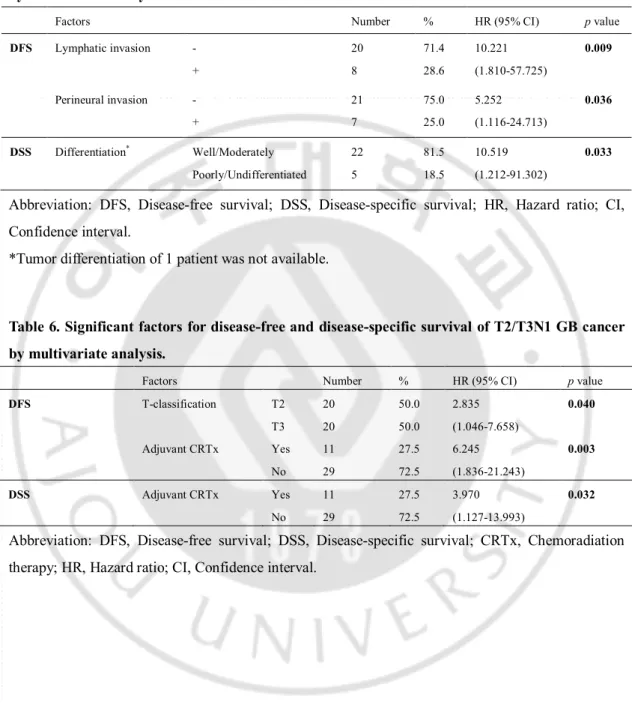
F. Prognostic factor analysis for lymph node positive T2/T3 stage (T2/T3N1, n = 40) For the prognostic factor analysis of DFS in patients with lymph node positive T2/T3
GB cancer, age > 60 (P = 0.039), T3-classification (P = 0.047) and NCRTx (P < 0.001) were
significant by univariate analysis. By multivariate analysis, T3-classification and NCRTx
were proved to be independent prognostic factors (Table 6).
For the prognostic factor analysis of DSS, age > 60 (P = 0.047) and NCRTx (P =
0.003) were significant by univariate analysis. By multivariate analysis, only NCRTx was
15
Table 5. Significant factors for disease-free and disease-specific survival of T2/T3N0 GB cancer by multivariate analysis.
Factors Number % HR (95% CI) p value
DFS Lymphatic invasion - + 20 8 71.4 28.6 10.221 (1.810-57.725) 0.009 Perineural invasion - + 21 7 75.0 25.0 5.252 (1.116-24.713) 0.036 DSS Differentiation* Well/Moderately Poorly/Undifferentiated 22 5 81.5 18.5 10.519 (1.212-91.302) 0.033
Abbreviation: DFS, Disease-free survival; DSS, Disease-specific survival; HR, Hazard ratio; CI, Confidence interval.
*Tumor differentiation of 1 patient was not available.
Table 6. Significant factors for disease-free and disease-specific survival of T2/T3N1 GB cancer by multivariate analysis.
Factors Number % HR (95% CI) p value
DFS T-classification T2 T3 20 20 50.0 50.0 2.835 (1.046-7.658) 0.040 Adjuvant CRTx Yes No 11 29 27.5 72.5 6.245 (1.836-21.243) 0.003 DSS Adjuvant CRTx Yes No 11 29 27.5 72.5 3.970 (1.127-13.993) 0.032
Abbreviation: DFS, Disease-free survival; DSS, Disease-specific survival; CRTx, Chemoradiation therapy; HR, Hazard ratio; CI, Confidence interval.
16
IV. DISCUSSION
Even after radical resection of GB cancer, not only locoregional recurrence but also
distant metastasis is common, especially in advanced stages (Kresl et al., 2002; Jarnagin et
al., 2003; de Aretxabala et al., 2006; Gourgiotis et al., 2008; Hueman et al., 2009). Thus, the
multidisciplinary approach in addition to surgical resection may be necessary to treat
advanced GB cancers. For these reasons, a combination of adjuvant systemic chemotherapy
with radiation therapy is mandatory to decrease locoregional recurrence and distant
metastasis after surgical resection of GB cancer.
In a previous retrospective analysis of the SEER database, adjuvant radiation therapy
was associated with good survival only in patients with locally advanced GB cancer or GB
cancer accompanied by a regional disease (Mojica et al., 2007). Another study of SEER
database made a prediction model which predicts that adjuvant radiation therapy provides a
survival benefit in node-positive or ≥ T2 GB cancer (Wang et al., 2008). Some previous
studies on concurrent chemoradiation therapy for GB cancer have been reported (Kresl et al.,
2002; Czito et al., 2005; Gold et al., 2009). However, there is still no established standard
regimen of adjuvant chemotherapy for GB cancer (Treadwell and Hardin, 1976; Smoron,
1977; Hanna and Rider, 1978; Kopelson and Gunderson, 1983). Traditionally,
5-fluorouracil-based chemotherapy regimen has been used as an adjuvant therapy for biliary
tract malignancies including GB cancer (Kresl et al., 2002; Takada et al., 2002; de
Aretxabala et al., 2006; Reid et al., 2007). Recently, gemcitabine-, capecitabine, or
17
results from GB cancer (Penz et al., 2001; Ueno et al., 2004; Alberts et al., 2005; Knox et al.,
2005; Koeberle et al., 2008). However, few comparative analyses of treatments between
detailed TNM stages of GB cancer have been reported. Well designed prospective
randomized case-control studies are needed to confirm the results of these new trials.
Because of a relative paucity of GB cancer and lack of specific symptoms or signs in
its early stage of it, surgical resection was performed on only 100 patients for 9 consecutive
years at the National Cancer Center of Korea. Patients with stages less than T2N0M0 did not
undergo adjuvant therapy. None of the patients with T1 classification showed lymph node
metastasis in our series, and the number of patients with stages more than T3N1M0 was too
small to be enrolled for this comparative analysis. Sixty-eight patients (68.0%) showed T2 or
T3 classification without distant metastasis.
We evaluated patients’ survival using disease-specific survival rather than overall
survival to minimize selection bias effect from the following characteristics: (1) most of GB
cancers developed in patients at an old age, (2) many of them had comorbidities such as
other malignancies, cardiovascular disease and/or diabetes mellitus, and (3) the candidates
for adjuvant CRTx should be under an appropriate general condition in order to receive this
cytotoxic therapy. For the evaluation of prognostic factors, the TNM-specific 4 stages (T2N0,
T2N1, T3N0 and T3N1) were reassembled into 2 stages according to the lymph node
metastasis regardless of T-classification (T2/T3N0 and T2/T3N1), since the numbers of
patients in each stage were relatively small. In patients with lymph node-positive T2/T3
18
factor for both DFS and DSS, while adjuvant chemoradiation therapy did not gain any
survival benefit in lymph node negative T2/T3 (T2/T3N0) GB cancers.
Although T3N0M0 tumors belong to stage IIA of the sixth AJCC/UICC staging
system and T2N1M0 tumors belong to stage IIB, the overall prognosis was poorer for
T3N0M0 tumors (stage IIA) than for T2N1M0 tumors (stage IIB) in our series.
Neither DFS nor DSS showed any significant difference between the 2 groups in
lymph node-negative T2N0M0 and T3N0M0 stages (Tables 1 and 3). For T2N1M0 stage,
both DFS (P = 0.001) and DSS (P = 0.002) were higher in the CRTx group than in the
NCRTx group (Table 2). However, for T3N1M0 stage, only DFS was higher in the CRTx
group than in the NCRTx group (P = 0.006), and DSS did not show significant difference (P
= 0.071) (Table 4).
Clinicopathologic profiles were not different between the 2 groups in any stage
(Tables 1, 2, 3 and 4). Our study demonstrated that adjuvant chemoradiation therapy
conferred no DFS or DSS benefit on patients with lymph node-negative T2 or T3 GB cancer.
However, adjuvant chemoradiation therapy conferred DFS benefit on patients with lymph
node-positive T2 and T3. Our adjuvant therapy reduced recurrences of GB cancer following
resection in lymph node–positive patients. However, adjuvant chemoradiation therapy
conferred DSS benefit on patients with T2N1M0 tumors but not on those with T3N1M0
19
V. CONCLUSION
In summary, effective adjuvant therapy is necessary to improve treatment
outcome of GB cancer following resection. Most patients (87.5%) in this study
received 5-fluorouracil-based chemotherapy. The results of this study suggest that
adjuvant chemoradiation therapy may be effective in the treatment of lymph
node-positive T2/T3 GB cancers after surgical resection. Further randomized controlled
studies with a larger sample size and with a new chemotherapy regimen are needed
to confirm similar therapeutic effects on other stage tumors.
20
REFERENCES
1. Alberts SR, Al-Khatib H, Mahoney MR, Burgart L, Cera PJ, Flynn PJ, Finch TR,
Levitt R, Windschitl HE, Knost JA, Tschetter LK: Gemcitabine, 5-fluorouracil, and
leucovorin in advanced biliary tract and gallbladder carcinoma: a North Central
Cancer Treatment Group phase II trial. Cancer 103: 111-118, 2005
2. Czito BG, Hurwitz HI, Clough RW, Tyler DS, Morse MA, Clary BM, Pappas TN,
Fernando NH, Willett CG: Adjuvant external-beam radiotherapy with concurrent
chemotherapy after resection of primary gallbladder carcinoma: a 23-year
experience. Int J Radiat Oncol Biol Phys 62: 1030-1034, 2005
3. de Aretxabala X, Roa I, Berrios M, Hepp J, Gallardo J, Cordova A, Roa JC, Leon J,
Maluenda F: Chemoradiotherapy in gallbladder cancer. J Surg Oncol 93: 699-704,
2006
4. Duffy A, Capanu M, Abou-Alfa GK, Huitzil D, Jarnagin W, Fong Y, D'Angelica M,
Dematteo RP, Blumgart LH, O'Reilly EM: Gallbladder cancer (GBC): 10-year
experience at Memorial Sloan-Kettering Cancer Centre (MSKCC). J Surg Oncol 98:
485-489, 2008
5. Furlan A, Ferris JV, Hosseinzadeh K, Borhani AA: Gallbladder carcinoma update:
multimodality imaging evaluation, staging, and treatment options. AJR Am J
21
6. Furuse J: Postoperative adjuvant treatments for biliary tract cancer. J Hepatobiliary
Pancreat Surg 15: 463-467, 2008
7. Gold DG, Miller RC, Haddock MG, Gunderson LL, Quevedo F, Donohue JH,
Bhatia S, Nagorney DM: Adjuvant Therapy for Gallbladder Carcinoma: The Mayo
Clinic Experience. Int J Radiat Oncol Biol Phys, 2009
8. Gourgiotis S, Kocher HM, Solaini L, Yarollahi A, Tsiambas E, Salemis NS:
Gallbladder cancer. Am J Surg 196: 252-264, 2008
9. Hanna SS, Rider WD: Carcinoma of the gallbladder or extrahepatic bile ducts: the
role of radiotherapy. Can Med Assoc J 118: 59-61, 1978
10. Henson DE, Albores-Saavedra J, Corle D: Carcinoma of the gallbladder. Histologic
types, stage of disease, grade, and survival rates. Cancer 70: 1493-1497, 1992
11. Hueman MT, Vollmer CM, Jr., Pawlik TM: Evolving treatment strategies for
gallbladder cancer. Ann Surg Oncol 16: 2101-2115, 2009
12. Jarnagin WR, Ruo L, Little SA, Klimstra D, D'Angelica M, DeMatteo RP, Wagman
R, Blumgart LH, Fong Y: Patterns of initial disease recurrence after resection of
gallbladder carcinoma and hilar cholangiocarcinoma: implications for adjuvant
therapeutic strategies. Cancer 98: 1689-1700, 2003
13. Jemal A, Siegel R, Ward E, Hao Y, Xu J, Thun MJ: Cancer statistics, 2009. CA
22
14. Knox JJ, Hedley D, Oza A, Feld R, Siu LL, Chen E, Nematollahi M, Pond GR,
Zhang J, Moore MJ: Combining gemcitabine and capecitabine in patients with
advanced biliary cancer: a phase II trial. J Clin Oncol 23: 2332-2338, 2005
15. Koeberle D, Saletti P, Borner M, Gerber D, Dietrich D, Caspar CB, Mingrone W,
Beretta K, Strasser F, Ruhstaller T, Mora O, Herrmann R: Patient-reported outcomes
of patients with advanced biliary tract cancers receiving gemcitabine plus
capecitabine: a multicenter, phase II trial of the Swiss Group for Clinical Cancer
Research. J Clin Oncol 26: 3702-3708, 2008
16. Kopelson G, Gunderson LL: Primary and adjuvant radiation therapy in gallbladder
and extrahepatic biliary tract carcinoma. J Clin Gastroenterol 5: 43-50, 1983
17. Kresl JJ, Schild SE, Henning GT, Gunderson LL, Donohue J, Pitot H, Haddock MG,
Nagorney D: Adjuvant external beam radiation therapy with concurrent
chemotherapy in the management of gallbladder carcinoma. Int J Radiat Oncol Biol
Phys 52: 167-175, 2002
18. Mojica P, Smith D, Ellenhorn J: Adjuvant radiation therapy is associated with
improved survival for gallbladder carcinoma with regional metastatic disease. J Surg
Oncol 96: 8-13, 2007
19. Murakami Y, Uemura K, Hayasidani Y, Sudo T, Hashimoto Y, Ohge H, Sueda T:
Indication for postoperative adjuvant therapy in biliary carcinoma based on analysis
23
20. Penz M, Kornek GV, Raderer M, Ulrich-Pur H, Fiebiger W, Lenauer A, Depisch D,
Krauss G, Schneeweiss B, Scheithauer W: Phase II trial of two-weekly gemcitabine
in patients with advanced biliary tract cancer. Ann Oncol 12: 183-186, 2001
21. Reid KM, Ramos-De la Medina A, Donohue JH: Diagnosis and surgical
management of gallbladder cancer: a review. J Gastrointest Surg 11: 671-681, 2007
22. Smoron GL: Radiation therapy of carcinoma of gallbladder and biliary tract. Cancer
40: 1422-1424, 1977
23. Takada T, Amano H, Yasuda H, Nimura Y, Matsushiro T, Kato H, Nagakawa T,
Nakayama T: Is postoperative adjuvant chemotherapy useful for gallbladder
carcinoma? A phase III multicenter prospective randomized controlled trial in
patients with resected pancreaticobiliary carcinoma. Cancer 95: 1685-1695, 2002
24. Treadwell TA, Hardin WJ: Primary carcinoma of the gallbladder. The role of
adjunctive therapy in its treatment. Am J Surg 132: 703-706, 1976
25. Ueno H, Okusaka T, Ikeda M, Takezako Y, Morizane C: Phase II study of S-1 in
patients with advanced biliary tract cancer. Br J Cancer 91: 1769-1774, 2004
26. Wang SJ, Fuller CD, Kim JS, Sittig DF, Thomas CR, Jr., Ravdin PM: Prediction
model for estimating the survival benefit of adjuvant radiotherapy for gallbladder
24 - 국문요약 -