Vol. 44 / No. 4 / July 2017
351
Management of a Recurrent
Ischial Sore Using a 3-Flap
Technique
Jae Hyun Lee, Hee Chang Ahn
Department of Plastic and Reconstructive Surgery, Hanyang University College of Medicine, Seoul, Korea
Correspondence: Hee Chang Ahn
Department of Plastic and Reconstructive Surgery, Hanyang University College of Medicine, 222-1 Wangsimni-ro, Seongdong-gu, Seoul 04763, Korea Tel: +82-2-2290-8560 Fax: +82-2-2295-7671
E-mail: [email protected]
No potential conflict of interest relevant to this article was reported. Received: 12 Apr 2017 • Revised: 16 Jun 2017 • Accepted: 22 Jun 2017 pISSN: 2234-6163 • eISSN: 2234-6171
https://doi.org/10.5999/aps.2017.44.4.351 Arch Plast Surg 2017;44:351-352
Copyright 2017 The Korean Society of Plastic and Reconstructive Surgeons This is an Open Access article distributed under the terms of the Creative Commons Attribution Non-Commercial License (http://creativecommons.org/licenses/by-nc/4.0/) which permits unrestricted non-commercial use, distribution, and reproduction in any medium, provided the original work is properly cited.
As the quality of rehabilitation has improved (e.g., through the increased use of wheelchairs), ischial sores have become one of the top 3 most common types of sores in terms of location, with an annually increasing number of patients [1]. Even after musculocutaneous or perforator flaps are performed to treat pressure sores, complications such as ulcer recurrence and wound dehiscence still remain common [2].
A 48-year-old man underwent surgery to treat a lumbar spinal cord tumor in 2003. In 2005, due to his bedridden state, he experienced a left ischial sore. In
the same year, he was treated with bursectomy, a rotation flap, and a local flap. After a rehabilitation period that allowed him to ambulate and sit, the ischial sore recurred in 2017. After treating him with negative-pressure wound therapy, we performed a bursectomy, packed the dead space with a
semitendinosus muscle flap that had no effect on the patient’s ambulatory ability, and covered the skin area with a local flap. However, after surgery, we observed abrasions and seroma in the ischial region (Fig. 1). To fix this problem, we performed a complete
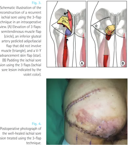
bursectomy, repositioned the semitendinosus muscle flap to apply more padding to the ischial tuberosity, packed the remaining dead space with an inferior gluteal artery pedicled adipofascial flap that did not involve muscle [3] to maintain the patient’s ambulatory ability, and covered the skin using a V-Y advancement flap (Figs. 2, 3).
Considering the patient’s ambulatory ability and the shortage of tissue due to the recurrence of the sore, we believe the usage of the 3-flap technique presented above was an appropriate treatment (Fig. 4). Thus, we must consider each patient’s condition
IMAGES
Images
Fig. 1.
A 48-year-old man with a recurrent ischial sore on the left buttock showing abrasions and seroma (incision line drawn with a violet marker).
Fig. 2.
Intraoperative photographs of the reconstruction of a recurrent ischial sore using the 3-flap technique. (A) After performing complete bursectomy, we repositioned the semitendinosus muscle flap that was used in the previous operation to apply more padding to the ischial tuberosity (circle). We also elevated an inferior gluteal artery pedicled adipofascial flap that did not involve muscle (triangle) and a V-Y advancement skin flap (star). (B) We packed the remaining dead space with the inferior gluteal artery pedicled adipofascial flap that did not involve muscle to maintain the patient’s ambulatory ability.
B A
352
and the availability and efficacy of various tissue types to increase the diversity of flap reconstruction.
References
1. VanGilder C, Amlung S, Harrison P, et al. Results of the 2008-2009 International Pressure Ulcer Prevalence Survey and a 3-year, acute care, unit-specific analysis. Ostomy Wound Manage 2009;55:39-45.
2. Bamba R, Madden JJ, Hoffman AN, et al. Flap reconstruction for pressure ulcers: an outcomes analysis. Plast Reconstr Surg Glob Open 2017;5:e1187. 3. Lin H, Hou C, Xu Z, et al. Treatment of ischial pressure
sores with double adipofascial turnover flaps. Ann Plast Surg 2010;64:59-61.
Fig. 3. Schematic illustration of the reconstruction of a recurrent ischial sore using the 3-flap technique in an intraoperative view. (A) Elevation of 3 flaps: a semitendinosus muscle flap (circle), an inferior gluteal artery pedicled adipofascial flap that did not involve muscle (triangle), and a V-Y advancement skin flap (star). (B) Padding the ischial sore lesion using the 3 flaps (ischial sore lesion indicated by the violet color).
Fig. 4. Postoperative photograph of the well-healed ischial sore lesion treated using the 3-flap technique.
A B
Images
Usefulness of the Versajet
Hydrosurgery System for the
Removal of Foreign Body
Granuloma
Min Choi, Kyung Min Son, Woo Young Choi, Ji Seon Cheon
Department of Plastic and Reconstructive Surgery, Chosun University College of Medicine, Gwangju, Korea
Correspondence: Kyung Min Son
Department of Plastic and Reconstructive Surgery, Chosun University College of Medicine, 309 Pilmun-daero, Dong-gu, Gwangju 61452, Korea Tel: +82-62-220-3180, Fax: +82-62-225-0996
E-mail: [email protected]
No potential conflict of interest relevant to this article was reported. Received: 4 Apr 2017 • Revised: 31 May 2017 • Accepted: 7 Jun 2017 pISSN: 2234-6163 • eISSN: 2234-6171
https://doi.org/10.5999/aps.2017.44.4.352 Arch Plast Surg 2017;44:352-353
Copyright 2017 The Korean Society of Plastic and Reconstructive Surgeons This is an Open Access article distributed under the terms of the Creative Commons Attribution Non-Commercial License (http://creativecommons.org/licenses/by-nc/4.0/) which permits unrestricted non-commercial use, distribution, and reproduction in any medium, provided the original work is properly cited.
Materials used for cosmetic/reconstructive purposes can elicit foreign body reactions, resulting in granulomas. Foreign body granulomas are treated with intralesional corticosteroid injections and excisional surgery [1].
A 44-year-old woman presented with irregularities and areas of hardness across the forehead, glabella, and temple. She had undergone a cosmetic procedure involving the injection of an unknown material into these sites 10 years before. We diagnosed the case as foreign body granuloma. We administered 2 intralesional triamcinolone injections (20 mg/mL) at 1-month intervals, but the discomfort persisted. Therefore, we performed surgery using the Versajet hydrosurgery system. The patient was administered anesthesia via propofol, followed by local anesthesia with lidocaine. After 1-cm incisions in both suprabrow areas and 2-cm incisions in the temple area were made, dissection was performed subcutaneously (Fig. 1). We approached the target areas with the 15° Versajet handpiece to remove approximately 5 mL of granuloma fluid (Fig. 2). A postoperative compression dressing was maintained for 3 days to prevent hematoma. The swelling persisted for 1 month. After 3 months of follow-up, the irregularities had improved, and the patient was satisfied with the cosmetic outcomes (Fig. 3).