Characteristics of Resting-State Functional
Connectivity in HIV-Associated
Neurocognitive Disorder
Hea Won Ann1,2, Suhnyoung Jun3, Na-Young Shin4, Sanghoon Han3, Jin Young Ahn1,2, Mi
Young Ahn1,2, Yong Duk Jeon1,2, In Young Jung1, Moo Hyun Kim1, Woo Yong Jeong1, Nam Su Ku1,2, June Myung Kim1,2, Davey M. Smith5,6, Jun Yong Choi1,2*
1 Department of Internal Medicine, Yonsei University College of Medicine, Seoul, South Korea, 2 Department of AIDS Research Institute, Yonsei University College of Medicine, Seoul, South Korea, 3 Department of Psychology, Yonsei University, Seoul, South Korea, 4 Department of Radiology, Ewha Womans University School of Medicine, Seoul, South Korea, 5 Department of Medicine, University of California San Diego, La Jolla, CA, United States of America, 6 Veterans Affairs San Diego Healthcare System, San Diego, CA, United States of America
*seran@yuhs.ac
Abstract
Background
HIV-associated neurocognitive disorder (HAND) can occur in patients without prior AIDS defining illness and can be debilitating. This study aimed to evaluate the difference in the patterns of intrinsic brain activity between patients with or without HAND for deepening our understanding of HAND.
Methods
We evaluated 24 HIV-infected individuals, 12 with previously diagnosed HAND and 12 pre-viously diagnosed without HAND, and 11 seronegative individuals. These individuals then underwent repeat NP testing and a functional brain MRI scan. For functional MRI analysis, seed-based analysis with bilateral precuneus cortex seed was applied.
Results
Among the 12 individuals with previously diagnosed HAND, 3 showed improvement of their neurocognitive function and 1 was excluded for worsening liver disease. Among the 12 patients who previously had normal neurocognitive function, 2 showed neurocognitive impairment. Overall, the HAND group, who had impaired cognitive function at the time of MRI scan, showed significant decrease of resting status functional connectivity between bilateral precuneus and prefrontal cortex (PFC) compared with nonHAND group, those who had normal neurocognitive function (CorrectedP<0.05). The functional connectivity with the right inferior frontal operculum and right superior frontal gyrus was positively correlated with memory and learning ability.
a11111
OPEN ACCESS
Citation: Ann HW, Jun S, Shin N-Y, Han S, Ahn JY, Ahn MY, et al. (2016) Characteristics of Resting-State Functional Connectivity in HIV-Associated Neurocognitive Disorder. PLoS ONE 11(4): e0153493. doi:10.1371/journal.pone.0153493 Editor: Marco Colasanti, University of Rome, ITALY Received: December 3, 2015
Accepted: March 30, 2016 Published: April 22, 2016
Copyright: © 2016 Ann et al. This is an open access article distributed under the terms of theCreative Commons Attribution License, which permits unrestricted use, distribution, and reproduction in any medium, provided the original author and source are credited.
Data Availability Statement: Data are within the paper and available upon request to the authors due to the presence of identifying information. Please contactseran@yuhs.acfor data access.
Funding: This research was supported by the Basic Science Research Program through the National Research Foundation of Korea (NRF) funded by the Ministry of Education, Science and Technology (NRF-2013R1A1A2005412), a Chronic Infectious Disease Cohort grant (4800-4859-304-260) from the Korea Centers for Disease Control and Prevention, and BioNano Health-Guard Research Center funded by the Ministry of Science, ICT, and Future Planning of Korea as a Global Frontier Project (Grant
H-Conclusions
This cross-sectional study found a significant difference in fMRI patterns between patients with and without HAND. Decreased functional connectivity between precuneus and PFC could be possible functional substrate for cognitive dysfunction in HIV patients, which should be characterized in a longitudinal study.
Introduction
HIV associated neurocognitive disorder (HAND) is a general term that incorporates a number of neurological impairments with a range of severity including asymptomatic neurocognitive impairment (ANI), mild neurocognitive disorder (MND), and HIV associated dementia (HAD) based on self-report about interference with daily functioning [1]. Impairments in daily functioning associated with HAND include employment, taking medications, financial man-agement, cooking etc [2,3]. It is a quite common medical condition even during effective anti-retroviral therapy (ART) with the prevalence in HIV-infected individuals between 16–52% [1,
4,5]. Elaborate neuropsychological (NP) examinations are required to diagnose HAND [1], and there is no consensus on how to detect HAND via imaging tools.
Recent developments in functional magnetic resonance imaging (fMRI) make it an attrac-tive option is getting help in understanding pathophysiology of HAND, since it can evaluate connectivity and communications between brain regions [6]. The dysfunctions in these com-munications between brain regions are likely associated with the mechanisms underlying HAND pathogenesis [7]. In this study, we evaluated whether brain fMRI can identify a pattern associated with HAND.
Methods
Study design
Twelve male patients who diagnosed with HAND in 2011 [5] and 12 male patients without HAND (also confirmed in 2011, same test procedure with HAND patients) were randomly recruited from HIV cohort. As a control group, 11 age- and sex- matched seronegative subjects were also recruited. Potential participants were not considered for enrollment if they had: (1) recent and/or significant traumatic brain injury, (2) a neurological disorder not related to HIV infection, (3) infections that can affect the CNS, (4) a significant CNS opportunistic infection based upon history and/or neuromedical examination (5) a current or past psychotic disorder, (6) significant substance use (more than three alcoholic drinks per day over the last month or recreational drug use more than once per week during the last month), (7) symptoms of a cur-rent, active infection, a body temperature of>38.5°C at the time of recruitment or current treatment for a serious, systemic infection within 3 months, (8) colour blindness, (9) a hearing deficit that appears to affect auditory comprehension [1]. All subjects provided informed con-sent and received standardized neurological, NP, and functional assessments at enrollment. This study was approved by the Institutional Review Board of the hospital (IRB #4-2013-0499).
Demographic and clinical data
Age, sex, duration of education, performance status [8], body mass index, hemoglobin level, estimated glomerular filtration ratio, antiretroviral regimen, CNS penetration effectiveness (CPE) [9], initial CD4 T-cell count, pre-cART CD4 T-cell count, current CD4 T-cell count,
GUARD_2013M3A6B2078953) and a grant from the Ministry of Health & Welfare, Republic of Korea (grant number: HI14C1324). DMS was supported by the San Diego Veterans Affairs Healthcare system and National Institutes of Health grants AI100665, MH062512 and AI036214.
Competing Interests: The authors have declared that no competing interests exist.
lowest CD4 T-cell count, initial viral load (VL), pre-cART VL and current VL and highest VL were available and included in the analysis.
Neuropsychological tests
The NP exam and fMRI scans were carried out within a month of study enrollment for each participant. The neurocognitive status assessed six ability domains. (1) speed of information processing using the Korean version of Wechsler Adult Intelligence Scale(K-WAIS) digit sym-bol subtest, trail making test part A; (2) memory (learning and recall) using the Korean version of auditory verbal learning test and complex figure test; (3) abstraction/executive function using the Wisconsin Card Sorting Test, K-WAIS similarity subtest and trail making test part B; (4) attention/working memory using the K-WAIS digit span subtest; (5) sensory perception/ motor skills using the Grooved Pegboard Test; (6) verbal/language using the K-WAIS vocabu-lary subtest. The decision of functional impairment was made when the score of a patient show below 1 standard deviation (SD) of demographically corrected normative means.
To evaluate functional status, eight questions designed by Kuet al. [5] (based on the sugges-tions of Antinoriet al. [1]) were used. The questions can be translated as below:‘Is it hard to take medication in the correct dosages at the correct time?’; ‘Is it hard to manage financial mat-ters independently (budgeting, writing cheques, paying rent and bills, going to the bank)?’; ‘Is it hard to perform household tasks alone or with occasional assistance?’; ‘Do you have trouble managing your daily schedule?’; ‘Do you make more mistakes in your working?’; ‘Do you need more time than before to do the same amount of work?’; ‘Do you find it more difficult than before to carry out tasks successfully?’; ‘Are you less able to produce your best work?’.
The Frascati criteria [1] were used to diagnose HAND, and HAND was sorted to asymp-tomatic neurocognitive impairment (ANI), mild neurocognitive disorder (MND) and HIV-associated dementia (HAD).[1]
Functional MRI data acquisition
All participants took a resting-state fMRI on a 3T MR scanner (MAGNETOM Tim Trio, Sie-mens Healthcare, Erlangen, Germany) and functional data was acquired using a gradient-echo planar pulse sequence. For each subject, 150 axial volume scans were obtained with the follow-ing parameters: TR = 3000 msec, TE = 30 msec, FOV = 192 x 192 mm2, voxel size = 3 x 3 x 3 mm3, slice number = 50 (interleaved). In order to securely support the head and minimize head movement, vacuum molded cushions and soft pads were used. Total scan time was 7 min 39 sec.
Functional MRI data analyses
All functional images underwent preprocessing using Statistical parametric mapping software (SPM8, Wellcome Department of Imaging Neuro-Science, London, U.K.) and the SPM8-based pipeline implemented in the Data Processing Assistant for Resting-State fMRI (DPARSF, ver-sion 2.2) [10]. The first 4 volumes were not analyzed to allow magnetization stabilization. The remaining 146 volumes were corrected by resampling all slices relative to the middle slice in temporal order. The functional images were realigned and spatially normalized to the Montreal Neurological Institute (MNI) template provided with SPM8. Images were resampled into 3-mm cubes, followed by spatial smoothing with a 8-mm full-width, half-maximum (FWHM) isotropic Gaussian kernel. REST implemented in the DPARSF was used for linear regression analysis to remove possible spurious variables: six parameters obtained by rigid body correc-tion of head mocorrec-tion, mean signals from cerebrospinal fluid, averaged white matter signals, and averaged signals from whole brain. Unless stated otherwise, all statistical analyses were
corrected based on Monte Carlo simulation that helps determine the cluster extent threshold in a specificp value. After 10,000 iterations, the probability of each cluster extent was calculated and the cluster extent that yielded similar toP < 0.05 (i.e., 15 contiguous resampled voxels) was selected for use in voxel extent thresholding with an individual voxel threshold of P = 0.005 [11].
To perform functional connectivity (FC) analysis, the average blood-oxygen level dependent (BOLD) time series (TS) from the regions-of-interest (ROIs) in the left and right precuneus were calculated. The ROIs comprised the bilateral precuneus, as such regions are considered to be a structural and functional hub in the brain connectome.[12,13] FC between BOLD TS of all voxels and the BOLD TS of ROIs were calculated and generated voxel-wise partial correla-tion coefficients (GCs) to create FC pattern maps.
Statistical analyses
An independent t-test orχ2 test was used to assess differences in each variable between partici-pants with and without HAND. Two-sampled t-tests were performed to find differences in FC patterns between the groups as a second-level random-effects analysis, and two factors (i.e., age and sex) were used as covariates. To define FC related with behavioral performance, we per-formed correlation analysis with FC and NP test results. The 3-by-3-by-3 cube masks centering the peak coordinates which showed significantly decreased FC with precuneus seed in HAND and nonHAND were defined as ROIs. The mean correlation value of each mask was calculated for each HIV subject. Correlation analyses compared the list of mean correlation values with the NP test scores examining significantly impaired cognitive domains in HAND compared to nonHAND.
Results
Characteristics of study participants
Among the 12 participants who were diagnosed with HAND previously [5], 3 had improve-ment of their neurocognitive function and 8 continued to demonstrate impaired neurocogni-tive function at the time of enrollment. One individual was excluded because of his liver cirrhosis progression. Among the 12 participants who showed normal neurocognitive function previously [5], 10 remained normal and 2 showed worsened neurocognitive function. There-fore, at the time of this study, 10 participants had impaired neurocognitive function (HAND group) and 13 had normal neurocognitive function (nonHAND group). In HAND group, 2 had asymptomatic neurocognitive impairment and 8 had mild neurocognitive impairment. No participants had HAD. All of the participants, including seronegative control group, were male. There were no differences in age and years of education between the three groups. There were also no differences in an CD4 count and CPE measures, and ART regimens between the HAND and nonHAND groups, but the HAND group did have a higher peak HIV RNA levels and HIV RNA levels at enrollment than the nonHAND group (p<0.05) (Table 1). Irrespective of group, functional impairment was demonstrated mostly in abstraction/executive (n = 9) and sensory perception/motor skills (n = 10) domains. All of the participants in seronegative con-trol group showed normal cognitive function (Table 2).
Brain fMRI
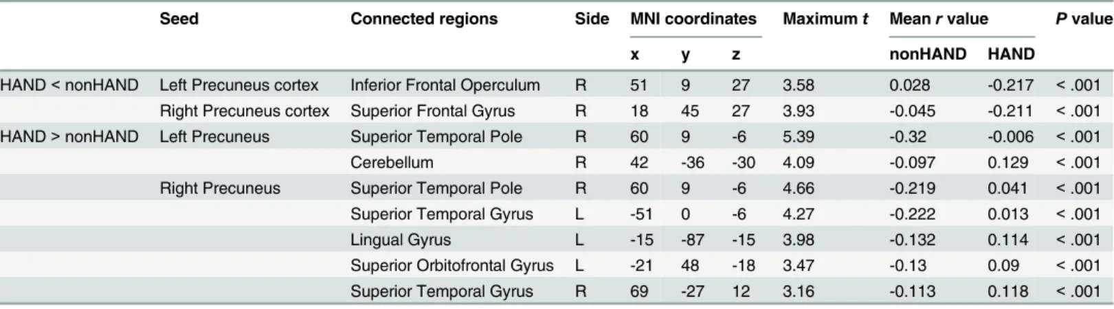
The intragroup FC patterns of each ROI were revealed using one-samplet-tests (Fig 1). In comparison with HAND and nonHAND groups, we found a decreased resting state FC between both precuneus with prefrontal cortex (PFC) regions (CorrectedP<0.05;Fig 2and
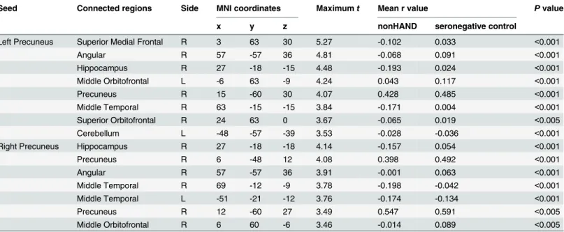
Table 3). Interestingly, when comparing the nonHAND and seronegative group, bilateral fron-tal areas, temporal and cerebellum also showed significantly decreased FC (Table 4).
Correlation Analyses between FC and NP test results
The right inferior frontal operculum and right superior frontal gyrus which had significantly decreased FC with precuneus in HAND compared to nonHAND were selected as ROIs for the correlation analysis. Among the NP tests examining memory, abstraction/executive, and sen-sory perceptual/motor skills domains which were significantly impaired in HAND relative to nonHAND, only tests examining memory domain were positively correlated with FC between precuneus and above-mentioned ROIs: both higher FC between left precuneus and right
Table 1. Baseline characteristics of study participants.
HAND (n = 10) nonHAND (n = 13) Control (n = 11) p-value
Sex(male) 100% 100% 100% Age(years) 56.0±8.2 52.8±6.2 54.7±4.2 0.47 BMI(kg/m2) 23.6±2.0 22.3±2.4 25.2±2.0 0.018 education (years) 13.1±4.4 12.7±3.3 12.7±1.6 0.949 Hemoglobin 14.2±1.5 14.0±1.4 14.3±2.2 0.931 eGFR 80.7±15.3 82.5±10.6 85.6±7.5 0.585
CD4 at diagnose (cells/uL, mean±SD) 203.8±220.7 284.1±239.1 0.441
VL at diagnose (log10 copies/mL) (mean± SD) 5.5±5.5 4.3±4.3 0.019
pre ART CD4 (cells/uL, mean±SD) 201.8±151.9 224.5±91.5 0.722
Pre ART VL (log10 copies/mL) (mean± SD) 5.2±5.5 4.8±4.9 0.304
current CD4 (cells/uL, mean±SD) 525.8±252.2 697.9±194.2 0.078
current VL (log10 copies/mL) (mean± SD) 1.4±1.0 1.3±0.0 0.264
Nadir CD4 (cells/uL, mean±SD) 188.9±183.1 216.8±147.9 0.69
Highest VL (log10 copies/mL) (mean± SD) 5.4±5.4 4.8±4.9 0.034
ART regimen (n, %) no treatment 0 0 2NRTIs+PI 6(60) 5(38) 0.327 2NRTIs+NNRTI 3(30) 4(31) 0.97 2NRTIs+II 1(10) 4(31) 0.251 CPE score(mean±SD) 7.60±1.35 7.92±0.76 0.474 Change in ART 5(50) 5(38) 0.6 Initial: at diagnose. doi:10.1371/journal.pone.0153493.t001
Table 2. Impairment of domains of neurocognitive function.
Domain Patients who showed impairment [n, (%)]
HAND (n = 10) nonHAND (n = 13) Seronegative Control (n = 11) p-value
Verbal/language 1 (10) 0 (0) 0 (0) 0.343
Speed of information processing 0 (0) 0 (0) 0 (0)
-Memory (learning and recall) 5 (50) 0 (0) 0 (0) 0.015
Abstraction/executive 8 (80) 1 (7.7) 0 (0) <0.001
attention/working memory 0 (0) 0 (0) 0 (0)
-Sensory perceptual/motor skills 8 (80) 2 (15.4) 0 (0) 0.001
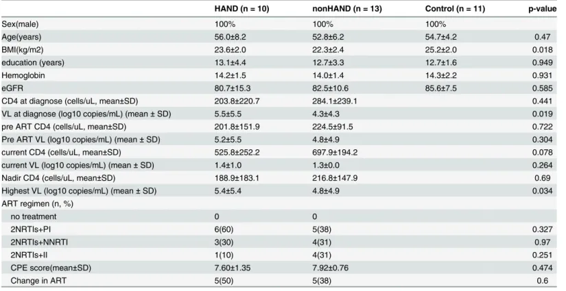
Fig 1. Renderdt maps of FC value. (A, B) show the FC maps of HAND with the seed located in the left and right precuneus, respectively. (C, D) show the FC maps of nonHAND group with the seed located in the left and right precuneus (P< .005, uncorrected).
inferior frontal operculum (r = .46, P = .025) and right precuneus and right superior frontal gyrus (r = .59, P = .003) were correlated with higher score in K-AVLT delayed recall test; higher FC between right precuneus and right superior frontal gyrus were correlated with higher score in K-AVLT delayed recognition test (r = .56, P = .006;Table 5).
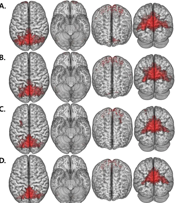
Fig 2. The differences of resting connectivity between HAND group and nonHAND group. Upper images display the regions showing the FC with the left precuneus (P< .05, corrected). Lower images display the regions showing the FC with the right precuneus (P < .05, corrected).
doi:10.1371/journal.pone.0153493.g002
Table 3. Decreased resting state functional connectivity in HAND group compared to nonHAND group.
Seed Connected regions Side MNI coordinates Maximumt Mean r value P value
x y z nonHAND HAND
HAND< nonHAND Left Precuneus cortex Inferior Frontal Operculum R 51 9 27 3.58 0.028 -0.217 < .001 Right Precuneus cortex Superior Frontal Gyrus R 18 45 27 3.93 -0.045 -0.211 < .001 HAND> nonHAND Left Precuneus Superior Temporal Pole R 60 9 -6 5.39 -0.32 -0.006 < .001
Cerebellum R 42 -36 -30 4.09 -0.097 0.129 < .001
Right Precuneus Superior Temporal Pole R 60 9 -6 4.66 -0.219 0.041 < .001 Superior Temporal Gyrus L -51 0 -6 4.27 -0.222 0.013 < .001
Lingual Gyrus L -15 -87 -15 3.98 -0.132 0.114 < .001
Superior Orbitofrontal Gyrus L -21 48 -18 3.47 -0.13 0.09 < .001 Superior Temporal Gyrus R 69 -27 12 3.16 -0.113 0.118 < .001 doi:10.1371/journal.pone.0153493.t003
Discussion
This study was designed to see the difference of FC in fMRI between HAND and nonHAND patients. To tackle this, we re-evaluated 24 HIV-infected individuals after 2.5 years, and found that 8.3% of these individuals demonstrated neurocognitive decline, 75% were stable, and 12.5% improved. Sensitivity, specificity, and inherent variability (in-patient variability) of NP testing need to be taken into account when interpretating HAND diagnosis and classification, especially in the setting of our small sample sizes. However, these observed changes are consis-tent with a previous report by Heaton et al. that showed 23% of patients had their cognitive function decline, 61% remained stable, and 17% showed improved cognitive function [14].
Overall, this study found differences in fMRI patterns between HIV-infected with and with-out HAND, and also between nonHAND and seronegative individuals. In particular, these results suggest that a pattern on fMRI can be used to distinguish HIV-infected people with and without HAND, mostly associated with differences in FC in left and right precuneus and pre-frontal brain regions.
Table 4. Decreased resting state functional connectivity in nonHAND group compared to seronegative control.
Seed Connected regions Side MNI coordinates Maximumt Mean r value P value
x y z nonHAND seronegative control
Left Precuneus Superior Medial Frontal R 3 63 30 5.27 -0.102 0.033 <0.001
Angular R 57 -57 36 4.81 -0.068 0.091 <0.001 Hippocampus R 27 -18 -15 4.48 -0.193 0.024 <0.001 Middle Orbitofrontal L -6 63 -9 4.24 0.043 0.117 <0.001 Precuneus R 15 -60 30 4.07 0.428 0.485 <0.001 Middle Temporal R 63 -15 -15 3.84 -0.171 0.004 <0.001 Superior Orbitofrontal R 24 63 0 3.67 -0.065 0.019 <0.005 Cerebellum L -48 -57 -39 3.53 -0.028 -0.036 <0.001
Right Precuneus Hippocampus R 27 -18 -18 4.14 -0.157 0.054 <0.001
Precuneus R 6 -48 12 4.08 0.398 0.492 <0.001 Angular R 57 -57 36 3.91 -0.001 0.063 <0.001 Middle Temporal R 69 -12 -9 3.78 -0.198 -0.042 <0.001 Middle Temporal L -51 -21 -12 3.76 -0.174 -0.134 <0.001 Precuneus R 12 -60 27 3.49 0.547 0.591 <0.005 Middle Orbitofrontal R 6 60 -6 3.46 -0.014 0.089 <0.005 doi:10.1371/journal.pone.0153493.t004
Table 5. Correlation analyses of decreased functional connectivity and neuropsychological function.
Regions of Interests [x y z] Seed NP tests
NP1 NP2 NP3 NP4 NP5 NP6
R Inferior Frontal Operculum [51 9 27] L precuneus 0.38 0.46* 0.25 0.23 0.25 0.28
R precuneus 0.46* 0.63** 0.59** 0.12 0.10 0.19
R Superior Frontal Gyrus [18 45 27] L precuneus 0.34 0.40 0.17 0.22 0.15 0.16
R precuneus 0.36 0.59** 0.56** 0.25 0.19 0.28
Note. Data are all Pearson’s r values. Values in the brackets are MNI coordinates. NP1: K-AVLT Total (Trials 1–5); NP2: K-AVLT Delayed Recall; NP3: K-AVLT Delayed Recognition; NP4: KCFT Copy; NP5: KCFT Immediate Recall; NP6: KCFT Delayed Recall.
* P < 0.05.
** Bonferroni-corrected P < 0.05. doi:10.1371/journal.pone.0153493.t005
Previous imaging studies have found that cerebral atrophy, decrease of baseline cerebral blood flow and cerebral signal intensity abnormalities are associated with HAND [15–17]. Fur-ther, Ances et al. reported decreased functional BOLD response of HIV–infected individuals compared with HIV-uninfected controls [18], and Küper et al. reported decrease of gray matter in the anterior cingulate and temporal cortices, along with longer duration of HIV infection or worse cognitive function [19]. Plessis et al. found that fronto-striatal dysfunction of HIV patients was associated with cognitive impairment by fMRI [20]. In our study, we found that there were significant FC differences between the nonHAND group patients and seronegative controls around that frontal and temporal areas, and this is concordant with several previous studies [16,17,19,20].
On correlation analysis, differences in FC in the frontal areas between HAND and non-HAND groups were related to performance capability. Among the brain regions that showed significant differences in the FC comparison between HAND and nonHAND, we selected the right inferior frontal operculum and right superior frontal gyrus as the ROIs of the correlation analysis. FC of the ROIs was positively correlated with scores from the NP test for memory, learning, and recall. Moreover, FC between precuneus and frontal areas were significantly cor-related with verbal memory/learning ability, keeping in line with previous studies [21]. There-fore, we speculated the altered resting-state FC within these areas might have critical role in cognitive dysfunction in HIV-infected patients.
This study has several limitations. First, while this study showed differences in fMRI between groups, this is an association and not a causation, and since this is a cross-sectional study, then prognostic and diagnostic validations cannot be determined. Second, the sample size was too small to analyze the risk factors of fluctuation in cognitive function. If we had fol-low up result of NP test of seronegative controls, we could adjust cognitive decline according to aging. Third, all the patients with HAND had asymptomatic or mild neurocognitive dysfunc-tion, so we could not assess the individuals with severe cognitive impairment, like HIV-associ-ated dementia. Further, the seronegative control group showed normal results in all 6
functional domains, which could bias the fMRI differences between groups. Lastly, all the par-ticipants were male, so these results are likely not generalizable to women.
In conclusion, there was significant difference between HAND and nonHAND groups on resting state FC in functional brain MRI. There was also significant difference between the nonHAND group and seronegative control group. Future studies will need evaluate these fMRI patterns prospectively in the diagnosis of people who will ultimately develop HAND, and if functional brain MRI may be useful to screen for HAND.
Supporting Information
S1 Dataset. Variables and data for analyses. (XLSX)
Acknowledgments
This research was supported by the Basic Science Research Program through the National Research Foundation of Korea (NRF) funded by the Ministry of Education, Science and Tech-nology (NRF-2013R1A1A2005412), a Chronic Infectious Disease Cohort grant (4800-4859-304-260) from the Korea Centers for Disease Control and Prevention, BioNano Health-Guard Research Center funded by the Ministry of Science, ICT, and Future Planning of Korea as a Global Frontier Project (Grant H-GUARD_2013M3A6B2078953) and a grant from the Minis-try of Health & Welfare, Republic of Korea (grant number: HI14C1324). Dr. Smith was
supported by the San Diego Veterans Affairs Healthcare system and NIH grants AI100665, MH062512 and AI036214.
Author Contributions
Conceived and designed the experiments: HWA NYS JYC. Performed the experiments: HWA SJ NYS JYC. Analyzed the data: HWA SJ NYS SH NSK JYC. Contributed reagents/materials/ analysis tools: SJ NYS SH. Wrote the paper: HWA SJ NYS SH JYA MYA YDJ IYJ MHK WYJ NSK JMK DMS JYC.
References
1. Antinori A, Arendt G, Becker JT, Brew BJ, Byrd DA, Cherner M, et al. Updated research nosology for HIV-associated neurocognitive disorders. Neurology. 2007; 69(18):1789–99. PMID:17914061 2. Heaton RK, Marcotte TD, Mindt MR, Sadek J, Moore DJ, Bentley H, et al. The impact of HIV-associated
neuropsychological impairment on everyday functioning. J Int Neuropsychol Soc. 2004; 10(3):317–31. PMID:15147590
3. Chernoff RA, Martin DJ, Schrock DA, Huy MP. Neuropsychological functioning as a predictor of employment activity in a longitudinal study of HIV-infected adults contemplating workforce reentry. J Int Neuropsychol Soc. 2010; 16(1):38–48. doi:10.1017/S1355617709990828PMID:19765357
4. Chan LG, Kandiah N, Chua A. HIV-associated neurocognitive disorders (HAND) in a South Asian pop-ulation—contextual application of the 2007 criteria. BMJ Open. 2012; 2(1):e000662. doi:10.1136/ bmjopen-2011-000662PMID:22331389
5. Ku NS, Lee Y, Ahn JY, Song JE, Kim MH, Kim SB, et al. HIV-associated neurocognitive disorder in HIV-infected Koreans: the Korean NeuroAIDS Project. HIV Med. 2014; 15(8):470–7. doi:10.1111/hiv. 12137PMID:24580888
6. van den Heuvel MP, Hulshoff Pol HE. Exploring the brain network: a review on resting-state fMRI func-tional connectivity. Eur Neuropsychopharmacol. 2010; 20(8):519–34. doi:10.1016/j.euroneuro.2010. 03.008PMID:20471808
7. Becker JT, Bajo R, Fabrizio M, Sudre G, Cuesta P, Aizenstein HJ, et al. Functional connectivity mea-sured with magnetoencephalography identifies persons with HIV disease. Brain Imaging Behav. 2012; 6(3):366–73. doi:10.1007/s11682-012-9149-4PMID:22328062
8. Mor V, Laliberte L, Morris JN, Wiemann M. The Karnofsky Performance Status Scale. An examination of its reliability and validity in a research setting. Cancer. 1984; 53(9):2002–7. PMID:6704925 9. Letendre SL, Ellis RJ, Ances BM, McCutchan JA. Neurologic complications of HIV disease and their
treatment. Top HIV Med. 2010; 18(2):45–55. PMID:20516524
10. Chao-Gan Y, Yu-Feng Z. DPARSF: A MATLAB Toolbox for "Pipeline" Data Analysis of Resting-State fMRI. Front Syst Neurosci. 2010; 4:13. doi:10.3389/fnsys.2010.00013PMID:20577591
11. Slotnick SD, Moo LR, Segal JB, Hart J Jr. Distinct prefrontal cortex activity associated with item memory and source memory for visual shapes. Brain Res Cogn Brain Res. 2003; 17(1):75–82. PMID:
12763194
12. Yang Z, Chang C, Xu T, Jiang L, Handwerker DA, Castellanos FX, et al. Connectivity trajectory across lifespan differentiates the precuneus from the default network. Neuroimage. 2014; 89:45–56. doi:10. 1016/j.neuroimage.2013.10.039PMID:24287438
13. Fransson P, Marrelec G. The precuneus/posterior cingulate cortex plays a pivotal role in the default mode network: Evidence from a partial correlation network analysis. Neuroimage. 2008; 42(3):1178– 84. doi:10.1016/j.neuroimage.2008.05.059PMID:18598773
14. Heaton RK, Franklin DR Jr, Deutsch R, Letendre SL, Ellis RJ, Casaletto K, et al. Neurocognitive change in the era of HIV combination antiretroviral therapy: A longitudinal CHARTER Study. Clin Infect Dis. 2014.
15. Hanning U, Husstedt IW, Niederstadt TU, Evers S, Heindel W, Kloska SP. Cerebral signal intensity abnormalities on T2-weighted MR images in HIV patients with highly active antiretroviral therapy: rela-tionship with clinical parameters and interval changes. Acad Radiol. 2011; 18(9):1144–50. doi:10. 1016/j.acra.2011.04.013PMID:21703882
16. Li C, Zhang X, Komery A, Li Y, Novembre FJ, Herndon JG. Longitudinal diffusion tensor imaging and perfusion MRI investigation in a macaque model of neuro-AIDS: a preliminary study. Neuroimage. 2011; 58(1):286–92. doi:10.1016/j.neuroimage.2011.05.068PMID:21658455
17. Towgood KJ, Pitkanen M, Kulasegaram R, Fradera A, Kumar A, Soni S, et al. Mapping the brain in younger and older asymptomatic HIV-1 men: frontal volume changes in the absence of other cortical or diffusion tensor abnormalities. Cortex. 2012; 48(2):230–41. doi:10.1016/j.cortex.2011.03.006PMID: 21481856
18. Ances B, Vaida F, Ellis R, Buxton R. Test-retest stability of calibrated BOLD-fMRI in HIV- and HIV+ sub-jects. Neuroimage. 2011; 54(3):2156–62. doi:10.1016/j.neuroimage.2010.09.081PMID:20932922 19. Kuper M, Rabe K, Esser S, Gizewski ER, Husstedt IW, Maschke M, et al. Structural gray and white
mat-ter changes in patients with HIV. J Neurol. 2011; 258(6):1066–75. doi:10.1007/s00415-010-5883-y PMID:21207051
20. Plessis SD, Vink M, Joska JA, Koutsilieri E, Stein DJ, Emsley R. HIV infection and the fronto-striatal system: a systematic review and meta-analysis of fMRI studies. Aids. 2014; 28(6):803–11. doi:10. 1097/QAD.0000000000000151PMID:24300546
21. Chang YL, Jacobson MW, Fennema-Notestine C, Hagler DJ Jr, Jennings RG, Dale AM, et al. Level of executive function influences verbal memory in amnestic mild cognitive impairment and predicts pre-frontal and posterior cingulate thickness. Cereb Cortex. 2010; 20(6):1305–13. doi:10.1093/cercor/ bhp192PMID:19776343