© 2016 The Korean Society of Pathologists/The Korean Society for Cytopathology
pISSN 2383-7837
Molecular Dimensions of Gastric Cancer: Translational and
Clinical Perspectives
Yoon Young Choi1
Sung Hoon Noh1,2
Jae-Ho Cheong1,2,3
1Department of Surgery, 2Brain Korea 21 PLUS
Project for Medical Science, 3Department of
Biochemistry and Molecular Biology, Yonsei University College of Medicine, Seoul, Korea
Gastric cancer is a global health burden and has the highest incidence in East Asia. This disease is complex in nature because it arises from multiple interactions of genetic, local environmental, and host factors, resulting in biological heterogeneity. This genetic intricacy converges on molec-ular characteristics reflecting the pathophysiology, tumor biology, and clinical outcome. There-fore, understanding the molecular characteristics at a genomic level is pivotal to improving the clinical care of patients with gastric cancer. A recent landmark study, The Cancer Genome Atlas (TCGA) project, showed the molecular landscape of gastric cancer through a comprehensive mo-lecular evaluation of 295 primary gastric cancers. The proposed momo-lecular classification divided gastric cancer into four subtypes: Epstein-Barr virus–positive, microsatellite unstable, genomic stable, and chromosomal instability. This information will be taken into account in future clinical trials and will be translated into clinical therapeutic decisions. To fully realize the clinical benefit, many challenges must be overcome. Rapid growth of high-throughput biology and functional validation of molecular targets will further deepen our knowledge of molecular dimensions of this cancer, allowing for personalized precision medicine.
Key Words: Stomach neoplasms; Translational medical research; Cancer genomics; Cancer genetics; Target therapy
Received: September 1, 2015 Accepted: September 9, 2015
Corresponding Author
Jae-Ho Cheong, MD, PhD
Department of Surgery, Yonsei University College of Medicine, 50-1 Yonsei-ro, Seodaemun-gu, Seoul 03722, Korea Tel: +82-2-2228-2094
Fax: +82-2-313-8289 E-mail: [email protected]
http://dx.doi.org/10.4132/jptm.2015.09.10
Gastric cancer (GC) is one of the most common and fatal dis-eases, and nearly two-thirds of the cases are concentrated in East
Asia.1 In Korea, GC is the second most common malignancy
after thyroid cancer. It was ranked as the third cause of cancer
mortality in 2012.2 An estimate of 35,000 people are expected
to be newly diagnosed, and about 7,500 patients will die in
2015.3 According to the National Cancer Screening Program
that began in 1999, the proportion of early gastric cancer (EGC) has increased, and the prognosis of EGC is favorable even with-out additional treatment after surgery.4,5 Radical D2 surgery with
adjuvant chemotherapy has been established as a standard treat-ment for locally advanced gastric cancer (AGC), and has
im-proved the prognosis of AGC.6-8 However, about half of patients
with AGC experience recurrence, and the proportion of patients
who benefit from adjuvant chemotherapy is around 20%,9-14 with
cases of metastasis being the least amenable to treatment.15,16
GC is a heterogeneous malignancy, which is the main reason for its different prognoses in patients with same clinical stage and for its diverse responses to the standard treatment. It is now widely appreciated that genomic complexity and heterogeneity
are fundamental causes of tumor phenotypic characteristics de-termining clinical outcomes. Therefore, understanding of the molecular and genetic characteristics is essential in effective and personalized management of GC.
GC has been classified according to histo-morphologic fea-tures. The World Health Organization (WHO) classifies GC into
papillary, tubular, mucinous, and poorly cohesive carcinomas,17
while the Lauren classification divides GC into intestinal, diffuse,
and mixed types.18 However, these classification systems do not
satisfactorily provide information relevant to clinical utilities and treatment guidelines. Over the past decades, the molecular landscape of GC has been shaped, and this review focuses on the molecular features of GC that can be translated into clinical use in order to guide precise therapeutic decisions. Details of surgi-cal, adjuvant or peri-operative chemotherapy, and radiotherapy are beyond the scope of this review and readers would be
direct-ed to other reviews dealing with these issues.19 The recent The
Cancer Genome Atlas (TCGA) study,20 a landmark study
divid-ing GC into four subtypes based on multi-dimensional profildivid-ing, (1) Epstein-Barr virus (EBV) tumor, (2) microsatellite unstable
(MSI) tumor, (3) genomically stable (GS) tumor, and (4) chro-mosomal instability (CIN), is used as a roadmap for this review. We also provide the current status of recent and ongoing clini-cal trials that focus on genomic alterations in molecular sub-types for targeted therapeutics (summarized in Table 1).15,16,21-34
EPSTEIN-BARR VIRUS–POSITIVE GASTRIC CANCER
The incidence of EBV-positive GC has been reported to be
around 10%20,35 and harbors a higher prevalence of DNA
hy-permethylation than other subtypes.20,36 The reason for
extraor-dinary DNA hypermethylation seems to be a cellular reaction to the viral infection.37 EBV-positive GC also has strong signatures
of interleukin 12-mediated signaling events, which reflects high immune cell infiltration.20,38 Intriguingly, tumors of this subtype
exhibit CD274 and PDCD1LG2 amplification of which pro-teins PD-L1 and PD-L2, respectively, are related to immune
sup-pressive functions, particularly immune checkpoints. Further, amplification at the 9p24.1 locus containing JAK2, which en-codes an oncogenic receptor tyrosine kinase (RTK), is also a po-tential therapeutic target. Those findings seemed to support the rationale that this type of GC would be a relevant target of RTK
and immune checkpoints inhibitors.20,37 A recent phase I study
showed that pembrolizumab (MK-3475), one of the anti–PD-1/2 immune checkpoint inhibitors, provided antitumor activity
in patients with AGC that expressed PD-L1.21 Predilection of
EBV-positive GC for PIK3CA mutation, which is related to the phosphoinositide 3-kinase (PI3K)/Akt signaling pathway, has
also been reported.20 Frequent PIK3CA mutations warrant
evalu-ation of PI3K inhibitors in EBV-positive GC. Although still in preclinical stages, BKM 120, which is a direct PIK3CA inhibi-tor, and BEZ235, a dual PIK3CA and mammalian target of ra-pamycin (mTOR) inhibitor, have been reported to reduce cell
viability and induce apoptosis in GC cell lines.22 Additionally,
the ARID1A mutation was detected in 10% of GC39 and was
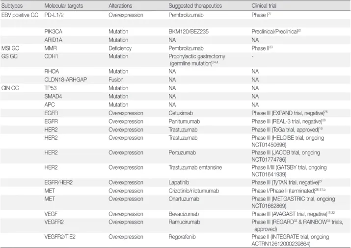
Table 1. Current status of targeted therapies based on molecular alterations according to GC subtype
Subtypes Molecular targets Alterations Suggested therapeutics Clinical trial
EBV positive GC PD-L1/2 Overexpression Pembrolizumab Phase I21
PIK3CA Mutation BKM120/BEZ235 Preclinical/Preclinical22
ARID1A Mutation NA NA
MSI GC MMR Deficiency Pembrolizumab Phase II23
GS GC CDH1 Mutation Prophylactic gastrectomy
(germline mutation)24,a
-RHOA Mutation NA NA CLDN18-ARHGAP Fusion NA NA CIN GC TP53 Mutation NA NA SMAD4 Mutation NA NA APC Mutation NA NA
EGFR Overexpression Cetuximab Phase III (EXPAND trial, negative)25
EGFR Overexpression Panitumumab Phase III (REAL-3 trial, negative)26
HER2 Overexpression Trastuzumab Phase III (ToGa trial, approved)16
HER2 Overexpression Trastuzumab Phase III (HELOISE trial, ongoing
NCT01450696)
HER2 Overexpression Pertuzumab Phase III (JACOB trial, ongoing
NCT01774786)
HER2 Overexpression Trastuzumab emtansine Phase II/III (GATSBY trial, ongoing
NCT01641939)
EGFR/HER2 Overexpression Lapatinib Phase III (TyTAN trial, negative)27
MET Overexpression Crizotinib/rilotumumab Phase I/Phase II (terminated)28-31,b
MET Overexpression Onartuzumab Phase III (METGASTRIC trial, ongoing
NCT01662869)
VEGF Overexpression Bevacizumab Phase III (AVAGAST trial, negative)15,32
VEGFR2 Overexpression Ramucirumab Phase III (REGARD33 & RAINBOW34 trials,
approved)
VEGFR2/TIE2 Overexpression Regorafenib Phase II (INTEGRATE trial, ongoing
ACTRN12612000239864) GC, gastric cancer; EBV, Epstein-Barr virus; N/A, not available; MSI, microsatellites unstable; GS, genomically stable; CIN, chromosomal instability.
aMainly in the Western countries; bAmgen-sponsored clinical trials of rilotumumab in advanced gastric cancer were terminated based on the pre-planned safety review by independent data monitoring committee.
found most frequently in EBV-positive GC.20 This mutation
encodes a component of SWI/SNF complex and acts as a tumor
suppressor in cancer.39 Recently, it was reported that EZH2
in-hibitor could be a novel therapeutic targeting
ARID1A-mutat-ed cancers.40 Therefore, the ARID1A mutation provides another
clinically actionable genetic alteration in EBV-positive GC that should be validated in a clinical study. Based on these findings, the molecular characteristics of EBV-positive GC are distinct from those of other GC subtypes, and some of the genetic alter-ations can be therapeutically exploited.
MICROSATELLITE UNSTABLE GASTRIC CANCER
MSI GC is related to the loss of function of mismatch repair (MMR) genes and is associated with older age, female gender, intestinal type, and less aggressive tumor stages.20,41,42 Because
the function of the MMR mechanism is defective mainly due to
MLH1 silencing by promoter hypermethylation,20 this subtype
has more mutations per megabase (Mb) compared to other types of GC. Intriguingly, MSI tumors possess common alterations in major histocompatibility complex (MHC) class I-related genes, including HLA-B and B2M. Since these MHC class I genes function in proper antigen presentation to the host immune sys-tem, these genomic alterations could provide hypermutated MSI GC with the selective advantage of immune surveillance evasion.
The incidence of MSI GC was previously reported to be 8.5%–
37.8%.43 While the prognosis of MSI GC was not assessed in
comparison with those of other molecular subtypes in a TCGA study,20 a meta-analysis43 and a recent study regarding the
mo-lecular classification of GC reported that MSI GC had the best overall prognosis with the lowest recurrence rate.44 Importantly,
the prognosis of MSI GC was prominent in the population treat-ed with surgery alone. Indetreat-ed, the prognosis of MSI GC with-out chemotherapy was similar to that of patients who received
chemotherapy after surgery,45 implying that the MSI subtype is
unresponsive to chemotherapy in an adjuvant setting,41 like MSI
colon cancer.46,47 Additionally, a recent phase II study that
eval-uated the clinical utility of pembrolizumab showed that MMR
status predicted the benefit of pembrolizumab,23 and a higher
mutational load was reported to be related to positive response
to anti-CTLA-4 in melanoma48 and PD-1 antibody in non-small
cell lung cancer.49 The legitimate explanation might be that
immune infiltrate related with mutation was directed at neoan-tigens, and recognition plays an important role in the
antitu-mor immune response.23 Consequently, MSI status is a promising
biomarker to predict the prognosis and responses to immune checkpoint inhibitor as well as chemotherapy in GC, as in colon cancer.50 There is no consensus on the definition of GC-specific
MSI in clinical settings at this time, and studies have used dif-ferent criteria to define MSI.20,43,45,51,52 Thus, it is necessary to
es-tablish appropriate analysis standards for MSI status in GC for precise detection and translation of “MSI-ness” for clinical ther-apeutic decisions.
GENOMICALLY STABLE GASTRIC CANCER
The GS subtype of GC is best represented as a diffuse type of GC, with lower mutation burden compared to other subtypes
and occurring at a relatively early age.20 CDH1 mutation is one
of the representative mutations in the GS subtype. CDH1 germ-line mutations are known to be related to hereditary diffuse GC. When patients harbor pathogenic hotspot mutations in CDH1,
prophylactic gastrectomy is recommended.24 However, only two
CDH1 mutations, neither of which is a pathogenic hotspot
mu-tation, were identified in a recent TCGA study.20 Another study
reported that somatic alterations of CDH1 were present in ap-proximately 30% of GC cases, and structural alterations in CDH1
were related to poor prognosis.53 In addition to CDH1
muta-tions, GS subtype tumors have RHOA mutations and
CLDN18-ARHGAP 6 or 26 fusions.20,54,55 RHOA is known to modulate
downstream Rho signaling, and its mutation imparts resistance
to anoikis, a form of programmed cell death.54 Also, RHOA acts
to control actin-myosin-dependent cell contractility and motil-ity;56,57 thus, its mutation might contribute to dispersed growth
and poorly cohesive patterns of diffuse type GC,20 which is
associ-ated with poor prognosis. Thus, the RHOA mutation could be a
good candidate for new approaches targeting GS subtype GC.37
CLDN18-ARHGAP6 or 26 fusions are mutually exclusive to RHOA and CDH1 mutation among GS tumors. The discovery
of recurrent interchromosomal translocation between CLDN18 and ARHGAP26 further implies biological significance of cell adhesion and deregulated Rho signaling in GS tumors since CLDN18 is involved in intercellular tight junction structure, and ARHGAP26, a GTPase-activating protein, imparts Rho sig-naling activation by facilitating the conversion of Rho GTPases to the GDP state. A recent study reported that this type of fu-sion in epithelial cells mediates epithelial disintegration and is
related to epithelial-mesenchymal transition (EMT).58
There-fore, the novel discoveries of RHOA mutation and
CLDN18-ARHGAP26 fusion could be exploited to develop new
to harbor the poorest prognosis of all GC tumors.44 However,
translating those new strategies to clinical practice is in the early stages and is largely lacking evidence of functional validity. Ad-ditionally, there have been no clinical trials to assess the efficacy of targeting those genomic alterations in GS subtype tumors.
CHROMOSOMAL INSTABILITY GASTRIC CANCER
CIN subtype GC is related to intestinal type histology,
fre-quent TP53 mutations, and amplification of RTKs.20 TP53
mu-tation is the most frequently detected mumu-tation in GC, occurring in up to 50% of all cases59 and 71% of cases of CIN subtype GC.20 TP53 mutation is associated with high levels of somatic copy
number variations in both chromosomal and focal gene regions.60
Also, other canonical tumor suppressor genes such as SMAD4
and APC have been reported to be mutated in GC.61 Since tumor
suppressor genes are regarded as poor candidates for targeted therapy development, alterations in RTKs will be discussed in this review.
EPIDERMAL GROWTH FACTOR RECEPTOR
The human epidermal growth factor receptors (HER) are a family of four transmembrane RTKs, ErbB1 (epidermal growth factor receptor, EGFR), ErbB2 (HER2), ErbB3
(HER3), and ErbB4 (HER4),62 that regulate diverse
down-stream signaling pathways and play an important role in GC development and progression. A biomarker study from Adjuvant
Chemotherapy Trial of S-1 for Gastric Cancer (ACTS-GC),14 a
phase III randomized controlled trial (RCT) that compared the effect of adjuvant S-1 over surgery alone in locally AGC, showed that EGFR overexpression was related to poor
progno-sis but was not found to be the case for HER2.63 There have
been two RCTs that investigated the benefit of EGFR inhibition
(EXPAND25 trial for cetuximab and REAL-3 trial26 for
panitu-mumab as first-line therapy); however, both trials failed to prove the additional clinical benefit of anti-EGFR antibody over stan-dard chemotherapy.
HER2
The success of a clinical trial that investigated the effects of trastuzumab targeting HER2 in HER2-overexpressed GC pa-tients resulted in changes in clinical practice. Addition of trastu-zumab to chemotherapy as the first-line treatment of metastatic
GC improved overall survival.16 However, an updated survival
analysis showed that the benefit of the trastuzumab decreased over time, the difference in median overall survival was reduced from 2.7 to 1.4 months, and the hazard ratio increased from 0.74
in primary analysis to 0.80.64 This raises a concern, requiring
further investigation to clarify the clinical benefit of trastuzum-ab in HER2-overexpressing GC patients. Indeed, two first-line therapy trials are underway to investigate the effect of addition of pertuzumab to a standard HER2 targeting regimen and the effect of two dose levels of trastuzumab (JACOB and HELI-OSE, respectively).
The frequency of HER2 mutation was reported to be 5% (9/180) in GC, and the relationship between HER2 mutation and responsiveness to trastuzumab has not yet been determined. Another clinically important issue regarding HER2 mutation and amplification might be derived from a recent EGFR bio-marker study in non-small cell lung cancer patients receiving gefitinib, where EGFR mutation and amplification correlated
with prolonged progression-free survival.65 Based on this, it
might be worthwhile to investigate the clinical benefit of HER2 inhibitor in HER2-mutated and amplified GC as alternative
candidates for HER2-targeted therapy.66 A phase III trial
(TY-TAN) was conducted to investigate the benefit of lapatinib, a dual inhibitor of EGFR and HER2, as a second-line therapy for AGC. Although overall survival was not significantly different,
post hoc analysis demonstrated that the HER2
immunohisto-chemistry 3+ subgroup showed statistically significant prolon-gation of overall survvial.27
KRAS
KRAS is one of the members of the RAS family, and its
muta-tion plays an important role in tumorigenesis by activating down-stream pathways such as PI3K and RAF. The frequency of KRAS mutations in GC was reported as 1.5%–5.8%, and most of
them were transversions.67 Overexpression of wild-type KRAS
seemed to be related to acquired resistance to inhibitors of other tyrosine kinase in GC cells.68 A phase II trial that evaluated the
efficacy of selumetinib, an inhibitor of MEK1/MEK2, down-stream of KRAS, for KRAS-mutant non-small cell lung cancer demonstrated promising efficacy and thereby warrants further clinical investigation.69 Thus, a MEK inhibitor could be a
poten-tial therapeutic agent for targeting KRAS-mutated GC; howev-er, evidence from a clinical trial is required.
MESENCHYMAL EPITHELIAL TRANSITION FACTOR
Mesenchymal epithelial transition factor (MET) amplifica-tion was not common (2%, 10/489) in GC it was reported to
be related with poor prognosis.28 However, two studies have
re-ported the possibility of targeted therapy for MET-positive GC. An expanded phase I cohort study showed that patients with
MET amplification had a favorable response to crizotinib
(PF-02341066), a MET/anaplastic lymphoma kinase tyrosine kinase
inhibitor.28 Furthermore, rilotumumab (AMG 102), a fully
hu-manized monoclonal antibody against hepatocyte growth fac-tor/MET, demonstrated favorable overall survival especially for
patients with MET-positive GC.29 Based on those results,
subse-quent trials were conducted (RILOMET-130 and NCT02137343);
however, all Amgen-sponsored clinical trials of rilotumumab in AGC were terminated based on a pre-planned safety review by the data monitoring committee due to an increase in death with
the study drug.31 Currently, a small-molecule MET inhibitor is
under investigation for MET-amplified GC. BRAF
BRAF mutations are related to tumorigenesis, and
dysregu-lated BRAF activity instigates abnormal cell growth and
prolif-eration through MEK and ERK pathways.70 The specific
muta-tion BRAFV600E is the most common type of BRAF mutation in
melanoma, and vemurafenib, a BRAF inhibitor, was found to
be beneficial in patients with BRAFV600E-mutated melanoma.71
BRAF mutations are rarely observed in GC, with only 2.2%
(7/319) of patients demonstrating BRAF mutation, most (five of
seven BRAF mutations) of which were BRAFV599M;72 in
addi-tion, there were no BRAF mutations among 167 patients in the
REAL-3 trial.26 Furthermore, only 0.2% of patients (1/508) with
GC had a BRAFV600E mutation in another study.67 Therefore, it
is not yet clear if BRAF mutation is a driver mutation in GC.
VASCULAR ENDOTHELIAL GROWTH FACTOR RECEPTOR
Vascular endothelial growth factor receptor (VEGFR) expres-sion is closely related to angiogenesis in tumorigenesis. Since an-giogenesis is critical for tumor growth and metastasis, it is a therapeutic target for many cancer types. Bevacizumab, a monoclonal antibody that inhibits VEGF (VEGF-A), showed
survival benefits in advanced colorectal cancer73 and non-small
cell lung cancer.74 Also, a phase II trial that showed efficacy of
bevacizumab for advanced gastro-esophageal cancer seemed to reinforce the success of targeted therapy against the VEGF
pathway in gastric cancer.75 Despite initial enthusiasm
regard-ing its use, bevacizumab combined with chemotherapy as a first-line therapy did not improve the overall survival of patients with GC in the AVAGAST trial. However, the more recent
RAIN-BOW34 and REGARD33 trials, which evaluated benefits of
ramucirumab (antibody targeting VEGFR2) as second-line and first-line therapies, respectively, reported improved overall sur-vival in GC patients. Intriguingly, subgroup analyses of the trials showed that benefit from VEGF-targeted therapy was observed mainly in non-Asian patients. Also, a subsequent biomarker study of AVAGAST showed that plasma VEGF-A and neuropi-lin-1 levels could be prognostic and predictive of bevacizumab treatment in a non-Asian population.32 Those findings imply that
GC is a complex and heterogeneous disease across the globe, which affects the response to anti-angiogenesis treatment and po-tentially other target therapies.37,76
CANCER STEM CELL-RELATED PATHWAYS
Cancer stem cells that initiate tumorigenesis through self-newal and differentiation are emerging concepts in cancer re-search. Such cells activate EMT, oncogenic pathways, and em-bryogenic pathways.77,78 Also, these cells are known to be resistant
to chemotherapy and radiotherapy, while Wnt, Notch, and Hedgehog pathways are crucial to the maintenance of cancer stem cells. Transcriptional factors such as Snail, Slug, Twist, and Zeb1/2 coordinate the EMT, while transforming growth factor β (TGF-β) is a central signaling pathway related to
transforma-tion into EMT.79,80 TGF-β can act as a proto-oncogene, driving
matrix deposit, stimulating EMT and stem cell renewal, and
in-hibiting apoptosis through transactivation of EGFR.81,82 Also,
its downstream signaling pathway, PI3K/Akt/mTOR, aids in cancer stem cell maintenance.83 Thus, targeting this pathway with
appropriate agents such as metformin would inhibit cellular transformation and selectively kill cancer stem cells, as previously
demonstrated in breast cancer.84 Also, a recent report showed
that patients with GC treated with metformin for diabetes mel-litus had better survival compared to those treated with
diabet-ic meddiabet-ications other than metformin.85 GC stem cells express
CD133, CD44, aldehyde dehydrogenase 1 (ALDH1), and ATP-binding cassette sub-family G member 2 (ABCG2). CD44 and ALDH1 have been reported to be related to resistance to
en-codes the GC stem cell marker CD44 was observed in a TCGA study,20 suggesting the potential of exploiting genomic alteration
in future development of cancer stem cell-directed therapies.
CONCLUSIONS AND FUTURE PERSPECTIVES
Through extraordinary efforts over the past decades, our kn-owledge on GC has advanced considerably, and standardized multidisciplinary treatment has improved the prognosis of GC. However, clinical development of targeted therapy in GC re-mains inferior to those of other cancer types such as lung, breast, and colon cancer in terms of genetic sequencing and molecular therapeutics.87 Most of the targeted therapies have been
investi-gated without patient selection based on a biomarker, and the results have been disappointing, with only a few targeted ag-ents16,33,34 showing benefit to patient survival at one year even
af-ter treatment for metastasis or recurrent GC. In the upcoming years, accumulation of genomic information and knowledge about molecular pathogenesis of GC will be accelerated through high-throughput systems biology, and the treatment will be focused on targeting specific GC subtypes based on specific molecular characteristics (e.g., somatic driver alterations and amplifica-tion). Presently, one of the hurdles in this achievement is the in-tegration of knowledge from various disciplines and its transla-tion into daily clinical practice. To achieve a sensible reductransla-tion in mortality due to this deadly disease, transdisciplinary cooper-ation among clinicians, pathologists, bioiformaticians, compu-tational biologists, and genomicists is required.
Conflicts of Interest
No potential conflict of interest relevant to this article was reported.
Acknowledgments
This research was supported by a grant of the Korea Health Technology R&D Project through the Korea Health Industry Development Institute (KHIDI), funded by the Ministry of Health & Welfare, Republic of Korea (HI3C2162), and by the National R&D Program for Cancer Control, Ministry of Health and Welfare, Republic of Korea (1020390, 1320360).
REFERENCES
1. Ferlay J, Soerjomataram I, Dikshit R, et al. Cancer incidence and mortality worldwide: sources, methods and major patterns in GLOBOCAN 2012. Int J Cancer 2015; 136: E359-86.
2. Jung KW, Won YJ, Kong HJ, et al. Cancer statistics in Korea: inci-dence, mortality, survival, and prevalence in 2012. Cancer Res Treat 2015; 47: 127-41.
3. Jung KW, Won YJ, Oh CM, et al. Prediction of cancer incidence and mortality in Korea, 2015. Cancer Res Treat 2015; 47: 142-8. 4. Lee KS, Oh DK, Han MA, et al. Gastric cancer screening in Korea:
report on the national cancer screening program in 2008. Cancer Res Treat 2011; 43: 83-8.
5. Suh M, Choi KS, Lee YY, Jun JK. Trends in cancer screening rates among Korean men and women: results from the Korean National Cancer Screening Survey, 2004-2012. Cancer Res Treat 2013; 45: 86-94. 6. Ajani JA, Bentrem DJ, Besh S, et al. Gastric cancer, version 2.2013:
featured updates to the NCCN Guidelines. J Natl Compr Canc Netw 2013; 11: 531-46.
7. Waddell T, Verheij M, Allum W, et al. Gastric cancer: ESMO-ESSO-ESTRO Clinical Practice Guidelines for diagnosis, treatment and follow-up. Ann Oncol 2013; 24 Suppl 6: vi57-63.
8. Japanese Gastric Cancer Association. Japanese gastric cancer treat-ment guidelines 2010 (ver. 3). Gastric Cancer 2011; 14: 113-23. 9. Songun I, Putter H, Kranenbarg EM, Sasako M, van de Velde CJ.
Surgical treatment of gastric cancer: 15-year follow-up results of the randomised nationwide Dutch D1D2 trial. Lancet Oncol 2010; 11: 439-49.
10. Wu CW, Hsiung CA, Lo SS, et al. Nodal dissection for patients with gastric cancer: a randomised controlled trial. Lancet Oncol 2006; 7: 309-15.
11. Bang YJ, Kim YW, Yang HK, et al. Adjuvant capecitabine and oxali-platin for gastric cancer after D2 gastrectomy (CLASSIC): a phase 3 open-label, randomised controlled trial. Lancet 2012; 379: 315-21. 12. Noh SH, Park SR, Yang HK, et al. Adjuvant capecitabine plus
oxali-platin for gastric cancer after D2 gastrectomy (CLASSIC): 5-year follow-up of an open-label, randomised phase 3 trial. Lancet Oncol 2014; 15: 1389-96.
13. Sakuramoto S, Sasako M, Yamaguchi T, et al. Adjuvant chemother-apy for gastric cancer with S-1, an oral fluoropyrimidine. N Engl J Med 2007; 357: 1810-20.
14. Sasako M, Sakuramoto S, Katai H, et al. Five-year outcomes of a randomized phase III trial comparing adjuvant chemotherapy with S-1 versus surgery alone in stage II or III gastric cancer. J Clin On-col 2011; 29: 4387-93.
15. Ohtsu A, Shah MA, Van Cutsem E, et al. Bevacizumab in combina-tion with chemotherapy as first-line therapy in advanced gastric cancer: a randomized, double-blind, placebo-controlled phase III study. J Clin Oncol 2011; 29: 3968-76.
16. Bang YJ, Van Cutsem E, Feyereislova A, et al. Trastuzumab in com-bination with chemotherapy versus chemotherapy alone for
treat-ment of HER2-positive advanced gastric or gastro-oesophageal junction cancer (ToGA): a phase 3, open-label, randomised con-trolled trial. Lancet 2010; 376: 687-97.
17. Bosman FT, Carneiro F, Hruban RH, Theise ND. WHO classification of tumours of the digestive system. 4th ed. Lyon: IARC Press, 2010. 18. Lauren P. The Two histological main types of gastric carcinoma: dif-fuse and so-called intestinal-type carcinoma: an attempt at a histo-clinical classification. Acta Pathol Microbiol Scand 1965; 64: 31-49. 19. Choi YY, Noh SH, Cheong JH. Evolution of gastric cancer treatment:
from the golden age of surgery to an era of precision medicine. Yonsei Med J 2015; 56: 1177-85.
20. Cancer Genome Atlas Research Network. Comprehensive molecular characterization of gastric adenocarcinoma. Nature 2014; 513: 202-9. 21. Muro K, Bang Y, Shankaran V, et al. A phase 1B study of pembroli-zumab (Pembro; MK-3475) in patients (pts) with advanced gastric cancer. Ann Oncol 2014; 25 Suppl 4: LBA15.
22. Mueller A, Bachmann E, Linnig M, et al. Selective PI3K inhibition by BKM120 and BEZ235 alone or in combination with chemother-apy in wild-type and mutated human gastrointestinal cancer cell lines. Cancer Chemother Pharmacol 2012; 69: 1601-15.
23. Le DT, Uram JN, Wang H, et al. PD-1 blockade in tumors with mis-match-repair deficiency. N Engl J Med 2015; 372: 2509-20.
24. Huntsman DG, Carneiro F, Lewis FR, et al. Early gastric cancer in young, asymptomatic carriers of germ-line E-cadherin mutations. N Engl J Med 2001; 344: 1904-9.
25. Lordick F, Kang YK, Chung HC, et al. Capecitabine and cisplatin with or without cetuximab for patients with previously untreated advanced gastric cancer (EXPAND): a randomised, open-label phase 3 trial. Lancet Oncol 2013; 14: 490-9.
26. Waddell T, Chau I, Cunningham D, et al. Epirubicin, oxaliplatin, and capecitabine with or without panitumumab for patients with previously untreated advanced oesophagogastric cancer (REAL3): a randomised, open-label phase 3 trial. Lancet Oncol 2013; 14: 481-9. 27. Satoh T, Xu RH, Chung HC, et al. Lapatinib plus paclitaxel versus
paclitaxel alone in the second-line treatment of HER2-amplified advanced gastric cancer in Asian populations: TyTAN: a random-ized, phase III study. J Clin Oncol 2014; 32: 2039-49.
28. Lennerz JK, Kwak EL, Ackerman A, et al. MET amplification iden-tifies a small and aggressive subgroup of esophagogastric adeno-carcinoma with evidence of responsiveness to crizotinib. J Clin On-col 2011; 29: 4803-10.
29. Iveson T, Donehower RC, Davidenko I, et al. Rilotumumab in com-bination with epirubicin, cisplatin, and capecitabine as first-line treatment for gastric or oesophagogastric junction adenocarcino-ma: an open-label, dose de-escalation phase 1b study and a double-blind, randomised phase 2 study. Lancet Oncol 2014; 15: 1007-18.
30. Doshi S, Gisleskog PO, Zhang Y, et al. Rilotumumab exposure-re-sponse relationship in patients with advanced or metastatic gastric cancer. Clin Cancer Res 2015; 21: 2453-61.
31. Amgen announces termination of all amgen-sponsored clinical studies of rilotumumab in advanced gastric cancer [Internet]. New York: PR Newswire Association LLC, 2014 [cited 2015 Oct 1]. Avail-able from: http://www.prnewswire.com/news-releases/amgen- announces-termination-of-all-amgen-sponsored-clinical-studies-of-rilotumumab-in-advanced-gastric-cancer-300000103.html. 32. Van Cutsem E, de Haas S, Kang YK, et al. Bevacizumab in
combi-nation with chemotherapy as first-line therapy in advanced gastric cancer: a biomarker evaluation from the AVAGAST randomized phase III trial. J Clin Oncol 2012; 30: 2119-27.
33. Fuchs CS, Tomasek J, Yong CJ, et al. Ramucirumab monotherapy for previously treated advanced gastric or gastro-oesophageal junction adenocarcinoma (REGARD): an international, randomised, multicentre, placebo-controlled, phase 3 trial. Lancet 2014; 383: 31-9. 34. Wilke H, Muro K, Van Cutsem E, et al. Ramucirumab plus
pacli-taxel versus placebo plus paclipacli-taxel in patients with previously treated advanced gastric or gastro-oesophageal junction adenocar-cinoma (RAINBOW): a double-blind, randomised phase 3 trial. Lancet Oncol 2014; 15: 1224-35.
35. Murphy G, Pfeiffer R, Camargo MC, Rabkin CS. Meta-analysis shows that prevalence of Epstein-Barr virus-positive gastric cancer differs based on sex and anatomic location. Gastroenterology 2009; 137: 824-33.
36. Kaneda A, Matsusaka K, Aburatani H, Fukayama M. Epstein-Barr virus infection as an epigenetic driver of tumorigenesis. Cancer Res 2012; 72: 3445-50.
37. Tan P, Yeoh KG. Genetics and molecular pathogenesis of gastric adenocarcinoma. Gastroenterology 2015; 149: 1153-62.e3.
38. Grogg KL, Lohse CM, Pankratz VS, Halling KC, Smyrk TC. Lym-phocyte-rich gastric cancer: associations with Epstein-Barr virus, microsatellite instability, histology, and survival. Mod Pathol 2003; 16: 641-51.
39. Wang K, Kan J, Yuen ST, et al. Exome sequencing identifies fre-quent mutation of ARID1A in molecular subtypes of gastric cancer. Nat Genet 2011; 43: 1219-23.
40. Bitler BG, Aird KM, Garipov A, et al. Synthetic lethality by target-ing EZH2 methyltransferase activity in ARID1A-mutated cancers. Nat Med 2015; 21: 231-8.
41. An JY, Kim H, Cheong JH, Hyung WJ, Kim H, Noh SH. Microsat-ellite instability in sporadic gastric cancer: its prognostic role and guidance for 5-FU based chemotherapy after R0 resection. Int J Cancer 2012; 131: 505-11.
out-comes of gastric cancers with microsatellite instability. Mod Pathol 2002; 15: 632-40.
43. Choi YY, Bae JM, An JY, et al. Is microsatellite instability a prognos-tic marker in gastric cancer? A systemaprognos-tic review with meta-analy-sis. J Surg Oncol 2014; 110: 129-35.
44. Cristescu R, Lee J, Nebozhyn M, et al. Molecular analysis of gastric cancer identifies subtypes associated with distinct clinical out-comes. Nat Med 2015; 21: 449-56.
45. Kim SY, Choi YY, An JY, et al. The benefit of microsatellite instability is attenuated by chemotherapy in stage II and stage III gastric can-cer: results from a large cohort with subgroup analyses. Int J Can-cer 2015; 137: 819-25.
46. Ribic CM, Sargent DJ, Moore MJ, et al. Tumor microsatellite-instabili-ty status as a predictor of benefit from fluorouracil-based adjuvant chemotherapy for colon cancer. N Engl J Med 2003; 349: 247-57. 47. Sargent DJ, Marsoni S, Monges G, et al. Defective mismatch repair
as a predictive marker for lack of efficacy of fluorouracil-based ad-juvant therapy in colon cancer. J Clin Oncol 2010; 28: 3219-26. 48. Snyder A, Makarov V, Merghoub T, et al. Genetic basis for clinical
response to CTLA-4 blockade in melanoma. N Engl J Med 2014; 371: 2189-99.
49. Rizvi NA, Hellmann MD, Snyder A, et al. Cancer immunology: mutational landscape determines sensitivity to PD-1 blockade in non-small cell lung cancer. Science 2015; 348: 124-8.
50. National Comprehensive Cancer Network. Clinical Practice Guide-lines in Oncology (NCCN guideGuide-lines): colon cancer, version 2.2015 [Internet]. Fort Washington: National Comprehensive Cancer Net-work, 2014 [cited 2015 Oct 1]. Available from: http://www.tri-ko-be.org/nccn/guideline/colorectal/english/colon.pdf.
51. Suraweera N, Duval A, Reperant M, et al. Evaluation of tumor mic-rosatellite instability using five quasimonomorphic mononucleotide repeats and pentaplex PCR. Gastroenterology 2002; 123: 1804-11. 52. Buhard O, Cattaneo F, Wong YF, et al. Multipopulation analysis of
polymorphisms in five mononucleotide repeats used to determine the microsatellite instability status of human tumors. J Clin Oncol 2006; 24: 241-51.
53. Corso G, Carvalho J, Marrelli D, et al. Somatic mutations and dele-tions of the E-cadherin gene predict poor survival of patients with gastric cancer. J Clin Oncol 2013; 31: 868-75.
54. Wang K, Yuen ST, Xu J, et al. Whole-genome sequencing and com-prehensive molecular profiling identify new driver mutations in gastric cancer. Nat Genet 2014; 46: 573-82.
55. Kakiuchi M, Nishizawa T, Ueda H, et al. Recurrent gain-of-function mutations of RHOA in diffuse-type gastric carcinoma. Nat Genet 2014; 46: 583-7.
56. Ridley AJ, Schwartz MA, Burridge K, et al. Cell migration:
integrat-ing signals from front to back. Science 2003; 302: 1704-9.
57. Thumkeo D, Watanabe S, Narumiya S. Physiological roles of Rho and Rho effectors in mammals. Eur J Cell Biol 2013; 92: 303-15. 58. Yao F, Kausalya JP, Sia YY, et al. Recurrent fusion genes in gastric
cancer: CLDN18-ARHGAP26 induces loss of epithelial integrity. Cell Rep 2015; 12: 272-85.
59. Hanazono K, Natsugoe S, Stein HJ, Aikou T, Hoefler H, Siewert JR. Distribution of p53 mutations in esophageal and gastric carcino-mas and the relationship with p53 expression. Oncol Rep 2006; 15: 821-4.
60. Lei Z, Tan IB, Das K, et al. Identification of molecular subtypes of gastric cancer with different responses to PI3-kinase inhibitors and 5-fluorouracil. Gastroenterology 2013; 145: 554-65.
61. Grabsch HI, Tan P. Gastric cancer pathology and underlying mo-lecular mechanisms. Dig Surg 2013; 30: 150-8.
62. Jimeno A, Hidalgo M. Blockade of epidermal growth factor recep-tor (EGFR) activity. Crit Rev Oncol Hematol 2005; 53: 179-92. 63. Terashima M, Kitada K, Ochiai A, et al. Impact of expression of
hu-man epidermal growth factor receptors EGFR and ERBB2 on sur-vival in stage II/III gastric cancer. Clin Cancer Res 2012; 18: 5992-6000.
64. U.S. Food and Drug Administration. Trastuzumab [Internet]. Of-fice of Medical Products and Tobacco [Internet]. Silver Spring: U.S. Food and Drug Administration, 2010 [cited 2015 Oct 1]. Available from: http://www.fda.gov/AboutFDA/CentersOffices/Officeof-MedicalProductsandTobacco/CDER/ucm230418.htm.
65. Fukuoka M, Wu YL, Thongprasert S, et al. Biomarker analyses and final overall survival results from a phase III, randomized, open-label, first-line study of gefitinib versus carboplatin/paclitaxel in clinically selected patients with advanced non-small-cell lung can-cer in Asia (IPASS). J Clin Oncol 2011; 29: 2866-74.
66. Lee J, Ou SH. Towards the goal of personalized medicine in gastric cancer: time to move beyond HER2 inhibition. Part II: Targeting gene mutations and gene amplifications and the angiogenesis path-way. Discov Med 2013; 16: 7-14.
67. van Grieken NC, Aoyama T, Chambers PA, et al. KRAS and BRAF mutations are rare and related to DNA mismatch repair deficiency in gastric cancer from the East and the West: results from a large international multicentre study. Br J Cancer 2013; 108: 1495-501. 68. Cepero V, Sierra JR, Corso S, et al. MET and KRAS gene
amplifica-tion mediates acquired resistance to MET tyrosine kinase inhibi-tors. Cancer Res 2010; 70: 7580-90.
69. Janne PA, Shaw AT, Pereira JR, et al. Selumetinib plus docetaxel for KRAS-mutant advanced non-small-cell lung cancer: a randomised, multicentre, placebo-controlled, phase 2 study. Lancet Oncol 2013; 14: 38-47.
70. Davies H, Bignell GR, Cox C, et al. Mutations of the BRAF gene in human cancer. Nature 2002; 417: 949-54.
71. Puzanov I, Amaravadi RK, McArthur GA, et al. Long-term out-come in BRAF(V600E) melanoma patients treated with vemu-rafenib: patterns of disease progression and clinical management of limited progression. Eur J Cancer 2015; 51: 1435-43.
72. Lee SH, Lee JW, Soung YH, et al. BRAF and KRAS mutations in stomach cancer. Oncogene 2003; 22: 6942-5.
73. Hurwitz H, Fehrenbacher L, Novotny W, et al. Bevacizumab plus irinotecan, fluorouracil, and leucovorin for metastatic colorectal cancer. N Engl J Med 2004; 350: 2335-42.
74. Sandler A, Gray R, Perry MC, et al. Paclitaxel-carboplatin alone or with bevacizumab for non-small-cell lung cancer. N Engl J Med 2006; 355: 2542-50.
75. Shah MA, Jhawer M, Ilson DH, et al. Phase II study of modified docetaxel, cisplatin, and fluorouracil with bevacizumab in patients with metastatic gastroesophageal adenocarcinoma. J Clin Oncol 2011; 29: 868-74.
76. Shah MA. Gastrointestinal cancer: targeted therapies in gastric can-cer-the dawn of a new era. Nat Rev Clin Oncol 2014; 11: 10-1. 77. Subramaniam D, Ramalingam S, Houchen CW, Anant S. Cancer
stem cells: a novel paradigm for cancer prevention and treatment. Mini Rev Med Chem 2010; 10: 359-71.
78. Tan DS, Gerlinger M, Teh BT, Swanton C. Anti-cancer drug resis-tance: understanding the mechanisms through the use of integra-tive genomics and functional RNA interference. Eur J Cancer 2010; 46: 2166-77.
79. Peinado H, Olmeda D, Cano A. Snail, Zeb and bHLH factors in tu-mour progression: an alliance against the epithelial phenotype? Nat Rev Cancer 2007; 7: 415-28.
80. Voutsadakis IA. The ubiquitin-proteasome system and signal trans-duction pathways regulating epithelial mesenchymal transition of cancer. J Biomed Sci 2012; 19: 67.
81. Bierie B, Moses HL. Tumour microenvironment: TGFbeta: the mo-lecular Jekyll and Hyde of cancer. Nat Rev Cancer 2006; 6: 506-20. 82. Watabe T, Miyazono K. Roles of TGF-beta family signaling in stem
cell renewal and differentiation. Cell Res 2009; 19: 103-15. 83. Zhou J, Wulfkuhle J, Zhang H, et al. Activation of the PTEN/
mTOR/STAT3 pathway in breast cancer stem-like cells is required for viability and maintenance. Proc Natl Acad Sci U S A 2007; 104: 16158-63.
84. Hirsch HA, Iliopoulos D, Tsichlis PN, Struhl K. Metformin selec-tively targets cancer stem cells, and acts together with chemothera-py to block tumor growth and prolong remission. Cancer Res 2009; 69: 7507-11.
85. Lee CK, Jung M, Jung I, et al. Cumulative metformin use and its impact on survival in gastric cancer patients after gastrectomy. Ann Surg 2015 Oct 22 [Epub]. http://dx.doi.org/10.1097/SLA.0000000000001086. 86. Takaishi S, Okumura T, Tu S, et al. Identification of gastric cancer
stem cells using the cell surface marker CD44. Stem Cells 2009; 27: 1006-20.
87. Wadhwa R, Song S, Lee JS, Yao Y, Wei Q, Ajani JA. Gastric cancer-molecular and clinical dimensions. Nat Rev Clin Oncol 2013; 10: 643-55.