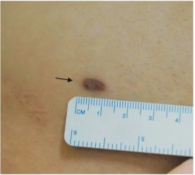
Hobnail 혈관종(hobnail hemangioma, targetoid hemosiderotic hemangioma)은 양성 혈관종양으로서 조직학적으로 얕은 진피에 서는 징 모양(“a short nail with a thick head”)의 hobnail 내피 세포로 구성된 확장된 혈관의 증식이, 깊은 진피에서는 보다 협 소한 종양 혈관의 증식과 아교질 박리(collagen dissection)가 관 찰되는 이상성 성장 양태(biphasic growth pattern)를 보인다1-3). 피부병변은 주로 젊은 남성의 다리, 팔, 몸통에 호발한다1,4). 육안 소견은 진피멜라닌세포모반(dermal melanocytic nevus), 비정형 색소모반(dysplastic nevus), 섬유종(fibroma), 섬유조직구종 (fibrous histiocytoma), 정맥기형(venous malformation, cavernous hemangioma), 혈관각화종(angiokeratoma) 등과 유 사하다3). 조직학적으로는 hobnail 내피세포의 양상을 보이는 다 양한 양성 및 악성 혈관종양들과 구분해야 하므로4,5) 본 종양은 병리조직학적 감별이 중요한 종양이라 하겠다. 본 질환은 국내 피부과 문헌에는 총 5례가 보고된 바 있다6-10). 저자들은 20세 남 성의 옆구리에 발생한 hobnail 혈관종을 경험하여 병리조직학적 감별에 대한 문헌 고찰과 함께 보고하는 바이다. 20세 남자가 1년 전부터 오른쪽 옆구리에 발생한 무증상의 피 부병변으로 내원하였다. 병변은 1 x 0.5 cm 크기의 연갈색을 띠 는 둥글고 융기된 단발성 종양으로 이를 에워싸는 암갈색의 테두 리가 존재하였다(Fig. 1). 진단 및 치료를 위해 외과적 절제술 및 조직생검을 시행하였다. 피부조직검사 상 저배율 시야에서 불규 칙한 크기의 내강을 가진 다수의 혈관들이 얕은 진피와 깊은 진 피에서 발견되었다(Fig. 2A). 얕은 진피에 존재하는 혈관들에서 는 현저히 확장된 내강과 이를 구성하는 뭉툭하게 돌출되어 징 모양을 보이는 단층의 내피세포들이 관찰되었다(Fig. 2B). 이들 내피세포는 크고 둥글며 균일한 핵과 희박한 세포질을 가지고 있 고 내강을 향해 유두모양으로 돌출(papillary projections)하는 양 상을 보였으며(Fig. 2C), 면역조직화학검사에서 종양성 내피세포 는 CD31 항원에 양성 반응을 나타냈다(Fig. 2D). 깊은 진피에서 는 얕은 진피에 비해 덜 확장된 혈관들이 증식하여 있었고, 얕은 진피층 혈관에서와 같은 hobnail 내피세포는 관찰되지 않았고, 편평한 내피세포에 의해 내강이 둘러싸여 있었다. 내원일로부터 6개월이 경과한 현재 환부의 종양은 완전히 제거된 상태로 환자 는 특별한 자각증상을 호소하지 않고 있다.
Hobnail 혈관종: 유사 조직소견을 동반한 혈관종양과의 감별 진단
박슬기
1, 천민석
2, 김재왕
2 1제주대학교 의학전문대학원, 2제주대학교 의학전문대학원 피부과학교실 (Received March 5, 2015; Revised March 12, 2015; Accepted March 20, 2015)Hobnail hemangioma: differential diagnosis of vascular tumors with similar
histopathologic findings
Seulgee Park
1, Minseok Cheon
2, Jae-Wang Kim
2 1Jeju National University School of Medicine,2Department of Dermatology, Jeju National University School of Medicine, Jeju, Korea.
Hobnail hemangioma(targetoid hemosiderotic hemangiomas) is a benign vascular tumor characterized histologically by a biphasic growth pattern of dilated vascular channels in the superficial dermis lined by prominent hobnail cells and rather narrow neoplastic vessels in deeper dermis. This tumor affects mainly the lower limbs or trunk of male adult. Histopathologically, hobnail hemangioma should be differentiated from a variety of benign to malignant vascular tumors. Herein, we represent a case of hobnail hemangioma which had been confirmed through the skin biopsy. (J Med Life Sci 2015;12(1):11-15)
Key Words
: Hobnail hemangioma서 론
Correspondence to : Jae-Wang Kim
Department of Dermatology, Jeju National University School of Medicine, 66 Jejudaehakno, 690-756, Jeju, Korea
E-mail : [email protected]
Abstract
Figure 1. Solitary brown-hued, rounded elevated
tumor(arrow) with peripheral dark rims on his right flank region.
Figure 2. (A) Low-magnification view of superficial and
deep blood vascular channels(arrows) with irregular congested lumens (H & E, ×50).
Figure 2. (C) Bland endothelial cells(arrows) with large
uniform, rounded nuclei and scanty cytoplasms forming intravascular papillary projections into the lumina in the superficial blood vessels (H & E, ×200).
Figure 2. (B) Dilated vascular spaces lined by prominent
hobnail endothelial cells(arrows) in the superficial dermis (H & E, ×100).
Figure 2. (D) Hobnail endothelial cells(arrows) showing
Hobnail 혈관종이란 고유의 명칭은 특유의 조직학적 소견에 의 해 불리어진 이름이다. 본 종양은 주로 20~70대(평균 32세) 남성 에서 다리, 등, 엉덩이, 가슴, 팔 등에 호발하며, 두경부나 손발은 잘 침범하지 않는다1-3). 대부분의 병변은 직경 2cm 이하의 적청 색, 자주색 혹은 연갈색의 단발성 무증상 구진으로 발견되며, 이 특징적인 색소침착은 깊은 진피의 출혈 및 혈철소(hemosiderin) 침착에 의한 것으로 추정한다1-3). 일부의 병변에서만 중심부 구진 의 바깥으로 창백하고 엷은 층(halo)과 출혈환(peripheral ecchymotic ring)이 에워싸서 표적(target) 형태를 나타내는데, 이 같은 육안 소견을 동반하는 전형적인 경우는 많지 않으므로 현재 는‘targetoid hemosiderotic hemangioma’란 명칭은 사용하지 않 는다2). 본 환자도 20세 남성으로 오른쪽 옆구리의 무증상 구진으 로 발견되었는데 암갈색의 테두리는 존재하였으나 명확한 표적 형태는 이루지 않았다. 조직소견으로는 얕은 진피에서 엷은 벽을 가지고 불규칙하게 확장된 혈관강(vascular spaces)의 증식이 관찰되어 저배율 시야 에서 쐐기 모양의 구조(wedge-shaped architecture)를 보인다1). 대부분에서 표피는 경도의 가시세포증(acanthosis)에 의한 과형성 (hyperplasia)을 나타낸다3). 얕은 진피에서 각 혈관강은 림프관과 유사하며, 특징적인 단층의 내피세포로 구성되어있는데 이들 세 포는 작고 뚱뚱하고, 세포질이 희박하며, 내강으로 돌출하는 크고 둥글며 균일한 과염색성 수포성 핵을 가져서 징 혹은 성냥개비 (matchstick-like)의 형태를 취한다1,2,5). 얕은 혈관의 내강에서는 섬유 중심(fibrous core)을 가진 미세한 유두모양 돌출이나 혈관 내 유두(intravascular papillae)가 관찰되거나1,2), 내피세포 사이에 가교(bridging)를 형성하기도 한다3). 혈관 내강에서는 섬유소 혈 전(fibrin thrombi)이 발견되거나 림프관종(lymphangioma, lymphatic malformation)에서처럼 창백한 호산성 단백액 (proteinaceous fluid)을 함유한다3). 반면 깊은 진피의 혈관들에서 는 전술한 내피세포의 변화들이 불명확해지거나 소실되며, 내강 이 좁아지고, 불규칙적으로 각이 지면서(angulated) 얇고 편평한 내피세포로 구성되어 아교질 다발을 박리하는 가성혈관육종 (pseudo-angiosarcomatous pattern)과 같은 양상이다2,3). 또한 때 로 풍부한 적혈구 혈관외유출, 림프구에 의한 경미한 염증성 침 윤, 광범위한 간질내(stromal) 혈철소 침착 및 섬유화 등이 동반 된다1-3). 오래 경과되고 성숙된 병변일수록 진피 섬유화가 현저하 고, 보다 협소한 혈관구조물이 많아진다3). 면역조직화학검사에서 는 내피세포가 CD31, CD34, factor VIII-related antigen 및 Ulex europaeus agglutinin-I 등에 다양하게 양성 혹은 음성을 나타내어 현재까지도 본 종양의 기원이 림프관인지, 혹은 혈관인 지 확정되지는 못했다3,6,7,8-10). 그러나 최근의 연구에 따르면 α-smooth muscle actin 양성의 혈관주위세포(pericytes)는 종양 혈 관의 일부에서만 발견되며, D2-40과 VEGFR-3(vascular endothelial growth factor receptor-3)에는 일관적으로 양성반응 을 보이는 것으로 보아 림프관 분화 세포일 가능성이 크다고 보 고되었다3,4). 다만, 종양성 림프관과 소혈관간의 미세우회로 (microshunts)가 존재하여 동맥류모양 미세구조, 혈관 내강 안팎 의 적혈구, 염증 변화, 섬유화, 간질내 혈철소 침착 등의 소견이 유발할 것으로 추정한다4). 본 증례에서는 상부 진피에서 얇은 벽 의 확장된 혈관 증식과 뭉툭한 hobnail 내피세포가 명확히 관찰 되었고, 하부 진피에선 좁은 혈관과 편평한 내피세포의 증식이 보여 hobnail 혈관종의 특징적인 이상성 성장을 확인할 수 있 었다. 다만 초기 병변이었기 때문에 현저한 적혈구 혈관외유출, 혈철소 침착, 진피 섬유화, 림프구 침윤 등은 명확히 관찰되지 않 았다. 본 종양과 유사하게 상피모양 내피세포(epithelioid endothelial cells)로 구성되어 조직학적 감별을 필요로 하는 다양한 악성도의 혈관종양들이 있다(Table 1). 먼저 반단계 Kaposi 육종(patch stage Kaposi’s sarcoma)은 기본적으로 비정형 방추세포로 구 성된 틈새 공간(slit-like spaces)의 증식이 깊은 진피 아래까지 완연하며, 확장된 얕은 혈관들과 hobnail 내피세포가 희박하고, 정상 혈관들과 피부부속기들이 새로 형성된 혈관 사이로 돌출하 는 ‘promontory(곶) sign’이 있으며, 들쭉날쭉한 테두리(jagged outlines)를 가진 혈관들이 아교질 다발을 분획시키며 증식하고, 육아종 조직에서처럼 각진 혈관과 두터운 벽의 혈관 줄기(cords), 형질세포의 침윤, 세포질내 봉입체(hyaline globules) 등이 발견된 다는 차이가 있다1-3). 그물모양혈관내피종(retiform hemangioendothelioma)은 hobnail 내피세포와 유두모양 돌출이 관찰되지만 침윤성 성장이 특징으로 그물진피 및 피하지방까지 침범하며, 정소와 같은 망 형태를 보이며 길게 연장되며 분지하는(arborizing) 혈관구조물이 있고, 혈관 내강 내부 및 간질에 현저한 림프구 침윤을 동반하며, 뭉툭한 방추세포 혹은 상피모양세포가 조밀하게 증식한 부위 (solid areas)가 존재한다1,2).
Dabska 종양(Dabska’s tumor, papillary intralymphatic angioendothelioma, malignant endovascular papillary angioendothelioma)도 hobnail 내피세포가 저명하지만 진피 전반 및 피하지방에 걸쳐 해면림프관종(cavernous lymphangioma, macrocystic lymphatic malformation)과 유사한 현저하게 확장된 얇은 벽의 혈관, 혈관 안팎의 림프구 침윤, 아교질 중심부 (collagenous cores)를 가진 혈관내 유두 등이 특징이다1,3). 반면 hobnail 혈관종은 보다 얕고 구획 지어져 있으며, hobnail 내피세 포 소견도 표피에 가까운 얕은 혈관들에서만 관찰된다는 차이가 있다. 상피모양혈관내피종(epithelioid hemangioendothelioma)는 침 윤성 성장이 특징으로 호산성 세포질과 수포성 핵을 가진 입방형 혹은 방추세포모양의 종양세포가 짧은 다발을 이루며 “indian-file” 양상으로 증식하며, 명확한 혈관구조물 대신 세포질공포 (intracytoplasmic vacuoles)가 현저하며, 연골모양(chondroid) 점 액성 간질이 풍부하다1). 상피모양혈관육종(epithelioid angiosarcoma)은 유사분열상이 뚜렷하고, 진피 전반에 걸쳐 호산성 세포질, 호산성 핵소체, 큰 수포성 핵을 가진 다형성 비정형 거대세포의 고형 증식(solid proliferation)이 저명하므로 감별이 가능하다1.
고 찰
Table 1. Histopathological differential diagnosis of hobnail hemangioma from other vascular tumors Hobnail endothelial cells ++ - + ++ + + - -Hobnail hemangioma Patch stage Kaposi’s sarcoma Retiform hemangioendoth elioma Dabska’s tumor Epithelioid hemangioendoth elioma Epithelioid angiosarcoma Acquired progressive lymphangioma Microvenular hemangioma Location of main changes Superficial dermis/mid-dermis Deep dermis /subcutis Deep dermis /subcutis Deep dermis /subcutis Deep dermis /subcutis Deep dermis /subcutis Deep dermis
/subcutis Deep dermis
Hemosiderin deposition + + - - - -Retiform structure + (Lower dermis) +/-++ (Normal rete testis-like) - - - - ++ Papillary projections/ Intravascular papilla + (Superficial dermis) +/- + ++ (Collagenous core) - - - -Dilated vascular spaces ++ (Superficial dermis) - - ++ - - ++ -Dermal fibrosis +/-(Lower dermis) +/- +/- +/- - - - -Lymphocytic infiltration +/-(Lower dermis) ++ ++ - - - - -Collagen dissection + (Lower dermis) ++ - - - ++ ++
-Table 1(Cont.). Histopathological differential diagnosis of hobnail hemangioma from other vascular tumors
Grade Benign Low-grade malignant vascular tumors High-grade malignant vascular tumors Benign Benign
Main features Circumscribed growth, Bland hobnail endothelial cells Atypical spindle cells, Slit-like spaces, Promontory sign, Vessels with jagged outlines, Dissecting collagen bundles, Plasma cells, Intracytoplasmic hyaline globules, Cords of angulated thick walled vessels Infiltrative growth pattern, Elongated arborizing vessels, Solid areas with spindle cells or epithelioid cells, Cavernous lymphangioma-like dilated vascular spaces, Prominent intravascular papilla with collagenous cores Infiltrative growth pattern, Variable cellular atypia, Cuboidal or spindle cells with eosinophilic cytoplasm & vesicular nuclei Nest or short fascicles forming indian-filing, Intracytoplasmic vacuoles(lumina) filled with RBCs, Mucoid to chondroid hyalinized stroma with sulfated mucopolysacch arides Prominent mitotic figures, Mimic melanoma with stromal hemorrhages, Solid proliferation of pleomorphic large cells with eosinophilic cytoplasm, eosinophilic nucleoli, larger vesicular nuclei Dilated thin-walled ectatic vascular spaces, Dissecting collagen bundles Small rounded or slit-like thinner vascular spaces Hobnail hemangioma Patch stage Kaposi’s sarcoma Retiform hemangioendoth elioma Dabska’s tumor Epithelioid hemangioendoth elioma Epithelioid angiosarcoma Acquired progressive lymphangioma Microvenular hemangioma
후천진행림프관종(acquired progressive lymphangioma, benign lymphangioendothelioma)는 단층 내피세포로 구성된 얇 고 확장된 혈관들이 가로로 배열하며 증식하며, 광범위한 아교질 박리 소견을 동반하지만 hobnail 내피세포, 혈관 내강으로의 유두 모양 돌출 및 혈철소침착은 없다2). 해면림프관종도 확장된 얇은 벽의 혈관강이 증식하지만 피하지 방까지 변화가 나타나며, 간질에서 풍부한 림프구 침윤 및 림프 구성 여포(lymphoid follicles)를 동반하지만 hobnail 내피세포, 유 두모양 돌출, 혈철소 침착 및 진피 섬유화는 저명하지 않다3).
미세세정맥혈관종(microvenular hemangioma)는 그물진피까지 작고 둥글거나 틈새와 같은 엷은 혈관들이 증식하되 확장된 얕은 혈관, 혈철소 침착, hobnail 내피세포가 적다는 점에서 차이가 있 다2).
끝으로 상피모양혈관종(epithelioid hemangioma, angiolymphoid hyperplasia with eosinophilia)은 저배율상 소엽 구조를 이루고, 호산구 및 림프구의 침윤이 현저하여 감별이 가능하다1). Hobnail 혈관종은 예후가 좋은 양성 종양이지만 조직학적으로 그물모양혈관내피종, Dabska 종양, 반단계 Kaposi 육종과 같은 중등도 악성 혈관종양이나, 상피모양혈관내피종, 상피모양혈관육 종 등 고도 악성 혈관종양과의 구분이 중요하므로 연속절편을 통 해 유사한 병리소견을 보이는 각 혈관종양과의 세밀한 감별 진단 이 필요할 것으로 사료된다.
1) Calonje E. Vascular tumors: tumors and tumor-like conditions of blood vessels and lymphatics, In: Elder DE, Elenitsas R, Johnson BL, Murphy GF, editors. Lever’s histopathology of the skin. 10th ed. Philadelphia: Lippincott Williams & Wilkins, 2009:1007-1056
2) Thompson LDR, Srivastava A, Wang SA, Comstock JM, Vega F, Wallentine JC, et al. Diagnostic pathology: neoplastic dermatopathology. 1st ed. Utah:Amirsys, 2012; I(8):20-21
3) Mentzel T, Partanen TA, Kutzner H. Hobnail hemangioma(targetoid hemosiderotic hemangiomas): clinicopathologic and immunohistochemical analysis of 62 cases. J Cutan Pathol 1999;26:279-286
4) Franke FE, Steger K, Marks A, Kutzner H, Mentzel T. Hobnail hemangioma(targetoid hemosiderotic hemangiomas) are true lymphangiomas. J Cutan Pathol 2004;31:362-367
5) Gutzmer R, Kaspari M, Herbst RA, Kapp A, Kiehl P. Absence of HHV-8 DNA in hobnail hemangiomas. J Cutan Pathol 2002;29:154-158
6) Yoon SY, Kwon HH, Jeon HC, Lee JH, Cho S. Congenital and multiple hobnail hemangiomas. Ann Dermatol 2011;23:539-543
7) Oh ST, Lee SD, Kim SW, Jang IG, Cho BK. A case of hobnail hemangioma. Ann Dermatol 2002;14:45-47 8) Roh BH, Whang KU, Kim YM, Cho MK, Park YL, Lee
JS. A case of childhood hobnail hemangioma. Korean J Dermatol 2007;45:979-982
9) Cho KM, Kim TJ, Whang KU, Lee JS, Lee SY, Park YL. A case of hobnail hemangioma. Korean J Dermatol 2003;41:1677-1680
10) Seo PG, Kang HA, Kim HO, Kim CW. A case of hobnail hemangioma. Korean J Dermatol 2001;39:1144-1147