www.issisglobal.org 13 Journal of International Society for Simulation Surgery 2017;4(1):13-16
Introduction
Oral cancer in the mandibular gingiva invades the lower jaw bone easily, especially in edentulous state. T4 grade tumor needs bone resection according to the extent of the invasion. When bone invasion is beyond inferior alveolar canal, partial seg-mental mandibulectomy is required. The fibular free flap is the flap of the choice in the mandibular reconstruction (1-5). The most common disadvantage of the fibular flap is short bone height to install dental implant. There are several methods to overcome this shortcoming such as double barrel flap, distrac-tion osteogenesis of the fibular flap and free bone graft over the vascularized fibular flap (6). However, distraction osteogenesis is technique sensitive and requires long-term treatment periods. Double barrel fibular flap has been tried, however, bulky flap in the oral cavity hinder its use (5).
Dental implants restoration is the ultimate goal for patients who needed jaw bone reconstruction (1, 2, 7). Implant fixture and crown ratio is important for long term stability and sur-vival of dental implants. The ideal implant to crown ratio is 1.5:1. When the fibular bone is located in the lower border of the mandibular bone defect, the implant to crown ratio could be 1: 2.5 or more. Even though dense bone quality of the fibular bone, the long-term successful results could not be obtained with unfavorable biomechanics.
Titanium reconstruction plate (R-plate) has been used simul-taneously with the free fibular flap to stabilize occlusion and to fix the fibular flap. The risk of skin perforation is decreased with using thin (1.8 mm) plate rather than 2.4 mm thickness R-plate.
In this study, a new trial has been applied in exclusive patients who was revealed thick skin flap with no neck metastasis. R-plate
O
riginal ArticleHow Can We Improve Crown-Implant Ratio in Reconstructed
Mandible with Fibular Free Flap?: A New Surgical Technique
Using 3D RP Model and Reconstruction Titanium Plates
Dong-Young Kim, D.D.S., Kang-Min Ahn, D.D.S., M.S.D., Ph.D
Department of Oral And Maxillofacial Surgery, College of Medicine, University of Ulsan, Asan Medical Center, Seoul, Korea
Fibular free flap reconstruction is the flap of the choice in long-span mandibular bone reconstruction. The most common dis-advantage of the fibular flap is short bone height to install dental implant. Double barrel fibular flap has been tried, however, bulky flap in the oral cavity hinder its use. Titanium reconstruction plate has been used simultaneously with the free fibular flap to stabilize occlusion and to fix the fibular flap. In this study, titanium reconstruction plate was fixed in the lower border of the mandible and the fibular free flap was fixed in the superior border of the titanium plate to improve implant-crown ratio. This new technique improved the longevity of the dental prosthodontics with dental implants.
Key WordsZZMandible ㆍReconstruction ㆍFree fibular flap ㆍ3D ㆍRapid prototype.
Received: May 9, 2017 / Revised: May 12, 2017 / Accepted: May 15, 2017 Address for correspondence: Kang-Min Ahn
Oral and Maxillofacial Surgery, College of Medicine, University of Ulsan, Asan Medical Center, 88 Olympic-ro, 43-gil, Songpa-gu, Seoul 05505, Korea Tel: 82-2-3010-5901, Fax: 82-2-3010-6967, E-mail: [email protected]
pISSN 2383-5389 / eISSN 2383-8116 https://doi.org/10.18204/JISSiS.2017.4.1.013
This is an Open Access article distributed under the terms of the Creative Commons Attribution Non-Commercial License (http://creativecommons.org/licenses/ by-nc/4.0/) which permits unrestricted non-commercial use, distribution, and reproduction in any medium, provided the original work is properly cited.
ORCID
Dong-Young Kim: orcid.org/0000-0002-2772-2519 Kang-Min Ahn: orcid.org/0000-0003-1215-5643
14
Journal of International Society for Simulation Surgery█ 2017;4(1):13-16
was used to maintain the facial contour and the fibular bone was fixed above the superior margin of the R-plate to idealize the implant-Crown ratio. The purpose of this study was to report a new surgical technique for fibular reconstruction with dental implant prosthetic restoration.
Material and Methods
The study was conducted in accordance with the Helsinki Declaration, and patients signed an informed consent. Ethical approval was gained from the IRB of Seoul Asan medical cen-ter. Institutional review board from our hospital issued an ex-emption to this study because of the use of collected existing data in such a manner that subjects cannot be identified di-rectly or indidi-rectly. A total of 36 patients underwent the fibular flap reconstruction between May 2006 and Dec 2016. Among the patients, two patients underwent operation with a new tech-nique to improve crown-implant ratio. The new techtech-nique in-volved reconstruction plate (Leibinger, San Diego, USA) in the lower border of the mandible and fibular bone fixation above the reconstruction plate. Fibular bone was fixed with microplates (Leibinger, San Diego, USA). Anastomosis was performed un-der microscope with one artery and two venae comitantes.
The indication for new technique was thick skin without neck metastasis. When neck node metastasis was suspicious during cancer work-up, conventional technique was applied. A new technique was performed in two patients who underwent left mandible partial mandibulectomy.
A new technique for fibular flap reconstruction
3D RP model fabrication and surgical stent preparation 3D RP model was fabricated according to the 3D facial CT
scan data. Invasion of the SCC into the mandibular bone was marked. Safety margin of 1.5 cm was drawn in the RP model. Extent of the mandibular bone resection was measured and cutting guide was fabricated. Surgical stent for cutting guide was made with acrylic resin (Fig. 1).
Prebent R-plate application before partial mandibulectomy R-plate was prebent according to the 3D RP model. Prebent R-plate was fixed to the mandible in both ends with three to four bicortical screws. R-plate was temporarily removed for partial mandibulectomy (Fig. 2).
Partial mandibulectomy
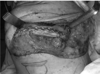
Partial mandibulectomy was performed and reposition of the R-plate was fixed with bicortical screws. R-plate was positioned in the inferior border of the mandible. R-plate was used to main-tain the facial contour (Fig. 3). Instead of using thick R-plate of 2.4 mm thickness, 1.8 mm thickness thin R-plate was used to
Fig. 1. Marking of the surgical resection margin in the 3D RP mod-el and fabrication of surgical stent for fibular reconstruction.
Fig. 2. Prebent reconstruction plate fixed to the mandible.
Fig. 3. Parital mandibulectomy and reposition of reconstruction plate.
Crown-Implant Ratio in Reconstructed Mandible with Fibular Free Flap █Kim DY and Ahn KM
www.issisglobal.org 15
prevent skin perforation.
Fibular flap fixation with miniplate
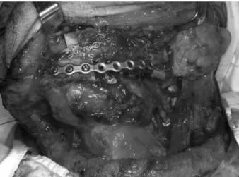
Fibular free flap was positioned above the R-plate and fixed with miniplates. Two miniplates as possible were used to fix the fibular bone to the residual mandible (Fig. 4). Instead of fixing in the lower border of the mandible, fibular bone was fixed in the level of alveolar bone to improve implant-crown ratio.
Postoperative radiograph
Postoperative panoramic radiograph showing fibular bone in the alveolar part (Fig. 5). Installation of the dental implant is much easier than the conventional methods.
Results
Among 36 patients, two patients underwent partial mandib-ulectomy and fibular flap reconstruction with a new technique (Fig. 6, 7). The other patients who required postoperative ra-diotherapy at the initial treatment planning were excluded for a new technique. Dental implants were installed three years af-ter cancer operation because magnetic inaf-terferences hinder
MRI interpretations during follow-up periods. The implant-crown ratios was 2:1 to 1.8:1 in patients with a new technique. Three years’ follow-up of a patient showed successful implant restoration.
Discussion
Mandibular reconstruction is one of the most challenging surgery in oral and maxillofacial area (4, 5, 8). Occlusion and facial contour should be restored during reconstruction proce-dure. Resection of the mandible usually involves teeth loss. Partial denture has been used for many decades, however, the retention of the partial denture was poor. Life of quality after mandibular resection was poor because of loss of teeth. Dental restoration is the utmost goal for patients who has mandibular bone defect.
Fibular bone has been used successfully for reconstruction of the mandible. Even though it has a straight topography, it could be osteotomized to fit the contour of the mandible (1-5, 8). The advantages of the fibular flap are long enough for mandibu-lar reconstruction, reliable vascumandibu-lar pedicle, long vessel length, least donor site morbidity and simultaneous harvest of soft and hard tissues (9-12). The disadvantages are short bone height to install dental implant. Traditionally fibular bone was fixed in the lower border of the mandible to stabilize mandibular bone defect. Inferior position of the fibular bone makes it difficult to install dental implant. And unfavorable crown-implant ratio
Fig. 4. Fibular flap located above R-plate to improve implant-crown ratio.
Fig. 5. Postoperative panoramic radiograph showing fibular bone fixed above R-plate.
Fig. 6. Postoperative panoramic radiograph showing fibular flap reconstruction in the alveolar part.
Fig. 7. Postoperative panoramic radiograph showing three dental implants in the fibular flap with favorable crown-implant ratio.
16
Journal of International Society for Simulation Surgery█ 2017;4(1):13-16
resulted in poor prognosis of the dental implants (1).
There are several methods to improve crown-implant ratio such as free onlay bone graft, distraction osteogenesis of the fib-ular bone and superior position of the fibula bone (6, 13-16). On-lay bone graft technique requires free bone graft over the vas-cularized fibular bone which is easily performed. However, the resorption rate of free bone graft is hard to predict and sometimes, complete resorption could be found. To overcome this shortcom-ing, double-barrel fibular flap has been developed (14). Howev-er, double barrel fibular flap is too bulky in the oral cavity. Pa-tients complained about the fullness of the oral cavity and biting of the skin island in the occlusal parts.
A new technique to overcome the disadvantages of these flap modification was performed in this study. R-plate was fixed in the lower border of the mandible to restore facial contour. Low-er bordLow-er continuity is the utmost important factor for facial symmetry. If the skin is thick and subcutaneous fat is enough, this new technique could be a good alternative for mandibular reconstruction. Patients who were diagnosed with neck metas-tasis were excluded in this study. Postoperative radiotherapy could be a risk factor for skin exposure in R-plate reconstruction area. Patients who were expected to receive postoperative ra-diotherapy were excluded in this study.
When the fibular flap was located in the alveolar part, soft tissue island was bitten by upper teeth during the healing peri-od. The soft tissue swelling was continued for several months. Three years’ follow-up showed resorption of soft tissue in the occlusal part. During the implant surgery, subcutaneous fat was removed and the bulky soft tissue was reduced. Implant was placed in the ideal position and the crown-implant ratio was excellent. Dental implants were successful with long-term follow-up. Patients were satisfied with the results and they could eat normal diet after dental implant restoration.
Conclusion
In this study, titanium reconstruction plate was fixed in the lower border of the mandible and the fibular free flap was fixed in the superior of the titanium plate to improve implant-crown ratio. This new technique improved the longevity of the dental prosthodontics with dental implants.
References
1. Kim DH, Cha HS, Ahn KM. Mandibular reconstruction with free
fibular flap and dental implant after ablative oral cancer surgery. J Int Soc Simul Surg 2014;1:90-94
2. Ahn KM, Kim JJ. Maxillary reconstruction with free fibular flap using 3d rp model. J Int Soc Simul Surg 2014;1:32-36
3. Kim YT, Yeom HR, Ahn KM, Myoung H, Hwang SJ, Seo BM, et al. Long term evaluation of volume change in free vascularized fib-ular flap mandible reconstruction. J Korean Assoc Oral Maxillofac Surg 2006;32:138-141
4. Ahn KM, Chung HJ, Ryom HR, Kim H, J., Kim YT, Hwang SJ, et al. Long-term analysis of reconstructed temporomandibular joint and mandible using free fibular flap. J Korean Assoc Oral Maxillofac Surg 2005;31:409-416
5. Lee JH, Kim MJ, Choi WS, Yoon PY, Ahn KM, Myung H, et al. Concomitant reconstruction of mandibular basal and alveolar bone with a free fibular flap. Int J Oral Maxillofac Surg 2004;33:150-156 6. Nocini PF, Wangerin K, Albanese M, Kretschmer W, Cortelazzi R.
Vertical distraction of a free vascularized fibula flap in a recon-structed hemimandible: Case report. Journal of cranio-maxillo-fa-cial surgery : officranio-maxillo-fa-cial publication of the European Association for Cranio-Maxillo-Facial Surgery 2000;28:20-24
7. Park JY, Ahn KM, Lee JH, Cha HS. Full mouth rehabilitation on a bilateral condylar fractured patient using orthognathic surgery and dental implant. J Adv Prosthodont 2011;3:51-55
8. Jeong WS, Choi JW, Choi SH. Computer simulation surgery for man-dibular reconstruction using a fibular osteotomy guide. Arch Plast Surg 2014;41:584-587
9. Lopez-Arcas JM, Arias J, Del Castillo JL, Burgueno M, Navarro I, Moran MJ, et al. The fibula osteomyocutaneous flap for mandible reconstruction: A 15-year experience. J Oral Maxillofac Surg 2010; 68:2377-2384
10. Shaha AR, Cordeiro PG, Hidalgo DA, Spiro RH, Strong EW, Zlotolow I et al. Resection and immediate microvascular reconstruction in the management of osteoradionecrosis of the mandible. Head Neck 1997;19:406-411
11. Shpitzer T, Neligan PC, Gullane PJ, Freeman JE, Boyd BJ, Rotstein LE, et al. Oromandibular reconstruction with the fibular free flap. Analysis of 50 consecutive flaps. Arch Otolaryngol Head Neck Surg 1997;123:939-944
12. Store G, Boysen M, Skjelbred P. Mandibular osteoradionecrosis: Reconstructive surgery. Clin Otolaryngol Allied Sci 2002;27:197-203
13. Friedrich RE, Schmelzle R. Distraction osteogenesis of a fibula free flap used for mandibular reconstruction: Preliminary report sergio siciliano, benoit lengere, henre reychler. Journal of cranio-maxillo-facial surgery : official publication of the European Association for Cranio-Maxillo-Facial Surgery 1999;27:398
14. Ch’ng S, Ashford BG, Clark JR. Alignment of the double-barrel fib-ula free flap for better cosmesis and bone height for osseointegrated dental implants. Plastic and reconstructive surgery 2013;132:688e-689e 15. Makiguchi T, Yokoo S, Hashikawa K, Miyazaki H, Terashi H. Eval-uation of bone height of the free fibula flap in mandible reconstruction. J Craniofac Surg 2015;26:673-676
16. Modabber A, Ayoub N, Mohlhenrich SC, Goloborodko E, Sonmez TT, Ghassemi M, et al. The accuracy of computer-assisted primary mandibular reconstruction with vascularized bone flaps: Iliac crest bone flap versus osteomyocutaneous fibula flap. Medical devices 2014;7:211-217