ISSN 2234-3806 • eISSN 2234-3814
http://dx.doi.org/10.3343/alm.2015.35.5.510
Routine Chromosomal Microarray Analysis is Necessary
in Korean Patients With Unexplained Developmental
Delay/Mental Retardation/Autism Spectrum Disorder
Saeam Shin, M.D.1, Nae Yu, M.D.1, Jong Rak Choi, M.D.1, Seri Jeong, M.D.2, and Kyung-A Lee, M.D.1
Department of Laboratory Medicine1, Yonsei University College of Medicine, Seoul; Department of Laboratory Medicine2, Kosin University College of Medicine, Busan, Korea
Background: All over the world, chromosomal microarray (CMA) is now the first tier diag-nostic assay for genetic testing to evaluate developmental delay (DD), mental retardation (MR), and autism spectrum disorder (ASD) with unknown etiology. The average diagnostic yield of the CMA test is known to be about 12.2%, while that of conventional G-banding karyotype is below 3%. This study aimed to assess the usefulness of CMA for the purpose of clinical diagnostic testing in the Korean population.
Methods: We performed CMA and multiplex ligation-dependent probe amplification (MLPA) tests in 96 patients with normal karyotype and unexplained DD, MR, or ASD. The CMA was conducted with CytoScan 750K array (Affymetrix, USA) with an average resolu-tion of 100 kb.
Results: Pathogenic copy number variations (CNVs) were detected in 15 patients by CMA and in two patients by MLPA for four known microdeletion syndromes (Prader-Willi/Angel-man syndrome, DiGeorge syndrome, Miller-Dieker syndrome and Williams syndrome) designated by National Health Insurance system in Korea. The diagnostic yield was 15.6% and 2.1%, respectively. Thirteen (13.5%) patients (excluding cases with patho-genic CNVs) had variants of uncertain clinical significance. There was one patient with a 17.1-megabase (Mb) region of homozygosity on chromosome 4q.
Conclusions: Our findings suggest the necessity of CMA as a routine diagnostic test for unexplained DD, MR, and ASD in Korea.
Key Words: Copy number variation, Chromosomal microarray, Developmental delay, Men-tal retardation, Autism spectrum disorders
Received: January 13, 2015 Revision received: March 18, 2015 Accepted: June 17, 2015
Corresponding author: Kyung-A Lee
Department of Laboratory Medicine, Yonsei University College of Medicine,
211 Eonju-ro, Gangnam-gu, Seoul 135-720, Korea
Tel: +82-2-2019-3531 Fax: +82-2-2019-4822 E-mail: [email protected]
© The Korean Society for Laboratory Medicine
This is an Open Access article distributed under the terms of the Creative Commons Attribution Non-Commercial License (http://creativecom-mons.org/licenses/by-nc/3.0) which permits unrestricted non-commercial use, distribution, and reproduction in any medium, provided the original work is properly cited.
INTRODUCTION
Clinical genetic testing plays an important role in evaluating pa-tients with developmental delay (DD), mental retardation (MR), and autism spectrum disorders (ASD) with unknown etiology. DD indicates deficits in learning and adaptive functioning at the expected age. MR, often referred to as “intellectual disability”, is a disorder with intellectual and adaptive deficits and can be di-agnosed after the age of five years [1]. MR and DD affect 1-3%
of general population [2]. ASD encompasses a group of disor-ders like autism, Asperger’s syndrome, and pervasive develop-mental disorder. ASD shows features like impairment in social communication and interactions, and repetitive and restrictive behaviors [1]. The frequency of occurrence of ASD is approxi-mately 1% of general population [1]. Genetic causes, including known genetic syndromes and chromosomal abnormalities, comprise the main etiology of these disorders. Approximately 17.4-47.1% of DD/MR and 10% of ASD can be explained by
genetic causes [3, 4].
The National Health Insurance system of the Republic of Ko-rea only permits multiplex ligation-dependent probe amplifica-tion (MLPA) tests for four microdeleamplifica-tion syndromes (Prader-Willi/ Angelman syndrome, DiGeorge syndrome, Miller-Dieker syn-drome, and Williams syndrome), conventional cytogenetics, and FISH. Thus, the medical institutions in Korea cannot perform other diagnostic genetic tests. Conventional cytogenetics allows for detection of numerical and structural chromosomal abnor-malities present in the entire genome, but has a limited resolu-tion of 5-10 megabases (Mb). Thus, submicroscopic aberra-tions cannot be detected, and interpretation of the test results remains subjective. FISH and MLPA can detect specific cytoge-netic aberrations with a higher sensitivity than conventional cy-togenetics; however, they cannot cover entire regions of chro-mosomes. Thus, it takes substantial effort to detect abnormali-ties involving multiple regions, and the diagnostic yield is de-creased when the clinical spectrum of the disease is variable. DD, MR, and ASD are difficult to define disease categories clini-cally owing to overlapping symptoms and comorbidities. Chromosomal microarray (CMA) is now recommended world-wide as the first-tier clinical diagnostic test for patients with DD, MR, and ASD of unknown causes [5]. CMA detects copy num-ber variations (CNVs) in the entire genome with a much higher resolution than conventional cytogenetics. Before the CMA test was adopted as a routine clinical diagnostic test, conventional cytogenetics and single gene tests such as fragile-X syndrome testing were done as initial tests for unexplained DD, MR, and ASD. However, the diagnostic yields of conventional cytogenet-ics and fragile-X syndrome testing in patients with DD/MR is be-low 3% and 1.2%, respectively [5, 6]. CMA detects pathogenic CNVs with an average diagnostic yield of 12.2% in 33 previous
studies involving 21,698 patients with neurodevelopmental dis-orders or multiple congenital anomalies [5].
We evaluated the utility of CMA as a routine clinical diagnostic test in the Korean population. In addition, we aimed to report a few interesting clinical cases confirmed by CMA test. We used Affymetrix single nucleotide polymorphism (SNP) array with an average resolution of 100 kb. The SNP array has been validated in a previous study indicating that it has sufficient resolution to detect single gene deletions [7]. Although CMA detects genomic imbalance with higher diagnostic yield than conventional cyto-genetics, it cannot detect balanced rearrangements and low-level mosaicism [8]. Thus, we selected clinically affected indi-viduals with normal karyotype.
METHODS
1. Patients
Genetic tests were requested by physicians from pediatrics, re-habilitation, neurology, and psychiatric departments for unex-plained DD, MR, and ASD with or without dysmorphism or sei-zures. Written informed consent approved by institutional review board was obtained from patients or patients’ parents for ge-netic analysis.
Conventional G-banded karyotype analysis from peripheral blood was performed as a part of initial screening tests, and 96 patients with normal karyotypes were included in this study. As we retrospectively performed CMA using the blood samples col-lected during MLPA studies, parental samples could not be ob-tained and were therefore, not available for analysis.
From March 2012 to April 2014, 96 patients with normal karyotype (67 males and 29 females, Table 1) were referred for further genetic testing. The median age at diagnosis was three Table 1. Number of patients with copy number variations according to demographic and clinical features
N of total patients (%) N of patients with pathogenic CNVs (%) N of patients with VOUS (%)* N of patients with benign CNVs (%)† Sex Male 67 (69.8) 11 (16.4) 8 (11.9) 1 (1.5) Female 29 (30.2) 4 (13.8) 5 (17.2) 4 (13.8) Clinical features ASD 34 (35.4) 0 - 2 (5.9) 2 (5.9) DD/MR 54 (56.3) 15 (27.8) 7 (13) 3 (5.6) ASD and DD/MR 8 (8.3) 0 - 4 (50) 0 -Total 96 (100) 15 (15.6) 13 (13.5) 5 (5.2)
*Patients with pathogenic CNVs were excluded; †Patients with pathogenic CNVs or with VOUS were excluded.
Abbreviations: CNV, copy number variation; VOUS, variant of uncertain clinical significance; ASD, autism spectrum disorder; DD, developmental delay; MR, mental retardation.
years (range: 7 months-22 yr). Thirty four patients were ASD, 54 patients were DD and/or MR, and 8 patients were ASD com-bined with DD and/or MR (Table 1).
2. DNA preparation
EDTA whole blood was used to extract DNA by using the Easy-DNA kit (Invitrogen Corporation, Carlsbad, CA, USA). The con-centration and quality of genomic DNA was analyzed by Nano-drop ND-1000 (Thermo Scientific, Wilmington, DE, USA).
3. MLPA
Microdeletion syndromes were screened by using SALSA MLPA P245 Microdeletion Syndromes probemix (MRC-Holland, Am-sterdam, The Netherlands) according to the manufacturer’s in-structions. The P245 probemix contains 49 different MLPA probes with amplification product sizes between 130 and 486 nucleotides. The probes target 40 causative genes implicated in 23 known microdeletion/microduplication syndromes. DNA was denatured at 98°C for five minutes and hybridized with the probe set overnight at 60°C. Ligation reaction with ligase-65 en-zyme was performed at 54°C for 15 min, followed by five min-utes at 98°C for heat inactivation of the enzyme. PCR was per-formed with the specific SALSA PCR primers for 35 cycles (95°C for 30 sec; 60°C for 30 sec; 72°C for one minute) by using the GeneAmp PCR System (Applied Biosystems, Foster City, CA, USA). MLPA fragment analysis data were generated by using the Applied Biosystems 3,500×L Genetic Analyzer (Applied Biosys-tems). The data were analyzed by using the GeneMarker soft-ware (SoftGenetics, State College, PA, USA). For confirmatory analysis of abnormal results found with the P245 screening probemix, an MLPA test with P372, P373, or P374 syndrome-specific probemix, or a P339 SHANK3 probemix (MRC-Holland) was performed by using the same protocol. Pathogenic CNVs detected by CMA that were smaller than 1 Mb were confirmed by using a P343 or P181 probemix.
4. Chromosomal microarray
CMA was performed with a CytoScan 750K array (Affymetrix, Santa Clara, CA, USA) according to the manufacturer’s recom-mendations. The platform is composed of 550,000 non-poly-morphic CNV probes and more than 200,000 SNP probes with an average resolution of 100 kb. The data were analyzed by us-ing Chromosome Analysis Suite v2.1 Software (Affymetrix) and Nexus Copy Number v.7.5 Software (BioDiscovery, El Segundo, CA, USA). The February 2009 human reference sequence (GRCh37/Hg19) was used for genomic assembly.
5. Interpretation of detected CNVs
Detected CNVs were classified as pathogenic, benign, or variant of uncertain clinical significance (VOUS) in accordance with the recommended guidelines from the International Standard Cy-togenomic Array and the American College of Medical Genetics [5, 9, 10]. The data were interpreted by using information avail-able in scientific literature, public databases and other general information about pathogenic or benign CNVs (size, content of Online Mendelian Inheritance in Man [OMIM] morbid genes or dosage sensitive genes, and type of dosage imbalance: duplica-tion or deleduplica-tion) [5, 11]. Genomic map from the UCSC Genome Browser (http://genome.ucsc.edu/cgi-bin/hgGateway) was used to map the locations of CNVs and gene distribution. The Data-base of Genomic Variants (DGV, http://dgv.tcag.ca/dgv/app/ home) provided catalogs of structural variations found in healthy controls. The dbVar (http://www.ncbi.nlm.nih.gov/dbvar/) data-base was also used to get information about CNVs from both normal and diseased populations. We also used the DatabasE of Chromosomal Imbalance and Phenotype in Humans using En-sembl Resources (DECIPHER, https://decipher.sanger.ac.uk/) as a reference for known microdeletion and microduplication syndromes, and the OMIM (http://omim.org/) for disease-caus-ing genes, their functions and inheritance patterns.
Besides excluding pathogenic CNVs overlapping with known genomic imbalance syndromes and benign CNVs reported as normal variant in healthy controls, VOUS should meet at least one of the three criteria: the CNV is not a common polymorphism, OMIM genes in the CNV interval are not known for dosage sensi-tivity, and are not associated with patient’s phenotypes.
RESULTS
1. Results of MLPA and CMA
We identified 42 CNVs in 33/96 (34.4%) patients. Among these, 17 CNVs were classified as pathogenic (40.5%), 15 as VOUS (35.7%), and 10 as benign (23.8%). One or more pathogenic CNVs were reported in 15 patients (Table 1). Therefore the di-agnostic yield of CMA was 15.6% (15/96) in our study. VOUS, excluding cases with pathogenic CNVs, were reported in 13/96 patients (13.5%). One or more benign CNVs, excluding cases with pathogenic CNVs and VOUS, were reported in five patients (5.2%).
Most of the 15 CNVs of unknown significance were not a com-mon polymorphism, except a 2.3 kb-sized duplication at 8p23.2 found in three patients (Table 2, Case 20, 21, and 22). The du-plicated region, which includes CSMD1 (MIM 608397) gene,
Table 2.
Summary of clinical features and genetic test results of patients with copy number variants or loss of heterozygosity
Case Age (yr) Sex Clinical features MLP A Microarray Size (kb) OMIM genes (N)
Critical genes or region
Classification Known microdeletion syndrome Reasons of classification* 1 1 M
DD, seizure, both pes planovalgus, cleft palate, simian crease on left hand, cryptorchidism on right
15q11.2(P181)x1 1q21.1(145,512,555-145,888,926)x1/1q44 (244,933,278-247,517,799)x1/15q11.2 (22,770,421-23,277,436)x1 376/ 2,584/ 507 8/ 9/ 4 -/ 1q44/15q11.2 Benign/ Pathogenic/ Pathogenic -/MIM 612337/MIM 615656 2/1/ 1 2 5 M
DD, seizure, short stature, microcephaly
Negative 3q27.3q29(186,874,563-194,994,615)x1 8,120 27 3q27.3 Pathogenic 3q27.3 deletion syndrome [36] 1 3 3 F DD, microcephaly ,
bulbous nose, low set ears, clinodactyly of the hands, pes planovalgus 10p14-15 (P245,P372)x3 4q35.1q35.2(185,230,774-190,837,600)x1/ 10p15.3p11.23(96,852-28,456,830)x3 5,606/ 28,360 16/92 CASP3, PDLIM3, KLKB1, F11, F AT1/GA TA3 Pathogenic/ Pathogenic 4q deletion syndrome [18]/ MIM 146255
1/1
4
3
M
DD, esotropia on left eye
5p15(P245)x1 5p15.33p15.31(113,576-7,183,668)x1 7,070 27 TER T Pathogenic
Cri du chat syndrome
1 5 6 F DD Negative 8p23.1(8,107,752-11,882,913)x1 3,775 17 GATA 4 Pathogenic 8p2 3.1 del etio n sy nd rom e [3 7, 3 8] 1 6 2 M DD Negative 8p23.3p23.1(158,048-6,590,060)x3/10q26.1 3q26.3(124,682,180-135,426,386)x1 6,432/ 10,744 8/40 -/DOCK1 VOUS/ Pathogenic -/MIM 609625 4, 5, 6/1 7 2 M
DD, torticollis, pes planovalgus, right deaf
ness , dow nsla ting palpebral fissures 22q13.33 (P245,P373)x1 9p24.3p22.3(208,454-16,099,709)x3/ 22q13.31q13.33(46,790,049-51,197,766)x1 15,891/ 4,407 40/31 -/ARSA, SHANK3, ACR VOUS/ Pathogenic -/Phelan-McDermid syndrome 4, 5/1 8 3 F DD Negative 10p15.3(100,047-2,969,599)x1 2,869 6 ZMYND11 Pathogenic 10p15.3 deletion syndrome [39] 1 9 1 M DD, lipo m en in go m ye loc ele Negative 10q26.2q26.3(130,158,704-135,426,386)x1 5,267 20 10q26 Pathogenic MIM 609625 1 10 4 M DD, seizure 15q11.2(P181)x3 15q11.2(22,770,421-23,625,785)x3 855 4 15p11.2 Pathogenic MIM 608636 1 11 1 F DD, seizure 15q11.2 (P245,P374)x1 15q11.2q13.1(22,770,421-29,015,025)x1 6,244 16 SNRPN, UBE3A Pathogenic Angelman syndrome 1 12 7 M MR 15q13.3(P343)x3 15q13.3(31,999,631-32,444,043)x3 444 1 CHRNA7 Pathogenic MIM 608636 1 13 2 M
DD, abnormality of the skin, pectus carinatum
Negative 16p13.11(15,450,289-16,497,406)x3 1,047 8 16p13.11 Pathogenic 16p13.11 duplication syndrome [40] 1 14 7 M
MR, full cheek, bullous nose, low set ears, narrow forehead 22q11 (P245,P372)x1 16p11.2(32,524,764-33,863,672)x3/ 22q11.21(18,916,842-21,800,471)x1 1,338/ 2,883 -/39 -/TBX1, CRKL Benign/ Pathogenic -/DiGeorge syndrome 2, 3/1
Case Age (yr) Sex Clinical features MLP A Microarray Size (kb) OMIM genes (N)
Critical genes or region
Classification Known microdeletion syndrome Reasons of classification* 15 2 M DD 22q13.33 (P245,P373)x1 22q13.31q13.33(48,398,495-51,154,658)x1 2,756 27 ARSA , SHANK3 Pathogenic Phelan-McDermid syndrome 1 16 5 M MR, ASD Negative 1q42.12(225,168,456-225,520,146)x1 351 1 -VOUS 4, 5, 6 17 1 M DD, seizure 2q11.1(P181)x3 2q11.1(95,518,497-96,326,105)x3 808 4 VOUS 4, 5, 6 18 12 F MR Negative 2q13(110,498,141-110,980,295)x1 482 4 -VOUS 4, 6 19 5 M ASD, DD, MR Negative 6q22.31(121,220,756-121,617,795)x1 397 1 -VOUS 4, 5 20 8 M DD, MR, pes planovalgus Negative 8p23.2(3,685,300-5,935,671)x3 2,250 1 -VOUS 5 21 4 F DD, ASD Negative 8p23.2(3,685,300-5,935,671)x3 2,250 1 -VOUS 5 22 1 M DD Negative 8p23.2(3,685,300-5,935,671)x3 2,250 1 -VOUS 5 23 3 M DD, MR Negative 11q13.4(70,993,028-71,745,766)x3 753 6 -VOUS 4, 5 24 2 F
DD, alternating exotropia, vesicoureteral reflux
Negative 11q21(95,576,015-96,054,843)x3 479 2 -VOUS 4, 5, 6 25 10 M ASD Negative 12p11.23(27,283,451-27,793,637)x3 510 3 -VOUS 4, 5, 6 26 3 M ASD, DD, MR Negative 14q12(25,529,986-26,282,808)x3 753 -VOUS 3, 4 27 7 F MR Negative 16p11.2(31,991,160-33,981,750)x3/ Xp21.1p14(37,046,232-37,682,073)x3 1,990/ 635 -/3 -/-Benign/VOUS 2, 3/ 4, 5, 6 28 12 F ASD Negative 19p13.3(2,940,905-3,957,892)x1 1,016 23 -VOUS 4, 5, 6 29 9m F
DD, hearing impairment, central hypotonia
Negative
4q32.1q34.1(158,700,137-175,792,268)x2 hmz
17,092
28
-*1.Overlapping with a known genomic imbalance syndrome; 2. In the category of genomic imbalance in healthy individuals as per p
ublic database; 3. No OMIM genes in the CNV interval; 4. CNV
is not a common polymorphism; 5. OMIM genes in the CNV interval are not known for dosage sensitivity; 6. OMIM genes in the CNV
interval are not associated with patient’
s phenotype.
Abbreviations: MLP
A, multiplex ligation-dependent probe amplification; VOUS, variant of uncertain clinical significance; ASD, autism spectrum dis
order; DD, developmental delay; MR, mental retar
-dation; m, months.
Table 2.
was also found in normal controls [12]. CSMD1 was shown to be interrupted by duplication in a patient with speech delay and au-tism in a previous study [13].
One patient showed a deletion in 2q13 (Case 18), which in-cluded four OMIM genes (RGPD6, RGPD5, MALL, and NPHP1). The four genes are not known to have any relation with this pa-tient’s phenotype, but homozygous deletion of NPHP1 (MIM 607100) is known to be associated with nephronophthisis [14]. TBC1D32 gene (MIM 615867) was deleted in one patient (Case 19). TBC1D32 has been reported to be mutated in a patient with multiple congenital anomalies [15]. A duplication at 11q13.4 in a patient (Case 23) includes six OMIM genes (DHCR7, NADSYN1, KRTAP5-9, FAM86C1, IL18BP, and NUMA1). Mutation of DHCR7 gene is associated with Smith-Lemli-Opitz syndrome (SLOS, MIM 270400), which is an autosomal recessive multiple congenital anomaly and intellectual disability syndrome [16]. Thus far, the only reported DHCR7 mutations associated with SLOS are point mutations or multiple exonic deletions [16,17]. Thus, it is difficult to interpret the significance of duplication in our patient. A 753 kb-sized duplication at 14q12 in a patient (Case 26) contains no OMIM gene, so the CNV is likely to have a benign nature. However, we interpreted it as VOUS because the duplicated region was not reported in databases for control pop-ulation [9].
Pathogenic CNVs were identified in 27.8% patients with DD and/or MR (Table 1). No pathogenic CNVs were seen in patients with ASD and ASD combined with DD and/or MR in our study. The size range of the pathogenic CNVs was 444 kb to 28.4 Mb. Most of the pathogenic CNVs (94.1%) were above 500 kb (Ta-ble 3). Six CNVs between 5-10 Mb and three CNVs above 10 Mb were not detected by conventional cytogenetics. All benign CNVs were below 5 Mb (Table 3).
MLPA microdeletion syndrome screening probemix for 23 chromosomal disorders detected six patients with known micro-deletion or microduplication syndromes with a diagnostic yield
of 6.3%. There were one patient with Angelman syndrome and one with DiGeorge syndrome. Therefore, the diagnostic yield of four microdeletion syndromes designated by National Health In-surance system in Korea was 2.1%.
2. Clinical case reports
1) Hypoparathyroidism, sensorineural deafness, and renal dysplasia syndrome
A 3-yr-old female (Table 2, Case 3) was referred for evaluation of developmental delay. G-banded karyotype was normal, while CMA revealed a 5.6 Mb deletion at 4q35.1q35.2 region and a 28.4 Mb duplication at 10p15.3p11.2 region.
Chromosome 4q deletion is known to be associated with intel-lectual disability, ASD, and craniofacial dysmorphism [18]. The critical region for ASD phenotype, containing MTNR1A, FAT1, and F11 at 4q35.2 [18], is identical to that in our patient. Dupli-cation of GATA3 gene at 10p14 can cause hypoparathyroidism, sensorineural deafness, and renal dysplasia syndrome (HDR syndrome, MIM 146255) [19]. DD and facial dysmorphism have also been described in a HDR syndrome patient [19]. A previous study reported unbalanced cryptic translocation der(4) (4;10)(q35;p15), similar to our case [20]. Parental study re-vealed that the genomic imbalance was originated from unbal-anced segregation of maternal balunbal-anced reciprocal translocation t(4;10)(q35;p15) [20]. Both patients showed learning disabili-ties, facial dysmorphisms, and immunodeficiency [20]. Our pa-tient’s karyotype was initially interpreted as normal, however, af-ter CMA results, a more detailed karyotype analysis revealed der(4)(4;10)(q35;p15), as previously described [20].
There were three patients with cryptic unbalanced transloca-tions involving two chromosomes in our study (Case 3, 6, and 7), suggesting inheritance from parental balanced translocation. In these cases, other family members were given genetic evalu-ation and proper counseling about the disorders and the recur-rence risk in family [9].
2) Patient with a region of homozygosity on chromosome 4 A 9-month-old female (Table 2, Case 29) was evaluated for de-layed development due to central hypotonia. CMA showed no CNV, but there was one 17.1-Mb region of homozygosity (ROH) on chromosome 4.
SNP- based array, unlike array-based CGH, can identify copy-number changes as well as ROH, which indicates uniparental disomy (UPD) or identity by descent (IBD) [21]. When the ROHs exist on multiple chromosomes, they usually suggest IBD, i.e. inheritance from a common ancestor [22]. In contrast, UPD is Table 3. Size distribution of pathogenic CNVs found in patients
Size (Mb) N of pathogenic CNVs (%) N of VOUS (%) N of benign CNVs (%)
<0.5 1 (5.9) 4 (26.7) 6 (60) 0.5-1 2 (11.8) 5 (33.3) 1 (10) 1-5 7 (41.2) 4 (26.7) 3 (30) 5-10 5 (29.4) 1 (6.7) 0 ->10 2 (11.8) 1 (6.7) 0 -Total 17 (100) 15 (100) 10 (100)
Abbreviations: Mb, megabase; CNV, copy number variation; VOUS, variant of uncertain clinical significance.
suspected when the large ROH is on a single chromosome [22]. ROH may have pathogenic implications in two ways: if there are imprinted genes within the region, or if it leads to homozygosity for deleterious alleles involved in autosomal recessive disorders [23]. In our case, 28 OMIM genes map to this 17.1 Mb ROH on chromosome 4. Previous reports included chromosome 4 UPD in a patient with mental disorder [23] and an interstitial deletion at 4q32q34, a region that overlaps with our case, in a patient with DD and dysmorphism [24]. However, multiple ROHs can be detected in a normal population. Therefore, caution should be exercised when reporting and interpreting ROH [22]. A pa-rental study and genetic counseling should be recommended in this instance.
3) Cri du chat syndrome
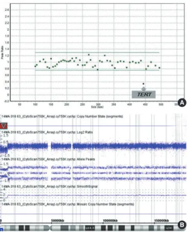
A 3-yr-old male (Table 2, Case 4) visited the psychiatric depart-ment for speech delay. He also had esotropia in his left eye and was diagnosed as having DD. MLPA with P245 probemix re-vealed deletion in only one probe site on exon 3 of TERT gene
(Fig. 1A), which is known to be involved in the phenotype of cri du chat syndrome (MIM 123450). CMA showed a deletion at 5p15.33p15.31(113,576-7,183,668) (Fig. 1B), confirming the MLPA result. A decreased signal for only one probe in MLPA ex-periment can indicate a false-positive result owing to polymor-phism in the probe ligation site [25]. Therefore, confirmation of MLPA finding by another method is essential in such cases [25]. The region involved in the cri du chat syndrome is known to be variable in size, and it can be a terminal or an interstitial deletion [26]; so confirmative tests like CMA can be of help.
DISCUSSION
We conducted CMA analysis in 96 unexplained DD, MR, and ASD patients with normal karyotype as assessed by conventional cytogenetics. The diagnostic yield of CMA was 15.6%, which is higher than the average rate from previous reports including chromosomal aneuploidies (12.2%) [5]. Our finding supports the necessity of implementing CMA as a routine diagnostic test in the Korean population. Moreover, first-tier use of CMA for clinical genetic evaluation of unexplained DD, MR, and ASD in the Korean population could be beneficial for patients, consider-ing the cost-effectiveness of CMA compared with current con-ventional cytogenetics and MLPA or FISH test strategy [27-29]. Owing to the significantly higher diagnostic yield of CMA (CMA 15.6% vs. MLPA for the four microdeletion syndromes 2.1%), an additional 13.5% of patients with genetic etiology can be di-agnosed through initial CMA testing and can save much time, cost, and effort from additional diagnostic tests.
Although we found no pathogenic CNV in patients with ASD and ASD combined with DD/MR, the possibility of later onset of symptoms of autistic features in these patients has to be consid-ered, with regard to the previous findings about high frequency of pathogenic CNVs in ASD patients [1, 30, 31].
It is known that the size of CNV is of limited importance in in-terpretation of pathogenicity [9]. The size distribution of patho-genic CNVs varied a lot in this study. Pathopatho-genic CNVs greater than 5 Mb were missed by conventional cytogenetics in six cases (Cases 2, 3, 4, 6, 9, and 11). One patient (Case 3) had a terminal deletion on chromosome 4 and a terminal duplication on chro-mosome 10. Initially, the karyotype of the patient was interpreted as normal; however, after referring to the CMA results, a reanaly-sis of the karyotype revealed der(4)(4;10)(q35;p15). Case 11 with Angelman syndrome had a 6 Mb-sized deletion downstream of the centromere. Four of these six cases (Cases 2, 4, 6, and 9) in-volved a terminal deletion. Although 5-6 Mb is usually considered
A
B
Fig. 1. Multiplex ligation-dependent probe amplification (MLPA) and microarray results of patient with Cri du Chat syndrome. (A) The MLPA scatter plot showing deletion of a probe site on TERT. (B) Microarray profile with a copy number loss on 5p15.33p15.31 (113,576-7,183,668) (red bar).
to be the detection limit of conventional G-banded karyotyping, some cases, such as chromosome abnormalities involving termi-nal or near-centromere regions, can be missed [11].
Among our cases, two patients (2/96, 2.1%) were diagnosed as having Phelan-McDermid syndrome (MIM 606232). These two patients had deletions in 22q13 region, involving ARSA and SHANK3 loci (Cases 7 and 15). Haploinsufficiency of the SHANK3 product is known to have a causative role in neurologic symptoms in Phelan-McDermid syndrome [32]. Deletions, trans-locations, or point mutations involving SHANK3 locus have been reported as pathogenic in ASD/DD [32-34]. Higher frequency of CNVs involving SHANK3 was reported in Chinese patients (1.7%), compared with 20 studies of European, American, and Australian populations (0.24%) [34]. The prevalence of imbal-ance involving SHANK3 in our study (2.1%) was also higher than that reported in a previous study of Caucasian population (0.24%) [34], suggesting higher frequency of Phelan-McDermid syndrome in DD/MR/ASD patients among East Asians.
The result of a parental study [5] is one of the most useful evi-dences of clinical significance of CNVs found in a patient. The interpretation of VOUS can be helped from the information whether it is inherited from a healthy parent or if it occurred de novo in the proband [5]. The absence of parental analysis is a limitation of our study. Although we could not validate VOUS with a parental study and only included patients with normal karyo-type, the detection rate of pathogenic CNVs in our study was higher than that in previous reports [5]. Parental studies will help not only the interpretation of clinical significance of CNVs, but also the genetic counseling and evaluation of recurrence risk of the genetic abnormality in families, when CMA is available as a routine diagnostic test in future in Korea.
One major obstacle in implementing CMA as a routine diag-nostic test is the current introduction system for medical technol-ogy in Korea [35]. All new diagnostic tests, including genetic tests, must go through a unified regulatory process. It is possible to apply to New Health Technology Assessment of Health Insur-ance Review & Assessment Service for a new medical device af-ter getting permission from Ministry of Food and Drug Safety in Korea. Rapidly changing technology in clinical genomic testing requires a flexible system for assessing newly introduced medi-cal procedures and health technology.
Authors’ Disclosures of Potential Conflicts of
Interest
No potential conflicts of interest relevant to this article were
re-ported.
Acknowledgments
This study was supported by a faculty research grant of Yonsei University College of Medicine for 2014 (6-2014-0145).
REFERENCES
1. American Psychiatric Association. Diagnostic and statistical manual of mental disorders: DSM-5. 5th ed. Washington, D.C.: American Psychi-atric Association, 2013.
2. Battaglia A, Doccini V, Bernardini L, Novelli A, Loddo S, Capalbo A, et al. Confirmation of chromosomal microarray as a first-tier clinical diag-nostic test for individuals with developmental delay, intellectual disabili-ty, autism spectrum disorders and dysmorphic features. Eur J Paediatr Neurol 2013;17:589-99.
3. Weiss LA, Shen Y, Korn JM, Arking DE, Miller DT, Fossdal R, et al. As-sociation between microdeletion and microduplication at 16p11.2 and autism. N Engl J Med 2008;358:667-75.
4. Moeschler JB, Shevell M; American Academy of Pediatrics Committee on Genetics. Clinical genetic evaluation of the child with mental retarda-tion or developmental delays. Pediatrics 2006;117:2304-16.
5. Miller DT, Adam MP, Aradhya S, Biesecker LG, Brothman AR, Carter NP, et al. Consensus statement: chromosomal microarray is a first-tier clinical diagnostic test for individuals with developmental disabilities or congenital anomalies. Am J Hum Genet 2010;86:749-64.
6. Rauch A, Hoyer J, Guth S, Zweier C, Kraus C, Becker C, et al. Diagnos-tic yield of various geneDiagnos-tic approaches in patients with unexplained de-velopmental delay or mental retardation. Am J Med Genet A 2006;140: 2063-74.
7. Wagenstaller J, Spranger S, Lorenz-Depiereux B, Kazmierczak B, Nath-rath M, Wahl D, et al. Copy-number variations measured by single-nu-cleotide-polymorphism oligonucleotide arrays in patients with mental retardation. Am J Hum Genet 2007;81:768-79.
8. Bi W, Borgan C, Pursley AN, Hixson P, Shaw CA, Bacino CA, et al. Com-parison of chromosome analysis and chromosomal microarray analysis: what is the value of chromosome analysis in today’s genomic array era? Genet Med 2013;15:450-7.
9. Kearney HM, Thorland EC, Brown KK, Quintero-Rivera F, South ST. American College of Medical Genetics standards and guidelines for in-terpretation and reporting of postnatal constitutional copy number vari-ants. Genet Med 2011;13:680-5.
10. Richards S, Aziz N, Bale S, Bick D, Das S, Gastier-Foster J, et al. Stan-dards and guidelines for the interpretation of sequence variants: a joint consensus recommendation of the American College of Medical Genet-ics and GenomGenet-ics and the Association for Molecular Pathology. Genet Med 2015;17:405-23.
11. Xiang B, Zhu H, Shen Y, Miller DT, Lu K, Hu X, et al. Genome-wide oli-gonucleotide array comparative genomic hybridization for etiological di-agnosis of mental retardation: a multicenter experience of 1499 clinical cases. J Mol Diagn 2010;12:204-12.
12. Pinto D, Marshall C, Feuk L, Scherer SW. Copy-number variation in con-trol population cohorts. Hum Mol Genet 2007;16 Spec No. 2:R168-73. 13. Glancy M, Barnicoat A, Vijeratnam R, de Souza S, Gilmore J, Huang S,
et al. Transmitted duplication of 8p23.1-8p23.2 associated with speech delay, autism and learning difficulties. Eur J Hum Genet 2009;17:37-43.
14. Parisi MA, Bennett CL, Eckert ML, Dobyns WB, Gleeson JG, Shaw DW, et al. The NPHP1 gene deletion associated with juvenile nephronophthi-sis is present in a subset of individuals with Joubert syndrome. Am J Hum Genet 2004;75:82-91.
15. Adly N, Alhashem A, Ammari A, Alkuraya FS. Ciliary genes TBC1D32/ C6orf170 and SCLT1 are mutated in patients with OFD type IX. Hum Mutat 2014;35:36-40.
16. Nowaczyk MJ and Irons MB. Smith-Lemli-Opitz syndrome: phenotype, natural history, and epidemiology. Am J Med Genet C Semin Med Genet 2012;160C:250-62.
17. Weaver DD, Solomon BD, Akin-Samson K, Kelley RI, Muenke M. Cyclo-pia (synophthalmia) in Smith-Lemli-Opitz syndrome: first reported case and consideration of mechanism. Am J Med Genet C Semin Med Genet 2010;154C:142-5.
18. Vona B, Nanda I, Neuner C, Schröder J, Kalscheuer VM, Shehata-Diel-er W, et al. TShehata-Diel-erminal chromosome 4q deletion syndrome in an infant with hearing impairment and moderate syndromic features: review of literature. BMC Med Genet 2014;15:72.
19. Bernardini L, Sinibaldi L, Capalbo A, Bottillo I, Mancuso B, Torres B, et al. HDR (Hypoparathyroidism, Deafness, Renal dysplasia) syndrome associated to GATA3 gene duplication. Clin Genet 2009;76:117-9. 20. Cingoz S, Bisgaard AM, Bache I, Bryndorf T, Kirchoff M, Petersen W, et
al. 4q35 deletion and 10p15 duplication associated with immunodefi-ciency. Am J Med Genet A 2006;140:2231-5.
21. Papenhausen P, Schwartz S, Risheg H, Keitges E, Gadi I, Burnside RD, et al. UPD detection using homozygosity profiling with a SNP genotyp-ing microarray. Am J Med Genet A 2011;155A:757-68.
22. Rehder CW, David KL, Hirsch B, Toriello HV, Wilson CM, Kearney HM. American College of Medical Genetics and Genomics: standards and guidelines for documenting suspected consanguinity as an incidental finding of genomic testing. Genet Med 2013;15:150-2.
23. Middleton FA, Trauzzi MG, Shrimpton AE, Gentile KL, Morley CP, Me-deiros H, et al. Complete maternal uniparental isodisomy of chromosome 4 in a subject with major depressive disorder detected by high density SNP genotyping arrays. Am J Med Genet B Neuropsychiatr Genet 2006; 141B:28-32.
24. Ramanathan S, Woodroffe A, Flodman PL, Mays LZ, Hanouni M, Modahl CB, et al. A case of autism with an interstitial deletion on 4q leading to hemizygosity for genes encoding for glutamine and glycine neurotrans-mitter receptor sub-units (AMPA 2, GLRA3, GLRB) and neuropeptide re-ceptors NPY1R, NPY5R. BMC Med Genet 2004;5:10.
25. Taylor CF, Charlton RS, Burn J, Sheridan E, Taylor GR. Genomic dele-tions in MSH2 or MLH1 are a frequent cause of hereditary non-polypo-sis colorectal cancer: identification of novel and recurrent deletions by MLPA. Hum Mutat 2003;22:428-33.
26. Zhang X, Snijders A, Segraves R, Zhang X, Niebuhr A, Albertson D, et al. High-resolution mapping of genotype-phenotype relationships in cri du chat syndrome using array comparative genomic hybridization. Am J Hum Genet 2005;76:312-26.
27. Wordsworth S, Buchanan J, Regan R, Davison V, Smith K, Dyer S, et al.
Diagnosing idiopathic learning disability: a cost-effectiveness analysis of microarray technology in the National Health Service of the United King-dom. Genomic Med 2007;1:35-45.
28. Trakadis Y and Shevell M. Microarray as a first genetic test in global de-velopmental delay: a cost-effectiveness analysis. Dev Med Child Neurol 2011;53:994-9.
29. Coulter ME, Miller DT, Harris DJ, Hawley P, Picker J, Roberts AE, et al. Chromosomal microarray testing influences medical management. Genet Med 2011;13:770-6.
30. Roberts JL, Hovanes K, Dasouki M, Manzardo AM, Butler MG. Chro-mosomal microarray analysis of consecutive individuals with autism spectrum disorders or learning disability presenting for genetic services. Gene 2014;535:70-8.
31. Nicholl J, Waters W, Mulley JC, Suwalski S, Brown S, Hull Y, et al. Cog-nitive deficit and autism spectrum disorders: prospective diagnosis by array CGH. Pathology 2014;46:41-5.
32. Bonaglia MC, Giorda R, Beri S, De Agostini C, Novara F, Fichera M, et al. Molecular mechanisms generating and stabilizing terminal 22q13 deletions in 44 subjects with Phelan/McDermid syndrome. PLoS Genet 2011;7:e1002173.
33. Bonaglia MC, Giorda R, Borgatti R, Felisari G, Gagliardi C, Selicorni A, et al. Disruption of the ProSAP2 gene in a t(12;22)(q24.1;q13.3) is as-sociated with the 22q13.3 deletion syndrome. Am J Hum Genet 2001; 69:261-8.
34. Gong X, Jiang YW, Zhang X, An Y, Zhang J, Wu Y, et al. High proportion of 22q13 deletions and SHANK3 mutations in Chinese patients with in-tellectual disability. PLoS One 2012;7:e34739.
35. Lee KA. The genetic testing system- Pending issues and future exten-sions. Medical Review 2014;11:22-8. http://mdzone.co.kr/news/view. php?idx=13923 (Updated on April 2014).
36. Thevenon J, Callier P, Poquet H, Bache I, Menten B, Malan V, et al. 3q27.3 microdeletional syndrome: a recognisable clinical entity associat-ing dysmorphic features, marfanoid habitus, intellectual disability and psychosis with mood disorder. J Med Genet 2014;51:21-7.
37. Pehlivan T, Pober BR, Brueckner M, Garrett S, Slaugh R, Van Rheeden R, et al. GATA4 haploinsufficiency in patients with interstitial deletion of chromosome region 8p23.1 and congenital heart disease. Am J Med Genet 1999;83:201-6.
38. Ballarati L, Cereda A, Caselli R, Selicorni A, Recalcati MP, Maitz S, et al. Genotype-phenotype correlations in a new case of 8p23.1 deletion and review of the literature. Eur J Med Genet 2011;54:55-9.
39. DeScipio C, Conlin L, Rosenfeld J, Tepperberg J, Pasion R, Patel A, et al. Subtelomeric deletion of chromosome 10p15.3: clinical findings and molecular cytogenetic characterization. Am J Med Genet A 2012;158A: 2152-61.
40. Ullmann R, Turner G, Kirchhoff M, Chen W, Tonge B, Rosenberg C, et al. Array CGH identifies reciprocal 16p13.1 duplications and deletions that predispose to autism and/or mental retardation. Hum Mutat 2007; 28:674-82.