ISSN 2234-3806•eISSN 2234-3814
225
http://dx.doi.org/10.3343/alm.2012.32.3.225 www.annlabmed.org
Ann Lab Med 2012;32:225-228
http://dx.doi.org/10.3343/alm.2012.32.3.225
Case Report
Clinical Microbiology
The First Korean Case of Candidemia due to
Candida
dubliniensis
Nae Yu, M.D., Hye Ryoun Kim, M.D., and Mi-Kyung Lee, M.D.
Department of Laboratory Medicine, Chung-Ang University College of Medicine, Seoul, Korea
Candidemia due to uncommon Candida spp. appears to be increasing in incidence. C. dubliniensis has been increasingly recovered from individuals not infected with HIV. Iden-tification of C. dubliniensis can be problematic in routine clinical practice due to its phe-notypic resemblance to C. albicans. We report the first case of C. dubliniensis candidemia in Korea, which occurred in a 64-yr-old woman who presented with partial seizure, drowsi-ness, and recurrent fever. Germ-tube positive yeast that was isolated from blood and cen-tral venous catheter tip cultures formed smooth, white colonies on sheep blood agar and Sabouraud agar plates, indicative of Candida spp. C. dubliniensis was identified using the Vitek 2 system (bioMerieux, USA), latex agglutination, chromogenic agar, and multiplex PCR. The blood isolate was susceptible to flucytosine, fluconazole, voriconazole, and am-photericin B. After removal of the central venous catheter and initiation of fluconazole treatment, the patient’s condition gradually improved, and she was cleared for discharge from our hospital. Both clinicians and microbiologists should be aware of predisposing factors to C. dubliniensis candidemia in order to promote early diagnosis and appropriate treatment.
Key Words: Candida dubliniensis, Candidemia, Latex agglutination, Multiplex PCR
Received: September 16, 2011 Revision received: October 18, 2011 Accepted: December 13, 2011 Corresponding author: Mi-Kyung Lee
Department of Laboratory Medicine, Chung-Ang University Hospital, 224-1 Heukseok-dong, Dongjak-gu, Seoul 156-755, Korea
Tel: +82-2-6299-2719 Fax: +82-2-6298-8630 E-mail: [email protected]
© The Korean Society for Laboratory Medicine. This is an Open Access article distributed under the terms of the Creative Commons Attribution Non-Commercial License (http://creativecom-mons.org/licenses/by-nc/3.0) which permits unrestricted non-commercial use, distribution, and reproduction in any medium, provided the original work is properly cited.
INTRODUCTION
The laboratory identification of pathogenic yeasts is very impor-tant because of the increasing incidence of opportunistic infec-tions and the rise in resistant strains of yeasts. The widespread use of immunosuppressive agents or broad-spectrum antibiotics predisposes patients to subsequent opportunistic fungal infec-tions. The majority of these infections are caused by Candida al-bicans, but in recent years, close to 50% of all episodes of can-didemia have been caused by non-albicans species [1-3]. The incidence of candidemia due to these uncommon Candida spp. appears to be increasing, and certain species, such as C. dubli-niensis, have been reported to be less susceptible or resistant to antifungal agents [4].
C. dubliniensis was first isolated in 1995 from the oral cavities of HIV-infected individuals and AIDS patients [5]. While the
clin-ical significance of C. dubliniensis seems to involve its associa-tion with HIV infecassocia-tion, C. dubliniensis has also been increasingly found in individuals who are not infected with HIV [6, 7]. Identifi-cation of C. dubliniensis can be problematic in routine clinical practice due to its phenotypic resemblance to C. albicans [8]. Because there have been no prior case reports of C. dubliniensis candidemia in Korea, the prevalence of this species among the Korean population remains unclear. In this article, we report the first case of C. dubliniensis candidemia in Korea, which was identified using both phenotypic and genotypic methods.
CASE REPORT
A 64-yr-old woman who presented with partial seizures, drowsi-ness, and recurrent fever was transferred to our hospital with a central venous catheter and L-tube. She had been treated for
Yu N, et al.
Candidemia due to Candida dubliniensis
226
www.annlabmed.org http://dx.doi.org/10.3343/alm.2012.32.3.225fever with ceftriaxone at another hospital, but had shown no clinical improvement. On admission to our hospital, the patient, who also had a medical history of hypertension, had a body tem-perature higher than 39°C. Viral meningitis and bacterial pneu-monia were immediately suspected. Laboratory investigation re-vealed a leukocyte count of 5,920/mm3 with 87% neutrophils, 10
g/dL of hemoglobin, and a platelet count of 136,000/mm3. The
serum level of C-reactive protein was elevated (270.72 mg/L). The patient underwent cerebrospinal fluid (CSF) tap, chest X-ray examination, and two sets of peripheral blood cultures, and was started on an empirical antibiotic combination of ticarcillin/ clavulanate for the bacterial pneumonia and on acyclovir for the suspected viral meningitis for 4 days. However, this treatment yielded no clinical improvement. More than 3 days after admis-sion, the patient’s fever persisted, and her mental status change was exacerbated by convulsions. The patient continued to have fevers of >39°C and was subsequently transferred to a medical intensive care unit (ICU).
The blood cultures obtained to evaluate the fever on the first day of admission were negative. Three days after admission, a second blood culture was performed and microbial growth was observed after 24 hr of incubation in the BacT/Alert 3D system (bioMerieux, Durham, NC, USA). The same microorganisms were found in culture sets of central venous catheter tips, in bile cultures, and in a third blood culture taken 3 days after the sec-ond blood culture. A subculture on sheep blood agar plates was performed for the peripheral blood and other specimens. After
24 hr of incubation on sheep blood agar plates at 35°C in the presence of 5% CO2, we observed smooth and white colonies,
suspected to be Candida spp. Isolates were subcultured on Sa-bouraud dextrose agar (SDA) plates for further phenotype test-ing. C. albicans ATCC 14053 and C. dubliniensis ATCC MYA-646
were used as reference strains.
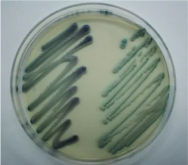
Identification was made on the basis of a combination of vari-ous phenotypic methods and confirmed by species-specific multiplex PCR. Isolates produced germ tubes in human pooled serum in 3 hr at 37°C (Fig. 1) and did not grow at 45°C for 96 hr on SDA plates. Isolates were cultured on CHROMagar Candida media (Becton Dickinson, Sparks, MD, USA) at 35°C for 48 hr. The resulting colonies were dark green in color, which differs from the light to medium green color of the more commonly ob-served C. albicans (Fig. 2). These isolates were identified as C. dubliniensis using the Vitek 2 ID-YST System (bioMerieux Inc., Hazelwood, MO, USA) (probability, 91.0%) and the latex aggluti-nation test using Bichro-Dubli Fumouze (Fumouze Diagnostics, France) (Fig. 3) [9]. To confirm this finding, we performed spe-cies-specific multiplex PCR using the Seeplex Candida
Detec-Fig. 2. Candida dubliniensis on CHROMagar Candida plate incu-bated at 35°C for 48 hr (left, C. dubliniensis; right, C. albicans).
Fig. 3. Latex agglutination test for Candida dubliniensis: left, C. dub-liniensis; middle, C. glabrata; right, C. albicans.
Fig. 1. (A) Candida dubliniensis and (B) Candida albicans ATCC 14053 showing germ tube formation in human pooled serum (×1,000).
Yu N, et al.
Candidemia due to Candida dubliniensis
227
http://dx.doi.org/10.3343/alm.2012.32.3.225 www.annlabmed.org
tion kit (Seegene, Seoul, Korea), which contained two sets of primer mixtures for the detection of 8 different Candida spp. [10]. The isolates were confirmed as C. dubliniensis.
An antifungal susceptibility test was performed using the Vi-tek 2 system (bioMerieux Inc.), and we determined that the iso-lates were susceptible to flucytosine, fluconazole, voriconazole, and amphotericin B. On the basis of the results of the suscepti-bility tests, the patient was treated with fluconazole for 4 weeks. In addition, the central venous catheter that was the suspected cause of the candidemia was removed immediately after C. dubliniensis was identified from the second blood culture. After initiation of antifungal therapy and removal of the central venous catheter, the patient’s clinical symptoms, including the fever, were gradually improved. However, due to other medical prob-lems including cholecystitis, gastric ulcer, and hypertension, the patient remained in the ICU for 93 days, after which she was discharged to another hospital.
DISCUSSION
The incidence of candidemia in the overall population ranges from 1.7 to 10 episodes per 100,000 inhabitants, and Candida is one of the 10 leading causes of bloodstream infections in devel-oped countries. An estimated 33-55% of all episodes of candi-demia occur in the ICU environment and are associated with mortality rates ranging from 5% to 71% [3]. Candidemia may have an endogenous or an exogenous origin, and in recent years, a growing proportion of episodes of candidemia have been caused by Candida spp. other than C. albicans. There are a number of conditions predisposing ICU patients to candidemia, including prior abdominal surgery, intravascular catheters, acute renal failure, malignancy, vancomycin treatment longer than 3
days, parenteral nutrition, broad-spectrum antibiotics, a pro-longed ICU stay, the use of corticosteroids, and mucosal coloni-zation with Candida spp. [3, 11, 12]. For the patient in this case, the predisposing factors included a central venous catheter, par-enteral nutrition, broad-spectrum antibiotics, and a prolonged ICU stay. Among these, the central venous catheter was the most likely explanation for nosocomial candidemia, which is supported by the clinical improvement of the patient after removal of the catheter. A central venous catheter has also been cited as a risk factor for nosocomial candidemia in several reports, and removal of catheters has been recommended to reduce the rate of cathe-ter-related bloodstream infection and to prevent nosocomial clus-ters in the ICU [13, 14].
C. dubliniensis has recently been described as an
opportu-nistic yeast that is primarily associated with oropharyngeal infec-tions in HIV-infected patients. Unlike C. albicans, C. dublinien-sis is rarely found in the oral microflora of normal healthy indi-viduals, and it is responsible for as few as 2% of cases of candi-demia (compared to approximately 65% for C. albicans) [15]. Phenotypically, C. dubliniensis and C. albicans have many similarities, including their microscopic morphology and ability to form germ tubes in serum [8]. Therefore, the objective of most previous studies has been to distinguish C. dubliniensis from C. albicans or other germ tube-positive Candida species [16, 17]. Tests based on different phenotypic characteristics, such as the inability of C. dubliniensis to grow at 45°C [18], and/ or in Sabouraud dextrose broth containing 6.5% sodium chlo-ride (NaCl) [19]; the production of abundant pseudohyphae and chlamydospores on Staib agar [20]; and the formation of dark green colonies on CHROMagar Candida medium [21], have been claimed to differentiate between the two species. However, these characteristics are not definitive, and identification of C. dubliniensis in routine laboratory testing remains a problem. Ad-ditional tests, such as the latex agglutination test and molecular assays, are helpful in accurately differentiating between the two species.
The latex agglutination test used for this case is a commer-cialized protocol for identifying C. dubliniensis isolates that uses particles coated with a monoclonal antibody, which allows for the specific detection of an antigen located on the surface of the C. dubliniensis blastoconidia [9]. The latex agglutination of C. dubliniensis is visible to the unaided eye. Molecular techniques employing PCR have also increased the accuracy of identifying uncommon Candida spp. Although 16S RNA gene sequencing is a useful and definitive method for most Candida spp., it re-quires a specialized instrument to analyze the product. Multi-plex PCR using a specific primer can serve as an alternative, easy to perform, and accurate diagnostic tool for the identifica-tion of C. dubliniensis [10, 22, 23].
Resistance to antifungal agents is rarely found in Candida spp. The fluconazole resistance rate was reported to be 2% in Korean patients with candidemia, and each of the Candida spp. that caused candidemia in that study showed susceptibility to voriconazole [24, 25]. However, uncommon Candida spp., such as C. guilliermondii, C. rugosa, and C. dubliniensis, had suscep-tible-dose-dependent minimum inhibitory concentration (MIC) values for fluconazole [4]. Resistance to fluconazole has been reported to occur only rarely in C. albicans (0.6%), while occur-ring more commonly in C. dubliniensis (3.1%) [2]. However, the vast majority of C. dubliniensis isolates identified to date are
Yu N, et al.
Candidemia due to Candida dubliniensis
228
www.annlabmed.org http://dx.doi.org/10.3343/alm.2012.32.3.225susceptible to all of the commonly used antifungal agents [13]. C. dubliniensis isolates in the present case showed susceptibil-ity to flucytosine, fluconazole, voriconazole, and amphotericin B, which corresponds with previous reports.
In this report, we describe the case of C. dubliniensis candi-demia in an ICU patient with pneumonia and meningitis. To our knowledge, this report is the first case of C. dubliniensis candi-demia in Korea and the first case identified phenotypically and subsequently confirmed by molecular methods. During the di-agnostic process, a C. dubliniensis infection should be distin-guished from infection caused by other Candida spp., particu-larly C. albicans. Confirmation through various methods, such as latex agglutination and multiplex PCR, is needed for the ac-curate identification of isolates. Both clinicians and microbiolo-gists should be aware of predisposing factors for C. dubliniensis candidemia to promote early diagnosis and appropriate treat-ment.
Authors’ Disclosures of Potential Conflicts of
Interest
No potential conflict of interest relevant to this article was re-ported.
Acknowledgement
This research was supported by a Basic Science Research Pro-gram through the National Research Foundation of Korea (NRF) funded by the Ministry of Education, Science and Technology (2009-0077675).
REFERENCES
1. Alangaden GJ. Nosocomial fungal infections: epidemiology, infection control, and prevention. Infect Dis Clin North Am 2011;25:201-25. 2. Arendrup MC, Bruun B, Christensen JJ, Fuursted K, Johansen HK,
Kjaeldgaard P, et al. National surveillance of fungemia in Denmark (2004 to 2009). J Clin Microbiol 2011;49:325-34.
3. Bouza E and Muñoz P. Epidemiology of candidemia in intensive care units. Int J Antimicrob Agents 2008;32(S2):S87-91.
4. Chen SC, Marriott D, Playford EG, Nguyen Q, Ellis D, Meyer W, et al. Candidaemia with uncommon Candida species: predisposing factors, outcome, antifungal susceptibility, and implications for management. Clin Microbiol Infect 2009;15:662-9.
5. Sullivan D and Coleman D. Candida dubliniensis: characteristics and identification. J Clin Microbiol 1998;36:329-34.
6. Mubareka S, Vinh DC, Sanche SE. Candida dubliniensis bloodstream infection: a fatal case in a lung transplant recipient. Transpl Infect Dis 2005;7:146-9.
7. Brandt ME, Harrison LH, Pass M, Sofair AN, Huie S, Li RK, et al. Can-dida dubliniensis fungemia: the first four cases in North America. Emerg Infect Dis 2000;6:46-9.
8. Gutiérrez J, Morales P, González MA, Quindós G. Candida dubliniensis, a new fungal pathogen. J Basic Microbiol 2002;42:207-27.
9. Sahand IH, Moragues MD, Robert R, Quindos G, Ponton J. Evaluation of Bichro-Dubli Fumouze to distinguish Candida dubliniensis from Can-dida albicans. Diagn Microbiol Infect Dis 2006;55:165-7.
10. Lee MK, Kim HR, Lee YJ. Identification of candida species by multiplex polymerase chain reaction. Korean J Clin Microbiol 2006;9:119-24. 11. Dimopoulos G, Ntziora F, Rachiotis G, Armaganidis A, Falagas ME.
Can-dida albicans versus non-albicans intensive care unit-acquired blood-stream infections: differences in risk factors and outcome. Anesth Analg 2008;106:523-9.
12. Zaoutis TE, Prasad PA, Localio AR, Coffin SE, Bell LM, Walsh TJ, et al. Risk factors and predictors for candidemia in pediatric intensive care unit patients: implications for prevention. Clin Infect Dis 2010;51:e38-45. 13. Asmundsdottir LR, Erlendsdottir H, Haraldsson G, Guo H, Xu J, Gott-fredsson M. Molecular epidemiology of candidemia: evidence of clus-ters of smoldering nosocomial infections. Clin Infect Dis 2008;47:e17-24. 14. Odds FC, Hanson MF, Davidson AD, Jacobsen MD, Wright P, Whyte JA,
et al. One year prospective survey of Candida bloodstream infections in Scotland. J Med Microbiol 2007;56:1066-75.
15. Sullivan DJ, Moran GP, Pinjon E, Al-Mosaid A, Stokes C, Vaughan C, et al. Comparison of the epidemiology, drug resistance mechanisms, and virulence of Candida dubliniensis and Candida albicans. FEMS Yeast Res 2004;4:369-76.
16. Odds FC, Van Nuffel L, Dams G. Prevalence of Candida dubliniensis isolates in a yeast stock collection. J Clin Microbiol 1998;36:2869-73. 17. Jabra-Rizk MA, Baqui AA, Kelley JI, Falkler WA Jr, Merz WG, Meiller TF.
Identification of Candida dubliniensis in a prospective study of patients in the United States. J Clin Microbiol 1999;37:321-6.
18. Pinjon E, Sullivan D, Salkin I, Shanley D, Coleman D. Simple, inexpen-sive, reliable method for differentiation of Candida dubliniensis from Candida albicans. J Clin Microbiol 1998;36:2093-5.
19. Alves SH, Milan EP, de Laet Sant’Ana P, Oliveira LO, Santurio JM, Co-lombo AL. Hypertonic Sabouraud broth as a simple and powerful test for Candida dubliniensis screening. Diagn Microbiol Infect Dis 2002;43: 85-6.
20. Staib P and Morschhäuser J. Chlamydospore formation on Staib agar as a species-specific characteristic of Candida dubliniensis. Mycoses 1999;42:521-4.
21. Kirkpatrick WR, Revankar SG, Mcatee RK, Lopez-Ribot JL, Fothergill AW, McCarthy DI, et al. Detection of Candida dubliniensis in oropharyn-geal samples from human immunodeficiency virus-infected patients in North America by primary CHROMagar candida screening and suscep-tibility testing of isolates. J Clin Microbiol 1998;36:3007-12.
22. Innings A, Ullberg M, Johansson A, Rubin CJ, Noreus N, Isaksson M, et al. Multiplex real-time PCR targeting the RNase P RNA gene for de-tection and identification of Candida species in blood. J Clin Microbiol 2007;45:874-80.
23. Kim TH, Park BR, Kim HR, Lee MK. Candida dubliniensis screening using the germ tube test in clinical yeast isolates and prevalence of C. dubliniensis in Korea. J Clin Lab Anal 2010;24:145-8.
24. Shin JH. Antifungal Resistance in Yeasts and Filamentous Fungi. Infect Chemother 2009;41:65-71.
25. Shin JH, Chae MJ, Song JW, Jung SI, Cho D, Kee SJ, et al. Changes in karyotype and azole susceptibility of sequential bloodstream isolates from patients with Candida glabrata candidemia. J Clin Microbiol 2007; 45:2385-91.