1)
Introduction
Kawasaki disease is an acute febrile vasculitis which involves small to medium-sized arteries, usually affecting children under 5 years1). Without treatment, 15% to 25% of affected children may subsequently develop coronary artery aneurysm2, 3). Atypical Kawasaki disease describes the con-dition of children who fail to meet the strict definition for classic Kawasaki disease but have compatible laboratory findings and no other explanation for their illness4). The prevalence of such presentation has been described as much as 7-10%5, 6). Unfortunately, children with atypical Kawasaki disease, particularly infants younger than 1 year, are at risk for developing coronary aneurysms and other
접수 : 2004년 9월 7일, 승인 : 2004년 10월 12일 책임저자 : 최재영, 연세의대 심장혈관센터 소아심장과 Correspondence : Jae Young Choi, M.D.
Tel : 02)361-7270 Fax : 02)312-9538 E-mail : [email protected]
cardiac complications5).
We report a rare case of a 3-month old infant with multiple giant coronary aneurysms, severe coronary artery stenosis and a large mural thrombus caused by Kawasaki disease. The patient had a prolonged, complicated, and re-current course in spite of every effort to minimize coronary complications including multiple doses of immunoglobulin (IVIG), steroid and oral methotrexate(MTX), and expired after subsequent surgical intervention.
Case Report Patient : ○○ Yoon, M/3 months
Chief complaint : Fever for 5 days, conjunctival injec-tion, strawberry tongue and maculopapular rashes for 2 days.
Past and family history : The patient was born at full-term through transvaginal delivery with a birth weight of 2.9 kg. He had no previous history of illness or
hospitaliza-A Case of Multiple Giant Coronary hospitaliza-Aneurysms with
Large Mural Thrombus due to
Kawasaki Disease in a Young Infant
Eun Na Choi, M.D., Jeoung Tae Kim, M.D., Yuria Kim, M.D.
Byung Won Yoo, M.D., Deok Young Choi, M.D., Jae Young Choi, M.D.
Jun Hee Sul, M.D., Sung Kye Lee, M.D., Dong Soo Kim, M.D.
and Young Hwan Park, M.D.
*Department of Pediatrics and Cardiovascular Surgery*, Cardiovascular Center, Yonsei University College of Medicine, Seoul, Korea
Kawasaki disease is an acute systemic vasculitis of unknown origin. Giant coronary aneurysm is one of the most serious complications, although peripheral artery vasculitis can produce life-threat-ening events. Myocardial ischemia and infarction can be caused by coronary artery stenosis, aneu-rysm, and stagnation of blood flow in coronary arteries which triggers thromboembolism. Atypical presentation in young infants often interferes with prompt diagnosis and timely treatment, resulting in poor outcomes. We describe a 3-month-old infant with multiple giant coronary aneurysms with flow stagnation, stenosis and large mural thrombus due to Kawasaki disease. He presented with a prolonged course of severe coronary involvement in spite of all measures to reduce coronary com-plications. Finally, surgical intervention was tried because of the worsening coronary artery abnor-malities. The patient died of acute cardiorespiratory failure shortly after weaning from cardiopulmo-nary bypass. (Korean J Pediatr 2005;48:321-326)
Key Words : Mucocutaneous lymph node syndrome, Coronary aneurysm, Coronary thrombosis, In-fant, Myocardial ischemia
tion. There was no related family history.
Present illness : This 3-month old infant was hospita-lized at another hospital with initial complaint of fever (39.6℃) of 1 day's duration. At initial onset, he was tenta-tively diagnosed with sepsis and treated with intravenous antibiotics without symptomatic relief. He was transferred to another hospital three days later.
After one day, he developed generalized maculopapular rash, bilateral conjunctival injections, strawberry tongue and erythema at the site of BCG vaccination. There was no evidence of cervical lymphadenopathy or changes in the extremities. He was diagnosed as atypical Kawasaki dis-ease and high-dose IVIG (2 g/kg) was given on 4th day of the illness. The fever persisted for 2 days after the initial IVIG therapy and he was referred to our institution.
Physical examination : On admission, he was acutely ill-looking. His body temperature was 38.3℃ with heart rate of 130 b.p.m. He presented with generalized skin rash, bilateral conjunctival injections, strawberry tongue and ery-thema at the site of BCG vaccination. Upon auscultation, his lung sounds were slightly coarse and his heart beat was regular without audible murmur. His abdomen was soft and no hepatosplenomegaly was detectable. Erythema-tous swelling of both hands, which was initially not appar-ent, was noted. There was no cervical lymphadenopathy.
Laboratory data : The patient presented with leukocy-tosis of 24,970/mm3 with 73% neutrophils, 21% lympho-cytes and platelet count of 285 k/mm3. His platelet count increased progressively until hospital day 31 (1,020 k/mm3). Hypochromic anemia (hemoglobin 6.5 g/dL, hematocrit 19.9 %) was noted. Initial erythrocyte sedimentation rate (ESR) and C-reactive protein (CRP) were elevated at 55 mm/hr
(normal 0-10 mm/hr) and 17.4 mg/dL (normal 0-0.8 mg/ dL) respectively. Cerebrospinal fluid exam showed a WBC count of 78/mm3 with 38% polymorphoneuclear cells and 62 % monocytes. Blood, urine and cerebrospinal fluid were sterile. On routine blood chemical studies, the total protein and albumin levels were 4.9 and 2.0 g/dL, respectively. As-partate aminotransferase (AST) and alanine aminotransfe-rase (ALT) levels were 25 and 21 U/L. The abdominal ul-trasonogram was normal.
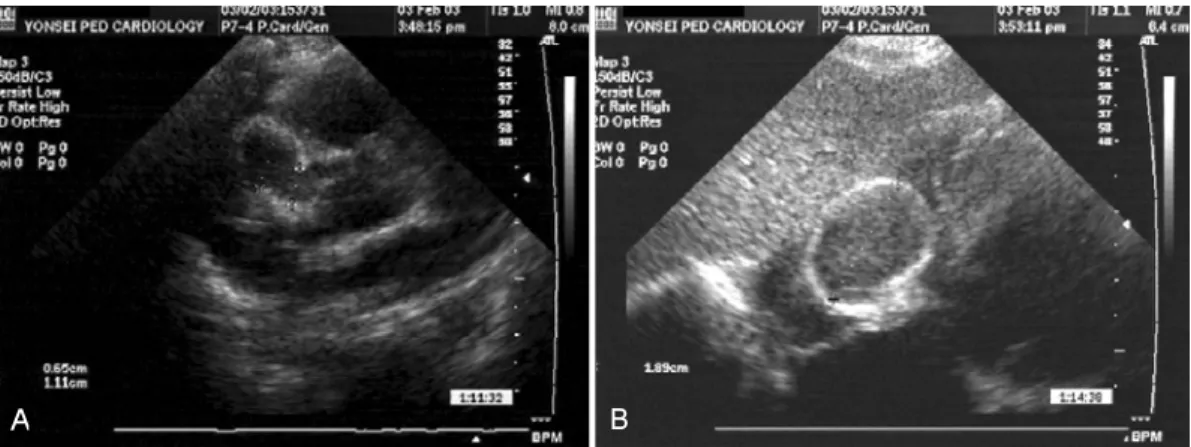
Progress and treatment : High-dose IVIG (2 g/kg) was given on day 4 of the illness before admission to our hospital. After transfer to our hospital, repeated doses of IVIG (2 g/kg) was given on day 6 of illness. Oral aspirin (100 mg/kg), dexamethasone (0.3 mg/kg/day, iv) and oral methotraxate (MTX, 3 mg/BSA, 1.5 mg, weekly) were also given because of the persisting fever. On the third day of admission to out hospital(8 total days of febrile illness), fever subsided. Aspirin dose was adjusted to 5 mg/kg dai-ly, and intravenous dexamethasone was tapered off. On hospital day 6, echocardiographic findings revealed mild dilatation of the main left coronary artery (LCA) (2.9 mm). After 10 day afebrile period, he was follwed-up with echo-cardiogram which revealed ongoing dilatation of both cor-onary areurysms. On hospital day 21, a giant aneurysm of right coronary artery (RCA) (6-11 mm) with large throm-bus (18-19 mm) was noted (Fig. 1). The ejection fraction was 70% and septal motion was normal. On EKG findings, there was no change of ST segment or T wave abnor-mality. On hospital day 22, the patient was irritable and developed a fever (38.3℃). In laboratory findings, CRP and ESR were elevated. No infection foci was found. Suspec-ting recurrence of Kawasaki disease, high-dose IVIG (2 g/
Fig. 1. Echocardiogram on day 21, showed giant aneurysm of right coronary artery (A) with a large mural thrombus (B) as a sequela of Kawasaki disease.
kg) was readministered and 30 mg/kg of oral aspirin was prescribed. Continuous intravenous heparin was started. In-travenous dexamethasone (0.3 mg/kg/day) and MTX (1.5 mg, orally, once a week) were administered. On hospital day 24, MIBI scan during resting state was performed. There was no perfusion defect. In follow-up echocardio-gram performed on day 26, multifocal aneurysmal dilata-tions of RCA (7-12 mm) with thrombus (20 mm in long axis) and LCA main dilatation (3.3 mm) were seen. On hospital day 33, the heart MRI showed left ventricular en-largement and severe hypokinesia on the anterior wall and septum. The anterior descending branch of left coronary artery (LAD) was not clearly visualized and marked sac-cular dilatations were noted in RCA and circumflex branch of left coronary artery(LCX). Particularly, the RCA showed a shape of an inflated balloon or sausage with a diameter of 8.5 mm in the proximal part and 21 mm in diameter distally. The maximal diameter of the LCX was 6 mm. Sluggish blood flow was demonstrated in the distal RCA, but thrombus was not clearly demonstrated(Fig. 2). EKG showed ST elevation and inverted T wave on lateral leads. Creatine kinase (CK) and CK-MB level were 51 IU/L (nor-mal 44-245 IU/L) and 4.9 ng/mL (nor(nor-mal 0-5 ng/mL). Tro-ponin-T level was elevated to 2.88 ng/mL (normal 0-0.1 ng/mL). On hospital day 34, intravenous dexamethasone was changed to oral prednisolone (1 mg/kg/day). Cardiac catheterization was performed on hospital day 38 to con-firm the coronary artery lesions, especially in the LAD. There was a main LCA aneurysm and stenosis in proximal LAD. Decreased blood flow to the LAD with associated left ventricular wall motion abnormality in LAD territory
was found (Fig. 3).
Aspirin was discontinued and warfarin was added to the anticoagulation therapy of IV heparin. A Beta-blocker (pro-pranolol, 1 mg/kg/d) was used to decrease tachycardia and increase effective coronary blood flow. His vital signs were stable and his general condition improved. On hospital day 43, EKG findings normalized and follow-up echocardiogram revealed no interval changes in coronary arteries. On hos-pital day 76, EKG showed non-specific ST elevation. A second coronary angiography was performed on hospital day 80 and multiple progressive coronary aneurysms in-volving the entire RCA and some segments of LCA were noted, with severe stenosis in the proximal LAD. There was stagnation of blood flow in RCA but no thrombus was visualized probably due to extremely poor blood flow.
On hospital day 88, surgical intervention was performed. The aneurismally dilated portion of the RCA was resected and Gore-tex tube graft was interposed from the proximal to distal RCA and each major branches of RCA was sep-arated at the branching portion with buttons and anasto-mosed to the Gore-tex tube. The left main coronary artery was incised longitudinally and a pericardial patch was placed on the incision. Weaning from the bypass was suc-cessful, but the patient developed acute cardiorespiratory failure from which he did not recover. On autopsy, a fresh blood clot was found in the Gore-tex tube.
Discussion
Kawasaki disease is an acute systemic vasculitis of un-known cause usually occurring in children younger than Fig. 2. On day 33, heart MRI was performed. The right coronary artery (RCA) showed a
shape similar to an inflated balloon measuring 21 mm in diameter (A) and circumflex branch of left coronary artery (LCX) was 6 mm in maximal diameter (B).
five years of age. Giant coronary aneurysm is one of the most serious form of complications. In previous study, at 1 to 3 months after the onset of Kawasaki disease, about 15% of patients had angiographic evidence of coronary ar-tery aneurysms and repeat angiography 5 to 18 months lat-er showed that the aneurysms had resolved in about 50% of the patients2). The most severe form of coronary artery lesions in Kawasaki disease is the giant aneurysm (internal luminal diameter of the coronary artery ≧8 mm).
Compared to smaller aneurysms, these lesions are less likely to resolve spontaneously and are known to be asso-ciated more frequently with severe complications such as thrombus, rupture or stenosis.
The diagnosis of Kawasaki disease in very young in-fants is challenging because of its rarity and high
inci-dence of atypical presentation (longer duration of illness before diagnosis, lower incidence of conjunctivitis, lower incidence of skin rash, lower incidence of extremity change, and lower C-reactive protein)4). The incidence of late car-diac sequelae has been reported to be much higher in in-fants, particularly in the first 6 months, than in older chil-dren with Kawasaki disease (64% and 9%, respectively)7). The case fatality for aneurysm is known to be highest in children under 1 year of age8). The predictors of risk for coronary artery aneurysm in infants is known to be resis-tance to IVIG treatment and prolonged duration of fever9). In young infants, Kawasaki disease is frequently subtle in manifestations, with a paucity of classic signs and symp-toms10). This may contribute to delay in diagnosis, inap-propriate therapy and development of cardiac sequelae. So Fig. 3. Coronary angiography on 38th day in the hospital, showed multiple giant
aneu-rysms on entire right coronary artery (RCA) (A, B) with severe stasis of blood flow and left coronary artery (LCA) with stenosis on proximal anterior descending branch of left coronary artery (LAD) (C, D). Left ventricular wall motion abnormality was ob-served on LAD territory.
A B
when possible, patients with questionable diagnosis should be referred to a pediatric facility with established expertise in the diagnosis and management of Kawasaki disease before therapy is initiated11).
Thrombosis is a major cause of ischemic heart disease in Kawasaki disease and is frequently associated with large coronary aneurysms.
This is thought to be a consequence of blood flow stag-nation and sudden reduction of shear stress in coronary aneurysms12). Endothelial damage induced by inflammatory changes associated with Kawasaki disease and increased wall stress, along with increase in coronary vessel diame-ter, may interfere with platelet adhesion and aggregation13). Stenosis, which occurs as a result of the healing process of the vessel walls, often leads to significant coronary ob-struction and myocardial ischemia.
For evaluation of coronary artery lesions, echocardio-graphy is adequate in early childhood. However in patients with anomalies in coronary vessels persisting over 6 months, follow-up coronary angiography, which is an inva-sive procedure, is usually required. Recently magnetic reso-nance angiography has been successfully used for the de-tection of aneurysms in Kawasaki disease14, 15).
If coronary obstruction is suspected and confirmed by coronary angiography, the therapeutic options of bypass grafting, balloon angioplasty, or other procedure to restore coronary blood flow should be considered. In cases of myo-cardial infarction related to thrombosis, acute thrombolytic therapy utilizing heparin, urokinase or TPA (tissue plas-minogen activator) may be necessary11, 16). Catheter inter-vention may be considered as the first-line treatment if coronary stenosis is relatively simple or limited to a single vessel17). Patients with Kawasaki disease and coronary ar-tery stenosis have ischemic events or sudden cardiac death probably due to thrombotic occlusion of the coronary ar-teries18). Unfortunately, predicting these ischemic events is difficult, which justifies preventive catheter interventions in selected patients with high risk. Coronary artery bypass grafting (CABG) may be indicated for children with multi-ple or commulti-plex lesions if there is evidence of ischemia, but often, this procedure is delayed due to dilated vessels. Re-cently, long-term follow-up studies of CABG have found a high bypass graft patency and growth of the bypass graft in length and diameter19). Regarding the material of the graft, autologous saphenous veins, the internal thoracic artery, the gastroepiploic artery or a combination of these
grafts have been utilized. Long-term patency of grafts pre-pared from saphenous veins is unsatisfactory at a young age because of a high occlusion rate and therefore the in-ternal thoracic artery is preferred as the graft of choice for very young infants, anticipating long-term patency and ad-aptation for rapid somatic growth19, 20). According to investi-gation conducted by Kitamura, the youngest patient who have undergone CABG was a child of 1 year and most pa-tients who receive grafts were 5-6 years of age20). In this case, coronary aneurysmectomy and long segment interpo-sition in the use of Gore-tex tube was performed due to the extreme complexity and severity of the lesions in the right coronary artery. Comparisons with other cases are limited, as no published literature exists on very young in-fants such as ours with coronary artery lesions that are severe enough to be refractory to medical treatment, mak-ing surgery the only curative option available. On review of the case, our thought is that this method may not be recommended because of the difficulty in restoring all coronary branches arising from the aneurysm and potential for thrombus formation within the Gore-tex tube.
국문 요약
가와사끼병 영아에서 발생한 혈전을 동반한
거대관상동맥류 1례
연세대학교 의과대학 심장혈관병원 소아심장과, 심장혈관외과* 최은나·김정태·김유리아·유병원·최덕영 최재영·설준희·이승규·김동수·박영환* 가와사끼병은 주로 소아연령에서 발생하는 급성 열성 혈관염 으로 관상동맥의 확장이 치명적 합병증으로 나타날 수 있다. 저 자들은 3개월된 영아에서 반복적인 정맥용 면역글로불린, 스테로 이드, 경구용 methotrexate의 치료에도 불구하고 진행되는 관상 동맥 병변으로 다발성 거대 관상류, 협착 및 관상동맥 내 혈전 을 보이며 심전도상 심근 허혈 소견과 함께 심장 자기공명영상 에서 좌심실의 확장과 전벽 및 중격의 심한 운동저하를 나타낸 증례를 보고한다. 환아는 보존적 요법에 반응하지 않는 관상동맥 병변으로 내원 88일째 수술적으로 우측 거대관상동맥류를 절개 하고 Gore-tex tube를 삽입하여 각각의 측부순환과 연결하였으 며, 좌측 관상동맥 협착부위는 세로로 절개한 후 심장막 반 (pericardial patch)을 삽입, 확장하였으나, 수술 후 심폐부전으 로 사망하였다.References
1) Kawasaki T. Acute febrile mucocutaneous syndrome with lymphoid involvement with specific desquamation of the fingers and toes in children. Jpn J Allergy 1967;116:178-222.
2) Kato H, Ichinose E, Yoshioka F, Takechi T, Matsunaga S, Suzuki K, et al. Fate of coronary aneurysms in Kawasaki disease : serial coronary angiography and long-term follow-up study. Am J Cardiol 1982;49:1758-66.
3) Suzuki A, Kamiya T, Kuwahara N, One Y, Kohata T, Ta-kahashi O, et al. Coronary arterial lesions of Kawasaki disease : cardiac catheterization findings of 1,100 cases. Pe-diatr Cardiol 1986;7:3-9.
4) Pfafferott G, Wirzfeld A, Permanatter B. Atypical Kawasaki syndrome : how many symptoms have to be present? Heart 1997;78:619-21.
5) Joffe A, Kabani A, Jadavji T. Atypical and complicated Kawasaki disease in infants. Do we need criteria? West J Med 1995;162:322-7.
6) Rosenfeld EA, Corydon KE, Shulman ST. Kawasaki dis-ease in infants less than one year of age. J Pediatr 1995; 126:524-9.
7) Nakamura Y, Fujita Y, Nagai M, Yanagawa H, Imada Y, Okawa S, et al. Cardiac sequelae of Kawasaki disease in Japan : statistical analysis. Pediatrics 1991;88:1144-7. 8) Asai T. Diagnosis and prognosis of coronary artery lesions
in Kawaski disease. Coronary angiography and the condi-tions for its application. Nippon Rinsho 1983;41:2080-5. 9) Sittiwangkul R, Pongprot Y, Thongsongkrit W, Silvilairat
S, Phornphutkul C. Kawasaki disease in Thai infants com-pared with older children. Ann Trop Paediatr 2004;24:59-63. 10) Hsiao JY, Chen MR, Huang FU, Kao HA, Sung TC. Clin-ical analysis of Kawasaki disease in infants below 6 months of age. Acta Paediatr Sin 1998;29:318-23.
11) Dajani AS, Taubert KA, Gerber MA, Shulman ST, Ferrieri P, Freed M, et al. Diagnosis and therapy of Kawasaki dis-ease in children. Circulation 1993;87:1776-80.
12) Kuramochi Y, Ohkubo T, Takechi N, Fukumi D, Uchikoba Y. Hemodynamic factors of thrombus formation in coronary aneurysms associated with Kawasaki disease. Pediatr Int 2000;42:470-5.
13) Sakariassen K, Niefelstein P, Coller B, Sixma J. The role of platelet membrane glycoprotein Ib and IIb-IIIa in platelet adherence to human artery subendothelium. Br J Haematol 1986;63:681-91.
14) Greil GF, Stuber M, Botnar RM, Kissinger KV, Geva T, Newburger TW, at al. Coronary magnetic resonance angi-ography in adolescents and young adults with Kawasaki disease. Circulation 2002;105:908-11.
15) Flacke S, Setser RM, Barger P, Wickline SA, Lorenz CH. Coronary aneurysms in Kawasaki's disease detected by magnetic resonance coronary angiography. Circulation 2000; 101:156-7.
16) Dajani AS, Taubert KA, Takahashi M, Bierman FZ, Freed MD, Ferrieri P, et al. Guidelines for long-term management of patientswith Kawasaki disease. Circulation 1994;89:916-22.
17) Kato H, Ishii M, Akaki T, Eto G, Iemura M, Tsutsumi T, et al. Interventional catheterization in Kawasaki disease. J Interven Cardiol 1998;11:355-61.
18) Kato H, Ichinose E, Kawasaki T. Myocardial infarction in Kawasakidisease : clinical analysis in 195 cases. J Pediatr 1986;108:923-7.
19) Yoshikawa Y, Yagihara T, Kameda Y, Taniguchi S, Tsuda E,Kawahira Y, et al. Result of surgical treatment in pa-tients withcoronary-arterial obstructive disease after Kawa-saki disease. Eur JCardiothorac 2000;17:515-9.
20) Kitamura S. The role of coronary bypass operation on chil-dren with Kawasaki disease. Coron Artery Dis 2002;13: 437-47.