DOI 10.3349/ymj.2008.49.6.1023
Purpose: IRF-5 is a direct transducer of virus-mediated and TLR-mediated signaling pathways for the expression of cytokines and chemokines which form homodimers or heterodimers with IRF-7. However, direct IRF-5-specific monoclonal antibodies (mAbs) are not available at present. These could be used to further evaluate the functions of IRF-5. In this study, we produced and characterized three mouse mAbs to human IRF-5. The binding of IRF-5 to nuclear import proteins was first identified using a mAb. Materials and Methods: His-tagged human IRF-5 protein spanning amino acid residues 193-257 was used as an antigen and three mAbs were produced. The mAbs were tested with ELISA, Western blot analysis (WB), immunofluorescent staining (IF), and immunoprecipi-tation (IP). In addition, the nuclear import protein which carried phosphorylated IRF-5 was identified using one of these mAbs. Results: MAbs 5IRF8, 5IRF10 and 5IRF24 which reacted with the recombinant His-IRF-5193-257 protein were
produced. All mAbs bound to human IRF-5, but not to IRF-3 or IRF-7. They could be used for WB, IF, and IP studies. The binding of phosphorylated IRF-5 to karyopherin- 1 and - 1α β was also identified. Conclusion: Human IRF-5-specific mAbs are produced for studying the immunologic roles related to IRF-5. Phosphorylated IRF-5 is transported to the nucleus by binding to nuclear import proteins karyopherin- 1 and - 1.α β Key Words: Human IRF-5, monoclonal antibody, nuclear import protein, karyopherins
INTRODUCTION
After first identifying interferon-regulatory factor 1 (IRF-1), nine additional IRF family proteins have been identified. IRF-1 binds to elements in the gene promoter that encodes for interferon-β (IFN- ).β 1 Extensive studies of IRF family of pro-teins have revealed that they are involved in regu-lating virus-IFN axis, antigen presentation, nitric oxide production, and the cell cycle.2,3 The IRF family of proteins also has an important role in the innate immune system, and the secondary response to cytokines. The signaling of toll-like receptors (TLRs) triggered by microbial components is important to the activation of innate and adaptive immune responses. For example, TLR-4 activation recruits the downstream adaptor molecule MyD88 and other signaling molecules for initial responses. IRF-3 and IRF-7 then interact with MyD88 and induce type I IFNs. In contrast, IRF-4 competes with IRF-5 and acts as a negative regulator for TLR signaling of the MyD88 interaction.4 This suggests that there are complex regulatory mechanisms involved in MyD88 signaling. IRF-3 and IRF-7 function as direct transducers of virus-mediated signaling, and play a crucial role in the expression of type I IFN.5-9
IRF-5 is a recently characterized member of the IRF family. It encodes a ~61-kDa protein which was originally identified as a regulator of type I
IFN gene expression.10 Recent studies have
indi-cated that it plays a role in host defense, including
Development of Monoclonal Antibodies Against Human IRF-5
and Their Use in Identifying the Binding of IRF-5 to Nuclear
Import Proteins Karyopherin- 1 and - 1
ɑ
β
Soo-In Yeon,1,3 Ju Ho Youn,1,2 Mi Hwa Lim,1,4 Hye Ja Lee,5 Young Mok Kim,5 Ji Eun Choi,6 Jae Myun Lee,1,2,3 and Jeon-Soo Shin1,2,3,4
1Department of Microbiology, 2Brain Korea 21 Project for Medical Science, 3Institute for Immunology and Immunological
Diseases,4NCRC for Nanomedical Technology, Yonsei University College of Medicine, Seoul;5ATGen Inc., Sungnam, Kyungkido; 6Department of Pediatrics, Seoul National University Borame Hospital, Seoul, Korea.
Received May 21, 2008 Accepted November 13, 2008
This study was supported by the Korea Health 21 R&D Project (A050260) of the Ministry of Health & Welfare, KOSEF through the NCRC (R15-2004-024-00000-0), and the Brain Korea 21 Project for Medical Sciences, Republic of Korea.
Reprint address: requests to Dr. Jeon-Soo Shin, Department of Microbiology, Yonsei University College of Medicine, 250 Seong-sanno, Seodaemun-gu, Seoul 120-752, Korea. Tel: 82-2-2228-1816, Fax: 82-2-392-7088, E-mail: jsshin6203@yuhs.ac
the induction of multiple cytokines.11 Similar to
IRF-3 and IRF-7, IRF-5 is a direct transducer of virus-mediated signaling. However, this only occurs with specific viruses such as the Newcastle disease virus, vesiculostomatitis virus, and herpes simplex type 1 virus.10,12 It also plays a role in the expression of cytokines and chemokines.10,12,13
IRF-5 is a direct target of pIRF-53. Its expression is modu-lated by p53,14and it has a role in the
p53-indepen-dent proapoptotic signaling pathway.15,16 Recent
studies have reported the association between IRF-5 and systemic lupus erythematosus. 17,18 In a gene chip study using overexpressed B cells which contained IRF-5 or IRF-7, the presence of IRF-5 was related to a strong immune response and adhesion genes. The presence of IRF-7, however, selectively upregulated the expression of mitochondrial genes and DNA repair genes.19 This suggests a distinct role for IRF-5. The IRF family of proteins resides in the cytoplasm of resting cells. They are activated by phosphorylation on the C terminus, and are transported to the nucleus after homo- or hetero-dimerization.10,13 IRF-5 dimerizes either with itself
or with IRF-3, and activates IFNA gene transcrip-tion.10,13 However, the heterodimerization of IRF-5
with IRF-7 represses IFNA transcription in virus-infected cells which were cotransfected with IRF-5 and IRF-7.10,13 Recently, IRF-5 was found to have
an important role in TLR signaling and the induction of proinflammatory cytokines such as interleukin (IL)-6, IL-12, and tumor necrosis factor (TNF)- . It is impaired in cells from IRF-5-deficientα mice,11suggesting that IRF-5 is generally involved
downstream of the TLR signaling pathway. IRF-5 associates with both MyD88 and TRAF-6, and is translocated to the nucleus in a MyD88-dependent
fashion.11 However, many of the downstream
mediators of the IRF-5 pathway need further iden-tification.
The proteins in the KAP family act as shuttling receptors. They bind to the NLS motifs of cargo proteins to facilitate their import into the nucleus.20 IRF-5 is phosphorylated by the stimulation of type I interferon and viral infections. It then enters the nucleus to regulate transcription.10,12 IRF-5 has two
nuclear localization signals (NLSs). These are found at residues 46 to 52 on a DNA binding domain, and on residues 448 to 454 on a transac-tivation domain. There is also one nuclear export
signal (NES) on residues 150 to 160.12,21 This
implies its tight control of nuclear transport. IRF-5 is localized to the cytoplasm in an unstimulated state. It moves to the cytoplasm in a CRM1-dependent pathway after it is dephosphorylated in the nucleus.21
In this study we developed and characterized monoclonal antibodies (mAbs) to the human IRF-5 protein and tested their applicability of IRF-5-specific mAbs. Our mAbs were found to bind to human IRF-5, but not to human IRF-3 or IRF-7. We demonstrated the usefulness of these mAbs in Western blot, immunocytochemical, and immuno-precipitation analyses, as well as the import of IRF-5 to the nucleus. In addition, it was identified that IRF-5 is transported into the nucleus with the aid of the carrier proteins karyopherin (KAP)- 1α and - 1.β
MATERIALS AND METHODS Cell cultures and transfection
The human macrophage line THP-1, the human embryonic kidney cell line HEK293 (ATCC), and NIH3T3 cells were cultured. The culture was done
at 37 C under 5% CO2 in RPMI1640 supplemented
with 10% FBS (Invitrogen Life Technologies, San
Diego, CA, USA), 100 U/mL penicillin, 100 g/μ
mL streptomycin, and 2 mM L-glutamine. For the transient transfection, FuGene6 (Roche Diagno-stics GmbH, Mannheim, Germany) was used for the HA-tagged human IRF-3, HA-tagged IRF-5, GFP-tagged IRF-5, and Flag-tagged IRF-7 expres-sions.
DNA constructs and protein purification
We cloned the gene which encodes six His-tagged human IRF-5 protein spanning residues of 65 amino acids into a pRSET vector (Invitrogen) (His-IRF-5192-256in NCBI accession No. AAU 12877
which corresponds to His-IRF-5176-240 in NCBI
accession No. NP_116032.1). This is a region which is located between a DNA-binding domain and an IRF-association domain. It seems to be the least homologous with IRF-3 and IRF-7. cDNA of human peripheral blood mononuclear cells were
amplified by PCR using the sense primer 5’-GCT TGCGGATCCCCGCCCACTCTG-3’, and the anti-sense primer 5’-AGACTGGAATTCTAGATCAGC AGGTCTGG-3’. The restriction enzyme sites BamHI and EcoRI are underlined, respectively. The con-struct was confirmed by DNA sequencing (Applied Biosystems). The protein was produced in E. coli BL21(DE3) pLysS (Novagen) containing ampicillin (50 g/mL) at 37μ C. Isopropyl- -D-1-thiogalactoβ -pyranoside (IPTG) was added to a final concen-tration of 0.5 mM when the OD 600 of the culture broth had reached 0.5 - 0.6. The cells were har-vested after 4 hours. The cell pellet was resus-pended in 100 mL of lysis buffer (20 mM Tris, 1 mM DTT, 10% glycerol, 1 mM EDTA, pH 8.0). The bacterial lysis was sonicated with a Sonosmasher followed by centrifugation. The supernatant was
loaded onto a Ni++-NTA agarose resin (Peptron,
Daejeon, Korea) which was equilibrated with a binding buffer (20 mM Tris pH 8.0, 5 mM imidazole, 0.5 M NaCl). The bound protein was washed with a washing buffer (20 mM Tris pH 8.0, 0.5 M NaCl, 20 mM imidazole) and eluted with an elution buffer (20 mM Tris pH 8.0, 0.5 M NaCl, 200 mM imidazole). For further purification of the protein, the eluent was concentrated with centricon (Vivaspin 20, VS2011) and applied to Sephacryl S200 gel chromatography (Amersham Biosciences, Uppsala, Sweden). The purity of the His-IRF-5193-257
protein was more than 90% using SDS-PAGE. The molecular weight was measured using MALDI-TOF (Applied Biosystems).
MAb production and purification
BALB/c mice were intraperitoneally immunized with 20 g of His-IRF-5μ 193-257 protein, which was emulsified in complete Freund's adjuvant and boosted twice with incomplete Freund's adjuvant at two week intervals. Finally, the mice were intra-venously injected with 50 g of His-IRF-5μ 193-257 protein three days before the fusion. Splenocytes were fused with P3-X63-Ag8.653 cells and selected with HAT medium (Sigma-Aldrich Co., St. Louis, MO, USA). The resulting hybridoma supernatants were screened using ELISA, as described below. The isotype of each mAb was determined using a mouse-hybridoma subtyping kit (Roche). The colonies which secreted anti-IRF-5 antibodies were
cloned with limited dilution. The three hybridoma clones, 5IRF8, 5IRF10, and 5IRF24, were purified for further characterization using a protein G-Sepharose affinity column (Amersham).
ELISA
Microtiter plates (Corning Inc., Corning, NY,
USA) were coated with 1 g/mL of His-IRF-5μ
193-257 for mAb screening. The plates were washed
with 0.05% Tween 20 in PBS (PBST) and blocked with 5% normal goat serum (NGS) in PBST. Hybridoma culture supernatants were added to the plates. The plates were then incubated for 1.5 hours at 37 C. After washing, horseradish perox-idase (HRP) labeled goat anti-mouse Ig (Sigma) was added. After 1 hour of incubation, o-pheny-lenediamine was added to the plates for color development. The ODs were measured at 490 nm.
Western blot analysis
Whole cell lysates (WCLs) from different cell lines were prepared using 1% Nonidet P-40 buffer which contained a protease inhibitor cocktail (Sigma). The protein concentrations were measured using a Bradford assay (Biorad) for the analysis of the WCLs. They were then electrophoresed in 10% SDS-PAGE. The proteins were transferred to a nitrocellulose membrane and probed with mAbs after blocking. HRP-conjugated rabbit anti-mouse Ig (Sigma) was used as a secondary antibody. The probe signals were revealed with enhanced chemi-luminescence (ECL, Labfrontier Co., Seoul, Korea). Mouse anti-HA and anti-Flag M2 (Sigma) were used to detect the corresponding tag proteins.
Immunoprecipitation
Cells were lysed with a protease inhibitor cock-tail. Cell homogenates were centrifuged at 20,000 g for 20 minutes and precleared by incubation with protein G-Sepharose (Amersham) at 4°C for 1 hour. The precleared extracts (500 g) were inμ -cubated with purified mAbs. Protein G-Sepharose was then added and the samples were incubated for 1.5 hours at 4°C. Immune complexes were collected by centrifugation and washed with PBS. The collected complexes were fractionated using
SDS-PAGE, transferred to membranes, and blotted with anti-GFP (Santa Cruz Biotechnology Inc., Santa Cruz, CA, USA). HRP-conjugated goat anti-mouse Ig (Sigma) was used to detect the probe signals.
Immunofluorescence and GFP imaging
To observe the applicability of our mAbs for use in an immunofluorescence assay, HEK293 cells which were transfected with an IRF-5-GFP plasmid were cultured in LabTek II chambers (Nunc). They were then fixed in 4% paraformaldehyde in PHEM buffer (60 mM PIPES, 25 mM HEPES, 10 mM
EGTA, 4 mM MgSO4, pH 7.0) for 10 minutes at
room temperature. After fixation, the cells were washed with PBS and incubated for 3 minutes at 4 C with a HEPES-based permeabilization buffer containing 300 mM sucrose and 0.2% Triton X-100. The cells were blocked with 0.2% BSA in PBS for 15 minutes. They were then incubated with anti-IRF-5 mAbs for 1 hour at room temperature. PE-con-jugated rabbit anti-mouse Ig (BD Pharmingen) was added after three washes. The cells were observed with a BX51 fluorescent microscope (Olympus). HEK 293 cells transfected with an IRF-5-HA plasmid were incubated for 24 hours and then treated with 20 nM of okadaic acid (Calbiochem) for 6 hours to investigate the movement of phosphory-lated IRF-5 using our mAbs, Next, the cells were stained with 5IRF10 and FITC-conjugated
anti-mouse Ig for tracing the movement of IRF-5 into the nucleus. The nuclei were stained with DAPI.
The binding of IRF-5 to KAP proteins
GST-KAP fusion proteins- 1, - 2, - 3, - 4, - 5, - 6ɑ ɑ ɑ ɑ ɑ ɑ , and - 1 were produced as previously described forβ the assay involving the KAP protein binding to the cargo protein.22HEK293 cells were transfected with
an HA-tagged IRF-5 plasmid. WCLs were pre-pared after 24 hours and allowed to bind to Sepharose 4B-immobilized GST-KAPs- 1, - 2, - 3,ɑ ɑ ɑ - 4, - 5, - 6, and - 1 at 4°C overnight. Unboundɑ ɑ ɑ β proteins were washed. Sepharose-bound proteins were dissolved in a Laemmli sample buffer, separated on 12% SDS-PAGE, and subjected to Western blotting. The membrane was blotted with 5IRF10 mAb and then reblotted with anti-GST to visualize the amount of Sepharose-immobilized GST-KAP proteins.
RESULTS
Generation of mAbs and antigen reactivity
Recombinant human His-IRF-5193-257 protein was
produced in E. coli and purified using an affinity column for immunogens. This region is located between a DNA-binding domain and an IRF-asso-ciation domain. The recombinant His-IRF-5193-257
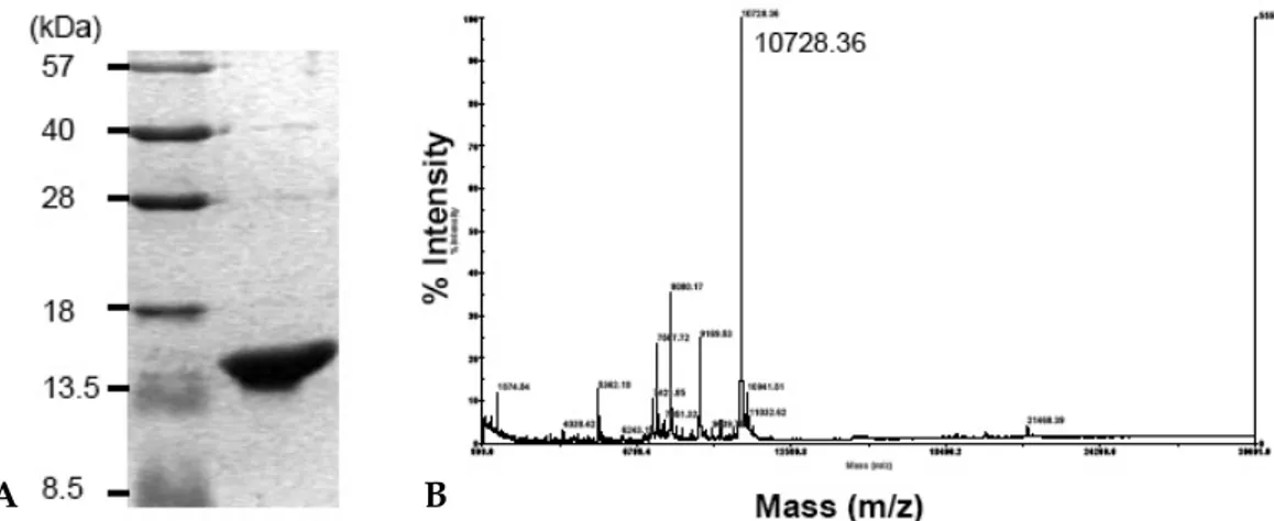
Fig. 1. SDS-PAGE and MALDI-TOF mass spectrum of recombinant His-IRF-5193-257. (A) SDS-PAGE analysis of purified His-IRF-5193-257. His-IRF-5193-257was eluted with 200 mM imidazole on a Ni++-NTA agarose resin and purifed by Sephacryl S-200 gel chromatography. (B) MALDI-TOF mass spectrum analysis of purified His-IRF-5193-257 protein. The molecular weight (MW) was determined to be 10728.36 Da using MALDI-TOF mass spectrum. This was very close to the theoretical MW of 10721.1 Da.
protein was purified and identified using SDS-PAGE (Fig. 1A). The molecular weight was deter-mined to be 10728.36 Da using MALDI-TOF mass spectrum (Fig. 1B). This was very similar to the theoretical molecular weight of 10721.1 Da, implying the exact fragment of IRF-5 protein.
We obtained clones of three hybridomas; 5IRF8, 5IRF10, and 5IRF24. The culture supernatants were tested for their specific antigen-binding capacity using ELISA. This was done by serially diluting the culture supernatant (Fig. 2A) or His-IRF-5193-257
antigen (Fig. 2B) to test whether these mAbs recog-nize the specific antigen. The serially diluted mAbs were shown to bind in a dose-dependent manner
at a fixed antigen coating concentration of 1 g/μ mL. MAbs also bound to antigen in a dose-depen-dent manner when the antigen coating concentra-tion was increased from 0.001 to 10 g/mL. Theseμ mAbs demonstrated no binding to irrelevant protein -synuclein at a concentration of 10ɑ μg/ mL. All isotypes of the mAbs were found to be IgG1 ( ).κ
Western blot analysis
We next tested the reactivity of our mAbs to His-IRF-5193-257 protein using Western blot analysis
to determine the antigen reactivity. All three mAbs
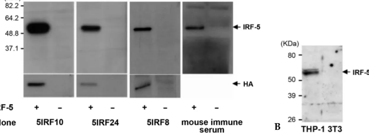
Fig. 3.Western blot analysis for antigen specificity. (A) An HA-tagged IRF-5 plasmid was transfected into HEK293 cells and the WCL was separated. The reactivity of mAbs to IRF-5 was tested. The expression of IRF-5 was tested with anti-HA. Mouse immune serum was used as a positive control Ab. (B) Detection of endogenous IRF-5. WCLs of THP-1 and NIH3T3 cells were separated and immunoblotted with 5IRF10 as a representative study. NIH3T3 cell lysate was used as a negative control.
Fig. 2. Binding curve between the anti-IRF-5 monoclonal antibodies and the His-IRF-5193-257 protein. (A) ELISA was performed to the wells. The wells were coated with 1 g/mL of His-IRF-5μ 193-257 protein using the various dilutions of culture supernatants of our three mAbs. (B) ELISA was performed with mAbs at a fixed dilution to the wells which were coated with various concentrations of the His-IRF-5193-257 protein. The bindings of all three mAbs to the control protein of -synuclein are shown inside the black triangle.ɑ
A
A B
detected the purified His-IRF-5193-257 protein (data
not shown). HEK293 cells were transfected with an HA-tagged IRF-5 plasmid, and WCLs were har-vested to determine whether these mAbs could recognize the wild type IRF-5 protein. All three mAbs readily recognized the wild type IRF-5 and did not bind to non-transfected control WCLs (Fig. 3A). IRF-5 is constitutively expressed by B cells, monocytes, and dendritic cells.10,23 We tested the
binding of 5IRF10 as a representative to the endo-genous IRF-5 using WCLs of human cell lines of THP-1 cells. 5IRF10 recognized two major IRF-5 bands at ~61 kDa (Fig. 3B).
Cross-reactivity analysis
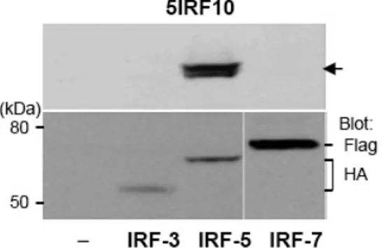
The spanning region of recombinant His-IRF-5193-257 protein was chosen because it has a very low amino acid homology with human IRF-3 and IRF-7. In order to exclude cross-reactivity with other proteins in the IRF family, we determined the binding specificity of all three mAbs to IRF-3 and IRF-7. For this study, HEK293 cells were transfected with each plasmid containing IRF-3, IRF-5, and IRF-7. The WCL proteins were separated using SDS-PAGE and immunoblotted with each mAb. This was followed by reblotting with anti-HA for IRF-3 and IRF-5, and with anti-Flag for IRF-7. All three mAbs showed specific binding to IRF-5 protein, but not to IRF-3 and IRF-7 proteins. This indicates that our mAbs bind specifically to IRF-5. We used 5IRF10 data as a representative
(Fig. 4).
Immunofluorescent analysis
Using immunofluorescent staining, three mAbs were examined for their ability to detect intracel-lular IRF-5 protein. HEK293 cells, which do not express the detectable endogenous IRF-5 through Western blotting (Fig. 4), were transfected with an IRF-5-GFP plasmid. Indirect immunofluorescent staining was also performed to observe the location of the IRF-5. This was done using each mAb and PE-conjugated anti-mouse Ig as the primary and secondary antibodies, respectively. Green fluor-escence was observed in the cytoplasm in an unstimulated state. IRF-5 protein stained inten-sively in the cytoplasm with all three mAbs (Fig. 5). When the two images were merged, the staining image of IRF-5 using each mAb was co-localized to the GFP expression image. This result demon-strates that our mAbs can be used for the immu-nofluorescent staining of IRF-5.
Immunoprecipitation
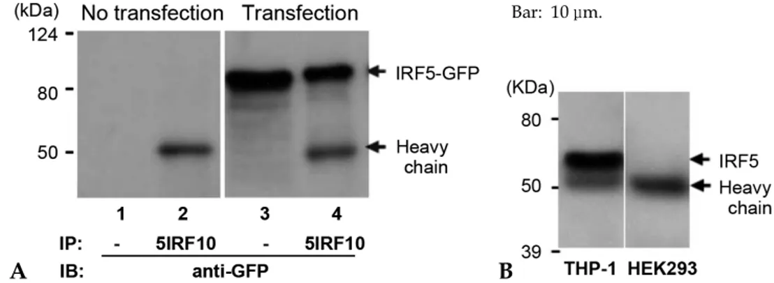
HEK293 cells were transfected with an IRF-5-GFP plasmid, and the WCLs were subjected to immunoprecipitation using our mAbs to identify the applicability of these mAbs to the immunopre cipitation procedure. The WCLs were immuno-precipitated with 5IRF10 mAb as a representative sample. The precipitates were analyzed with a Western blot using anti-GFP. IRF-5-GFP protein was observed at the expected molecular weight of ~88 kDa (Fig. 6A). The immunoprecipitation study was also performed against WCLs of THP-1 cells to determine whether 5IRF10 can immunopre-cipitate endogenous IRF-5 protein. The membrane was probed with anti-GFP. 5IRF10 could immuno-precipitate endogenous IRF-5 protein (Fig. 6B).
Import of phosphorylated IRF-5 into the nucleus
The nuclear import study of IRF-5 was perfor-med to further evaluate the functions of our mAbs. The movement of phosphorylated IRF-5 was detected after transfection. To do this, HEK 293 cells were transfected with an HA-IRF-5 plasmid, stained with 5IRF10. Okadaic acid is a type 1/2A
Fig. 4. Cross-reactive binding study. HA-tagged IRF-3, HA-tagged IRF-5, and Flag-tagged IRF-7 plasmids were transfected into HEK293 cells. WCLs were separated and the cross-reactivity of 5IRF10 to IRF-3 and IRF-7 was tested as a representative study. The membranes were reblotted with anti-HA or anti-Flag depending on the tagged protein. Arrow: IRF-5.
protein phosphatase inhibitor24 for forcibly
in-ducing phosphorylation of proteins. Okadaic acid was used at a low concentration of 20 nM for 6 hours to block entry into and the cell cycle.25IRF-5
was located in the cytoplasm in an unstimulated state and then moved to the nucleus after phos-phorylation (Fig. 7A). This confirms the results from a previous report.10
The KAP family proteins serve important func-tions as shuttling receptors. They bind to the NLS motifs of cargo proteins to facilitate their
move-ment into the nucleus.20 We produced the
GST-KAPs- 1, - 2, - 3, - 4, - 5, - 6, and - 1 inα α α α α α β E. coli
in order to investigate which KAP protein binds to IRF-5 to facilitate the transport of IRF-5 into the nucleus. WCLs were prepared from HEK 293 cells,
Fig. 5. Immunofluorescence staining of IRF-5. HEK293 cells were transfected with a GFP-tagged IRF-5 plasmid. The cells were fixed and stained with the indicated mAbs for the His-IRF-5193-257protein. PE-labeded anti-mouse Ig was added to the cells. The merged images are shown. Bar: 10 m.μ
Fig. 7. Import of phosphorylated IRF-5 to the nucleus. (A) The localization of phosphorylated IRF-5 to the nucleus. HEK293T cells and RAW264.7 cells were transfected with an IRF-5 plasmid and treated with 20 nM OA for 6 h after transfection. The cells were stained with 5IRF10, followed by FITC-conjugated anti-mouse Ig. DAPI was used for staining the nucleus. Bar: 10 m. (B) The binding of IRF-5 to KAP- 1 and - 1. GST-KAPs- 1, - 2, - 3, - 4, - 5, - 6 and - 1 fusionμ α β α α α α α α β proteins immobilized on glutathione-Sepharose 4B beads were incubated overnight at 4°C with WCLs of HEK293 cells transfected with an IRF-5 plasmid. Sepharose-bound proteins were separated and the membrane was blotted with 5IRF10. It was then reblotted with anti-GST.
Fig. 6.Immunoprecipitation. (A) HEK293 cells were transfected with a GFP-tagged IRF-5 plasmid. The WCL was immu-noprecipitated with 5IRF10. The bound proteins were separated and the membrane was blotted with anti-GFP. WCLs were used as controls (lanes 1 and 3). (B) Immunoprecipitation of endogenous IRF-5 protein. WCLs of human cell lines of THP-1 and HEK293 were immunoprecipitated with 5IRF10. They were separated and blotted with 5IRF24. HEK293 cell lysate was used as a negative control.
A B
which were transfected with HA-tagged IRF-5 plasmids, for an in vitro protein-protein interaction study. The WCLs were incubated overnight with Sepharose-immobilized GST-KAPs- 1, - 2, - 3, - 4,ɑ α α α - 5, - 6, and - 1 at 4α α β C. Sepharose-bound proteins were analysed using a Western blot. The mem-brane was blotted with 5IRF10. It was then reblotted with anti-GST to visualize the amount of Sepharose-immobilized GST-KAP proteins in the sample. We identified that KAP- 1 and - 1 were involved inα β the transport of the IRF-5 protein (Fig. 7B).
DISCUSSION
The His-IRF-5193-257 protein region was selected
to generate IRF-5-specific mAbs for the further functional study of IRF-5. This region was chosen because it belongs to a regulatory domain which has the least amino acid homology with IRF-3 and IRF-7. We produced three mAbs against human IRF-5. These mAbs are useful for performing Wes-tern blots, in addition to immunocytochemical, and immunoprecipitation analyses. They were very specific to His-IRF-5193-257 protein and recognized
the endogenous IRF-5 and did not show cross-reaction to IRF-3 and IRF-7 proteins. Using an overexpression study, there are at least nine reported human IRF-5 variant isoforms. Variants 1 through 6 encode ~61 kDa isoforms. Variants 7 and 8 encode ~47 kDa isoforms. Variant 9 encodes
a ~25 kDa isoform.17,23 Among these isoforms,
variants 1 through 7, but not variants 8 and 9, include the IRF-5193-257 protein region that was used
as an immunogen in this study. In our data, two major bands at ~61 kDa could be observed by Western blot using 5IRF10 in whole cell lysates of THP-1 cells. However, endogenous ~47-kDa protein could not be found. This may be due to the low expression of IRF-5 variant 7 since this expression
has only been studied in overexpressed cells.23
Human IRF-5 has about an 87% homology with mouse IRF-5. The region which codes for the His-IRF-5193-257 protein has a 72% (47/65) amino acid
sequence homology with mouse IRF-5. Using a Western blot, we observed that 5IRF10 bound to ~61 kDa mouse endogenous IRF-5 using mouse B cell lymphoma cell line A20.326(data not shown). Further evaluation is needed to confirm the
cross-reactivity with mouse IRF-5 by transfecting mouse IRF-5 genes.
IRF-5 is localized to the cytoplasm in an unsti-mulated state. It is moved to the nucleus by an inducible phosphorylation, such as a viral infec-tion.10,12 It is then relocalized to the cytoplasm in
a CRM1-dependent pathway.21 The transport of
IRF-3 into the nucleus is mediated by KAP- 3 andα KAP- 4α 27 and the transport out of the nucleus is mediated by a CRM1-dependent pathway. This study is the first to identify that IRF-5 is bound
to KAP- 1 and - 1 during transport into theα β
nucleus. This result suggests that the import of the IRF family of proteins into the nucleus has dif-ferent controls.
The importance of IRF-5 is increasing. It is one factor in the susceptibility to autoimmune diseases such as systemic lupus erythematosus17and rheu-matoid arthritis.28It functions as a tumor
suppres-sor and in antiviral immunity by inducing IRF-5-dependent apoptosis of virus-infected cells.15IRF-5
also plays an important role in TLR signaling and in the induction of the proinflammatory cytokines
IL-6 and IL-12.11 It is involved in Fas-induced
apoptosis.29 However, many of the downstream
mediators in the IRF-5 pathway need further iden-tification for the understanding of autoimmune diseases and host defense mechanisms and these mAbs may be helpful for the study.
In summary, we developed anti-IRF-5 mAbs for IRF-5-specific detection. We demonstrated that KAP- 1 and - 1 are the nuclear carrier proteins forα β IRF-5.
REFERENCES
1. Miyamoto M, Fujita T, Kimura Y, Maruyama M, Harada H, Sudo Y, et al. Regulated expression of a gene encoding a nuclear factor, IRF-1, that specifically binds to IFN-beta gene regulatory elements. Cell 1988; 54:903-13.
2. Lohoff M, Mak TW. Roles of interferon-regulatory factors in T-helper-cell differentiation. Nat Rev Immunol 2005;5:125-35.
3. Taniguchi T, Ogasawara K, Takaoka A, Tanaka N. IRF family of transcription factors as regulators of host defense. Annu Rev Immunol 2001;19:623-55.
4. Negishi H, Ohba Y, Yanai H, Takaoka A, Honma K, Yui K, et al. Negative regulation of Toll-like-receptor signaling by IRF-4. Proc Natl Acad Sci U S A 2005;102:
15989-94.
5. Sato M, Suemori H, Hata N, Asagiri M, Ogasawara K, Nakao K, et al. Distinct and essential roles of transcrip-tion factors IRF-3 and IRF-7 in response to viruses for IFN-alpha/beta gene induction. Immunity 2000;13:539-48.
6. Yeow WS, Au WC, Juang YT, Fields CD, Dent CL, Gewert DR, et al. Reconstitution of virus-mediated ex-pression of interferon alpha genes in human fibroblast cells by ectopic interferon regulatory factor-7. J Biol Chem 2000;275:6313-20.
7. Au WC, Yeow WS, Pitha PM. Analysis of functional domains of interferon regulatory factor 7 and its asso-ciation with IRF-3. Virology 2001;280:273-82.
8. Iwamura T, Yoneyama M, Yamaguchi K, Suhara W, Mori W, Shiota K, et al. Induction of IRF-3/-7 kinase and NF-kappaB in response to double-stranded RNA and virus infection: common and unique pathways. Genes Cells 2001;6:375-88.
9. Doyle S, Vaidya S, O'Connell R, Dadgostar H, Dempsey P, Wu T, et al. IRF3 mediates a TLR3/TLR4-specific antiviral gene program. Immunity 2002;17:251-63. 10. Barnes BJ, Moore PA, Pitha PM. Virus-specific
activation of a novel interferon regulatory factor, IRF-5, results in the induction of distinct interferon alpha genes. J Biol Chem 2001;276:23382-90.
11. Takaoka A, Yanai H, Kondo S, Duncan G, Negishi H, Mizutani T, et al. Integral role of IRF-5 in the gene induction programme activated by Toll-like receptors. Nature 2005;434:243-9.
12. Barnes BJ, Kellum MJ, Field AE, Pitha PM. Multiple regulatory domains of IRF-5 control activation, cellular localization, and induction of chemokines that mediate recruitment of T lymphocytes. Mol Cell Biol 2002;22: 5721-40.
13. Barnes BJ, Field AE, Pitha-Rowe PM. Virus-induced heterodimer formation between IRF-5 and IRF-7 modulates assembly of the IFNA enhanceosome in vivo and transcriptional activity of IFNA genes. J Biol Chem 2003;278:16630-41.
14. Mori T, Anazawa Y, Iiizumi M, Fukuda S, Nakamura Y, Arakawa H. Identification of the interferon regula-tory factor 5 gene (IRF-5) as a direct target for p53. Oncogene 2002;21:2914-8.
15. Yanai H, Chen HM, Inuzuka T, Kondo S, Mak TW, Takaoka A, et al. Role of IFN regulatory factor 5 transcription factor in antiviral immunity and tumor suppression. Proc Natl Acad Sci U S A 2007;104:3402-7. 16. Hu G, Mancl ME, Barnes BJ. Signaling through IFN regulatory factor-5 sensitizes p53-deficient tumors to DNA damage-induced apoptosis and cell death. Cancer Res 2005;65:7403-12.
17. Graham RR, Kozyrev SV, Baechler EC, Reddy MV, Plenge RM, Bauer JW, et al. A common haplotype of
interferon regulatory factor 5 (IRF5) regulates splicing and expression and is associated with increased risk of systemic lupus erythematosus. Nat Genet 2006;38:550-5. 18. Sigurdsson S, Nordmark G, Göring HH, Lindroos K, Wiman AC, Sturfelt G, et al. Polymorphisms in the tyrosine kinase 2 and interferon regulatory factor 5 genes are associated with systemic lupus erythematosus. Am J Hum Genet 2005;76:528-37.
19. Barnes BJ, Richards J, Mancl M, Hanash S, Beretta L, Pitha PM. Global and distinct targets of IRF-5 and IRF-7 during innate response to viral infection. J Biol Chem 2004;279:45194-207.
20. Macara IG. Transport into and out of the nucleus. Microbiol Mol Biol Rev 2001;65:570-94.
21. Lin R, Yang L, Arguello M, Penafuerte C, Hiscott J. A CRM1-dependent nuclear export pathway is involved in the regulation of IRF-5 subcellular localization. J Biol Chem 2005;280:3088-95.
22. Youn JH, Shin JS. Nucleocytoplasmic shuttling of HMGB1 is regulated by phosphorylation that redirects it toward secretion. J Immunol 2006;177:7889-97. 23. Mancl ME, Hu G, Sangster-Guity N, Olshalsky SL,
Hoops K, Fitzgerald-Bocarsly P, et al. Two discrete promoters regulate the alternatively spliced human inter-feron regulatory factor-5 isoforms. Multiple isoforms with distinct cell type-specific expression, localization, regulation, and function. J Biol Chem 2005;280:21078-90.
24. Bialojan C, Takai A. Inhibitory effect of a marine-sponge toxin, okadaic acid, on protein phosphatases. Specificity and kinetics. Biochem J 1988;256:283-90. 25. Cobb J, Cargile B, Handel MA. Acquisition of
com-petence to condense metaphase I chromosomes during spermatogenesis. Dev Biol 1999;205:49-64.
26. Kim KJ, Kanellopoulos-Langevin C, Merwin RM, Sachs DH, Asofsky R. Establishment and characterization of BALB/c lymphoma lines with B cell properties. J Immunol 1979;122:549-54.
27. Kumar KP, McBride KM, Weaver BK, Dingwall C, Reich NC. Regulated nuclear-cytoplasmic localization of interferon regulatory factor 3, a subunit of double-stranded RNA-activated factor 1. Mol Cell Biol 2000;20: 4159-68.
28. Dieguez-Gonzalez R, Calaza M, Perez-Pampin E, de la Serna AR, Fernandez-Gutierrez B, Castaneda S, et al. Association of interferon regulatory factor 5 haplo-types, similar to that found in systemic lupus erythe-matosus, in a large subgroup of patients with rheu-matoid arthritis. Arthritis Rheum 2008;58:1264-74. 29. Couzinet A, Tamura K, Chen HM, Nishimura K, Wang
Z, Morishita Y, et al. A cell-type-specific requirement for IFN regulatory factor 5 (IRF5) in Fas-induced apoptosis. Proc Natl Acad Sci U S A 2008;105:2556-61.