1371
ⓒ The Korean Society of Food Science and Technology
Anti-inflammatory Activities of
Chopi
(
Zanthoxylum piperitum
A.P.
DC)
Essential Oil: Suppression of the Inducible Nitric Oxide
Synthase and Cellular Adhesion
Je-Hyuk Lee1,Kyung-Mi Chang1, and Gun-Hee Kim1,2*
1Plant Resource Research Institute, Duksung Women’s University, Seoul 132-714, Korea 2Department of Food and Nutrition, Duksung Women’s University, Seoul 132-714, Korea
Abstract The aim of this study is to elucidate the anti-inflammatory activities of chopi (Zanthoxylum piperitum A.P. DC.)
essential oil. Essential oil (EO) of chopi was extracted by steam distillation method, and its major constituents were limonene and geranyl acetate. Chopi-EO decreased approximately 38% of nitrite production, as compared to the lipopolysaccharde (LPS)-induced nitrite production. However, chopi-EO and its components did not quench nitric oxide (NO) chemically in cell-free system, and markedly inhibited approximately 40.4% of inducible nitric oxide synthase (iNOS) mRNA transcription. In addition, the inhibition of E-selectin gene transcription by chopi-EO caused the suppression of cellular adhesion. These results suggest that chopi-EO may exert potential anti-immunological inflammatory activity.
Keywords:chopi, Zanthoxylum piperitum A.P. DC, essential oil, inflammation, interleukin, E-selectin
Introduction
Nitric oxide (NO) and inducible nitric oxide synthase (iNOS) have a significant pathophysiological role, being involved in the development and/or exacerbation of inflammation in many cells, including murine macrophages, hepatocytes, endothelial cells, and human monocytes/ macrophages (1). Expression of iNOS, which catalyzes the production of large amounts of NO from L-arginine and
molecular oxygen, istriggered by inflammatory cytokines such as interleukin (IL)-1β, tumor necrosis factor (TNF)-α,
and bacterial lipopolysaccharide (LPS) in murine macro-phages (2,3). NO, in turn, participates in the inflammatory response of macrophages (4). Therefore, inhibiting high-output NO production by blocking iNOS production or activity may be a useful strategy for treatment of inflammatory disorders.
Chopi (Zanthoxylum piperitum A.P. DC), belonging to Rutaceae family, has been mainly used for seasoning a food and medicines for strengthening of stomach, antiseptic, diuresis, and neuralgia in Korea, Japan, and China (5).
Chopi has been investigated mainly for the composition of volatile aroma, the stimulant effect, and the antimicrobial activity for Escherichia coli and Staphylococcus aureus.
Recently, the essential oils (EOs), which are volatile plant secondary metabolites, have been attracted attention for the usages in pharmacy, medicine, food, and beverages, cosmetics, perfumery, and aromatherapy. EOs have been shown to have the bactericidal, antioxidant (6), and anti-inflammatory effects on suppression of pro-anti-inflammatory mediator production (7). Additionally, it has been reported that pro-inflammatory cytokines, such as IL-1β, modulate
the expression of cyclooxygenase (COX)-2 through nuclear factor (NF)-κB, which is implicated in gene expression, in
human neuroblastoma cell and tracheal smooth muscle (8). Considering these findings, it can be speculated that a spinal cytokine/NF-κB/COX-2 pathway may play an important
role in the development of chronic pain following peripheral tissue inflammation. IL-6 is a multifunctional cytokine that play a central role in both innate and acquired immune responses, which is triggered by infection and inflammation.
In early stage of an immunological inflammatory disease, the adherence of monocyte to human umbilical vein endothelial cell (HUVEC) monolayer is elevated. Cellular adhesion molecules (CAMs) are stimulated by inflammatory mediators, such as TNF-α and ILs. Up-regulated expression
of CAMs increases the adhesion of monocyte to HUVEC monolayer, and is involved in rheumatoid arthritis (RA) and atherosclerosic responses (9).
So far, relatively little research has been carried out on anti-inflammatory activities of chopi-EO. To elucidate the mechanism for potential anti-inflammatory activities of
chopi-EO, it was investigated the EO composition of chopi
and the inhibitory effects on the production of NO, the cytokines, and E-selectin transcription level for cellular adhesion in this report.
Materials and Methods
Plant material and chemicals Chopi was purchased from Kyungdong herbal market (Seoul, Korea) in March 2007, and was authenticated by Prof. Gun-Hee Kim, Plant Resources Research Institute, Duksung Women’s University. The voucher specimen (No. LDU2007-052) was deposited at the Plant Resources Research Institute, Duksung Women’s University, Korea.
Dimethyl sulfoxide (DMSO), calcein O,O'-diacetate tetrakis (acetoxymethyl) ester (calcein-AM),
3-(4,5-*Corresponding author: Tel: +82-2-901-8663; Fax: +82-2-901-8661 E-mail: [email protected]
Received April 14, 2009; Revised August 3, 2009; Accepted August 8, 2009
dimethylthiazol-2-yl)-2,5-diphenyltetrazolium bromide (MTT), gelatin, and heparin were purchased from Sigma-Aldrich Inc. (St. Louis, MO, USA). Cell culture medium, fetal bovine serum (FBS), penicillin/streptomycin, endothelial cell growth supplement (ECGS), and trypsin-ethylenediamine tetraacetic acid (EDTA) were obtained from Gibco (Invitrogen Inc., Grand Island, NY, USA). TNF-α was purchased from BD Science (San Jose, CA,
USA). All other chemicals and solvents were of highest commercial grade.
Cell culture RAW 264.7 murine macrophage cells were obtained from the American Type Culture Collection (ATCC, Rockville, MD, USA). These cells were cultured in Dulbecco’s modified Eagle’s medium (DMEM) containing 10% FBS, penicillin (100 units/mL), and streptomycin (100µg/mL) in a 5% CO2 humidified incubator at 37oC.
RAW 264.7 cells were used at passage numbers 10-20 for LPS-induced nitrite production assay. Monocytic cell line, THP-1, was obtained from Korean Cell Line Bank (KCLB, Seoul, Korea). Monocytes were cultured in RPMI-1640 medium containing 10% FBS and 100 units/mL of penicillin/streptomycin at 37oC in a 5% CO
2 incubator
under controlled moisture. THP-1, the floating cell line, was subcultured after being collected by centrifugation at 2,090×g for 2 min, and were used for cell adhesion assay
at passage numbers 60-70. HUVECs (CRL-2480; ATCC) were cultured with F-12K nutrient mixture (Kaighn’s modification, Gibco, Invitrogen Inc.) containing 10% FBS, 100 units/mL of penicillin/streptomycin, 0.1 mg/mL of heparin, and 0.03 mg/mL of ECGS in a 5% CO2 humidified
incubator at 37oC. For subculture, RAW 264.7 murine
macrophage cells and HUVECs rinsed twice with phosphate buffered saline (PBS, pH 7.4) to remove all traces of serum (which can inhibit trypsin), and were subdivided using 0.05% trypsin with 0.53 mM EDTA. HUVECs were used at passage numbers 20-30 for cellular adhesion assay.
Extraction and separation of EO EO of chopi was
extracted by steam distillation method (10). Briefly, EO was collected from the air-dried and ground chopi for 3 hr
using a Clevenger-type apparatus (Hanil Labtech Ltd., Incheon, Korea), and was dried over anhydrous sodium sulfate for 24 hr.
Gas chromatography-mass spectrometry (GC-MS) analysis and identification of EO The constituents of essential oil were analyzed using an Agilent 6890 gas chromatography/5973 mass selective detector (Agilent Co., Palo Alto, CA, USA) equipped with a HP-5MS capillary column (30 m length×0.25 mm i.d.×0.25µm film thickness;
Aligent Co.). Helium was used as the carrier gas at the constant flow of 1.1 mL/min. The oven temperature was held at 40oC for 5 min, then programmed from 40 to 150oC
at the rate of 3oC/min, then held isothermal at 150oC for
5 min, raised to 220oC at the rate of 7oC/min, and held
finally at 220oC for 5 min. The injector and detector
(flame-ionization detector, FID) temperatures were kept at 250oC at a rate of 4oC/min, and then hold for 10 min. The
constituents of the volatile flavor were identified by comparison of the mass spectra with those in an on-line
computer library, Wiley 275 (Agilent Co.). Alkanes were used at reference points in the calculation of relative retention indices (RI). The RIs of compounds, determined using C8
-C22 as external references (11), were compared with the
published data (12). The quantification of each volatile component was carried out based on the ratio of the peaks obtained from a mass total ion chromatogram. Standard EOs (limonene, citronellal, and geranyl acetate) were purchased from Sigma-Aldrich and used for the test of potential anti-inflammatory activity as major constituents of chopi-EO.
Antioxidant assay For 1,1-diphenyl-2-picrylhydrazyl (DPPH) radical scavenging assay, a 0.2 mL of methanolic solution containing 500µg/mL of chopi-EO was mixed
with 4 mL of methanol, and a methanolic solution of DPPH (1 mM, 0.5 mL) was added. The mixture was vortexed for 15 sec, left to stand at room temperature for 30 min, and the absorbance was read at 517 nm.
The reducing power of chopi-EO was determined by
Fe3+ reduction. Chopi-EO (500µg/mL) in distilled water
were mixed with 2.5 mL of 0.2 M phosphate buffer (pH 6.6) and 2.5 mL of 1% K3Fe(CN)6. The mixture was
incubated at 50oC for 20 min. After that, 2.5 mL of 10%
trichloroacetic acid was added and centrifuged at 2,090×g
for 10 min. A 2.5 mL of supernatant layer was added to 2.5 mL of distilled water and 0.5 mL of 0.1% FeCl3. The
absorbance of the mixture was measured at 700 nm using UV-spectrophotometry (Agilent Technologies Inc., Santa Clara, CA, USA).
Cell viability assay RAW 264.7 cells and HUVEC monolayers were plated at a density of 1×10⁵ cells/well in
96-well tissue culture plate (Corning Inc., Corning, NY, USA), and were incubated at 37oC for 3-4 hr. Plated cells
were treated with indicated concentrations of chopi-EO.
After 24 hr incubation, MTT was added to all well at 0.5 mg/mL of concentration, and was incubated for 4 hr at 37oC. After discarding all medium from the plates, 100µL
of DMSO was added to the all well. The plates were placed for 5 min at room temperature with a shaking, so that complete dissolution of formazan was achieved. The absorbance of the MTT formazan was determined at 540 nm by UV-spectrophotometric plate reader (Emax, Molecular Devices Inc., Sunnyvale, CA, USA).
Nitrite assay RAW 264.7 cells were plated at a density of 2×10⁵ cells/well in a 96-well culture plate and incubated
for 3-4 hr in a 5% CO2 humidified incubator at 37oC.
Plated cells were treated with (1µg/mL) for stimulation of
nitrite-production and an indicated concentrations of
chopi-EOfor 24 hr incubation. LPS-stimulated nitrite-production from RAW 254.7 cells was measured by the Griess reaction (13). Briefly, 100µL of each supernatant was
mixed with 100µL of Griess reagent (1% sulfanilamide in
5% phosphoric acid and 0.1% N
-1-naphthylethylene-diamine dihydrochloride in distilled water), and the absorbance of the mixture was determined with a microplate reader (Emax) at 540 nm. In this experiment, 10
µM of L-NMMA, an iNOS inhibitor, was used as a
Extracellular NO scavenging assay Extracellular NO radical scavenging activity was measured by the modified protocol (14). In aqueous solution at physiological pH, NO generated from sodium nitroprusside interacts with oxygen to produce nitrite ions which were measured by Griess reaction. The reaction mixture (3 mL) containing sodium nitroprusside (10 mM in PBS) and chopi-EO or major
components (50µM) were incubated at 25oC for 150 min.
After incubation, 0.5 mL of the reaction mixture and 0.5 mL of Griess reagent were mixed. The absorbance of the chromophore formed was evaluated at 540 nm. Hemoglobin (Hb, 5µM) was used as a NO chemical scavenger (15).
Measurement of cytokines RAW 264.7 macrophage cells were cultured in 24-well culture plates. After reaching confluence, LPS-induced cells were treated with chopi-EO,
and then incubated in a humidified incubator at 37oC. After
48 hr incubation, the supernatant were collected, subdivided into portions and frozen at −74oC. Productions of IL-1β
and IL-6 were measured by enzyme-linked immunosorbent assay (ELISA) according to the manufacturer’s instruction on a monoclonal antibody based mouse cytokines microplate strip (Quantikine®M, R&D System, Minneapolis, MN, USA).
Cellular adhesion assay Prior to adhesion assay, monocytic THP-1 cells were fluorescent-labeled by the incubation with 5µM of calcein-AM in PBS (pH 7.4) for 30 min at
37oC. After loading calcein-AM, cells were washed 3
times with PBS to remove excess calcein-AM. Then THP-1 cells were resuspended in RPMI-THP-1640 medium for adhesion assay.
HUVECs were seeded at 1×10⁵ cells/well in 96-well
tissue culture plate (Corning 3603; Corning Inc.). After 24 hr incubation at 37oC, HUVEC monolayers were treated
with 50µg/mL of chopi-EOand 3 major constituents, and
then were stimulated with 10 ng/mL of TNF-α for 24 hr.
HUVEC monolayers were washed 3 times with PBS before cell adhesion assay (16). Calcein-AM labeled monocytes, THP-1, were cocultured at a density of 5×10⁵ cells/well
with HUVEC monolayers for 1 hr in a 5% CO2 humidified
incubator at 37oC. Non-adherent monocytes were removed
by 4 time-wash with PBS. Adherence of calcein-AM labeled monocytes was determined by fluorescent intensity, measured using a fluorescent plate reader (FL600; Bio-Tek Instruments Inc., Winooski, VT, USA). The excitation and emission wavelengths for the calcein-AM molecule were 485 and 530 nm, respectively.
For standardization of cell adhesion assay data, the amount of plated monolayer was measured by BCA protein assay (Pierce Inc., Rockford, IL, USA) using bovine serum albumin (BSA) as a standard, following solubilizing cells using 0.1 N NaOH and 1% of 3-(3-cholamidopropyl)dimethylammonio-1-propanesulfonate. Reverse transcription-polymerase chain reaction (RT-PCR) analysis Total RNA was isolated from RAW 264.7 cells and HUVEC monolayers using RNeasy kit (Qiagen Inc. Valencia, CA, USA) after treated with
chopi-EO. RT-PCR was performed using One-Step RT-PCR kit (Qiagen Inc.) and primers at a final concentration of 1µM.
For PCR of iNOS and E-selectin, the primers were used as
followed: mouse iNOS forward primer: 5'-CCCTTCC GAAGTTTCTGGCAGC-3', mouse iNOS reverse primer: 5'-GGCTGTCAGAGCCTCGTGGCTT-3', human E-selectin forward primer: 5'-ATCATCCTGCAACTTCA CC-3', and human E-selectin reverse primer: 5'-ACACCT CACCAAACCCTTC-3'. GAPDH primers were used for the PCR efficiency and the quantitation, as followed; mouse GAPDH forward primers, 5'-TGAAGGTCGGTGT GAACGGATTTGGC-3'; mouse GAPDH reverse primers, 5'-CATGTAGGCCATGAGGTCCACCAC-3', human GAPDH forward primer: 5'-ATGACAACAGCCTCAAG ATCATCAG-3', and human GAPDH reverse primer: 5'-CTGGTGGTCCAGGGGTCTTACTCCT-3'. For cDNA synthesis and predenaturation, 1 cycle of 50oC for 30 min
and 95oC for 15 min was performed on total RNA. The
PCR reaction of iNOS gene (494 bp-PCR product) was cycled 25 times between 94oC (denaturation) for 45 sec;
60oC (annealing), and GAPDH for 1 min; and 72oC
(extension) for 2 min (17,18). And PCRamplification of E-selectin gene (345 bp-PCR product) was performed by thermocycling 30 times between 95oC for a 1 min
denaturation,55oC for 2 min annealing, and 72oC for a 3
min extension (19,20). And the last 1 cycle was performed for the final extension at 72oC for a 10 min using Bio-Rad
thermal cycler (MJ Mini, Bio-Rad Inc., Hercules, CA, USA). And RT-PCR product was stored at 4oC until agarose
gel separation. Transcriptional changes were calculated using an electrophoresis image quantify program (Bio-Rad Inc.).
Statistical analysis All results were expressed as mean± standard deviation (SD), and were analyzed using one way analysis of variance (ANOVA) and Dunnett’s multiple comparison test for individual comparisons. Results were considered statistically significant when p-values were
*p<0.05.
Results and Discussion
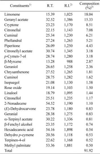
Composition of EO from chopi A colorless EO was
extracted from chopi. Among detected chemical compounds
of chopi-EO, the components (more than 0.5% amount,
91.92%) were listed in Table 1. Limonene (18.04) and geranyl acetate (15.33%) were the major constituents of
chopi-EO. Also, the considerable amounts of cryptone
(8.51), citronellal (7.08), phellandral (5.21), and piperitone (4.43%) were detected. This result was consistent with several reports on the analysis of chopi-EO (5,21,22).
However its composition varied with reports, due to the different plant part and place of origin. Although it is reported that 1,8-cineol, limonene, geranyl acetate, and myrcene were detected from chopi-EO (24), 1,8-cineol was
not detected in this report. Chopi-EO and citronellal,
geranyl acetate, and limonene, as major components, were investigated for a further study.
Antioxidant activities of chopi-EO Antioxidant activities
of chopi-EO were showed in Table 2. Chopi-EO had the
hydrogen donating activity to DPPH radical partly, and 3 EO constituents had the similar scavenging activities. DPPH radical scavenging of the reference antioxidant ascorbic acid (10µM) was found experimentally to be 92.21%. Although
all constituents in chopi-EOshowed antioxidant activities in
significant difference, it was much lower than that of ascorbic acid, a reference antioxidant. Therefore, chopi-EO
wasexpected to be partly electron donors and can react with free radicals to convert them to more stable products and terminate radical chain reactions. Also, the reducing power of chopi-EO was not detected at all.
Sometimes the cell and tissue injuries are due to the toxicity of reactive oxygen species (ROS) generated and released by activated phagocytes. NADPH oxidase in phagocytes is responsible for the production of superoxide anion and represents a major host defense mechanism of phagocytes against invading microorganisms. Superoxide initiates the formation of other ROS, such as hydroxyl radical and oxygen singlet, which are known as strong oxidant products, and hydrogen peroxide and hypochlorous acid (23). Free radical scavenging activity plays the critical role for quenching superoxide anions and maintaining the cellular redox homeostasis against harmful oxidants and free radicals in cells. In healthy blood, several enzymes, such as glutaredoxin (GRX) and glutathione reductase (GRD), and chemical compounds, such as ascorbic acid, vitamin E, and glutathione, play as antioxidant and reducing enzyme system for scavenging harmful superoxides and free radical compounds. From this result, the antioxidant activity of chopi-EO will help reduce the oxidative risk,
caused by harmful radical compounds.
Cytotoxicity of chopi-EO For murine macrophage cells
and HUVEC monolayers, chopi-EO had the significant
cytotoxicity over 50µg/mL concentration (IC50=236.1 and
411.5µg/mL, respectively) (Fig. 1). Limonene and geranly
acetate did not show any cytotoxic effect in tested concentration (0-500µg/mL) for both cell lines, whereas
citronellal affected a partly inhibition on cell viability (data not shown). However, it is reported that limonene has cytotoxicity on gastric cancer cells and leukemia cells, causing the apoptosis through the decrease of bcl-2 protein and the increase of p53 protein (24). In addition, limonene has been shown to have the carcinogenesis activity when bound to rat-specific α2u-globulin (25). From the above,
chopi-EO containing limonene and geranyl acetate is
presumed to exert the cell-specific cytotoxicity for human cancers and leukemia cells without any harmful effects on normal cells, such as macrophage cells and HUVEC monolayers.
Table 1. Chemical composition of chopi-EO
Constituents1) R.T. R.I.2) Composition
(%)3) Limonene 15.39 1,025 18.040 Geranyl acetate 32.52 1,386 15.330 Cryptone 23.23 1,170 8.51 Citronellal 22.15 1,143 7.08 Cuminal 25.34 1,230 6.21 Phellandral 27.24 1,263 5.21 Piperitone 26.09 1,250 4.43 Citronellyl acetate 30.74 1,345 3.18 p-Cymen-7-ol 28.74 1,280 3.18 β-Myrcene 13.28 0,988 2.87 Geraniol 26.65 1,258 2.36 Chrysanthenone 27.52 1,265 1.81 Cuminol 28.75 1,282 1.62 Isopuegol 21.08 1,130 1.58 Rose oxide 19.14 1,103 1.50 Linalool 18.79 1,095 1.44 Citronellol 25.51 1,235 1.17 2-Nonadecene 54.52 1,190 1.10 (E)-Dehydrocarvone 23.78 1,180 0.83 Geranial 28.38 1,275 0.83 α-Terpinyl acetate 30.22 1,336 0.81 β-Fenchyl alcohol 23.35 1,172 0.74 Hexadecanoic acid 54.16 1,898 0.54 Dehydro p-cymene 20.56 1,118 0.53 Terpinen-4-ol 22.62 1,168 0.52 Methyl palmitate 53.36 1,881 0.50 Total 91.920
1)Constituents having over 0.5% of peak area were showed in this
report.
2)Retention index were calculated against n-alkanes (C
8-C22) as
exter-nal references on HP-5MS capillary column.
3)Area composition is the average of the relative percentage of the
peak area in the MS total ion chromatogram (n=3).
Table 2. Antioxidant activity of chopi-EO and its constituents
DPPH radical scavenging
(% control) Reducing power (Abs700 nm)
Chopi-EO 018.3±2.65*1) 0.02±0.010
Citronellal 10.67±0.37* 0.01±0.001 Geranyl acetate 10.23±0.37* 0.01±0.001
Limonene 10.13±0.46* 0.01±0.001
Positive control 92.21±0.522)* 02.24±0.013)*
1)All samples were used in 500 µg/mL for antioxidant assay;
Signifi-cant difference at *p<0.05, as compared to the control.
2)Ascorbic acid (10 µM). 3)Pyrogallol (100 µg/mL).
Fig. 1. Effect of chopi-EO on cell viability. The control value for
cell viability was 99.98±5.56µg/mL. Significant difference at
*p<0.05 as compared to the control (untreated group by
Inhibition of chopi-EO on nitrite production Chopi-EO
inhibited partly LPS-induced nitrite production in a dose-dependent manner, as shown in Fig. 2. LPS stimulated nitrite production from macrophage cells, as compared to the basal. Also, L-NMMA (100µM), which is one of
selective NOS blockers, inhibited LPS-induced nitrite production significantly as a positive control. L-NMMA had suppressed approximately 82% of nitrite production of LPS-induced nitrite production to the basal. A 50µg/mL of chopi-EO showed approximately 38% decrease in nitrite
production, as compared to the LPS-induced nitrite production at a significant difference (p<0.05). Citronellal
and geranyl acetate (50µg/mL), except of limonene,
showed a partly inhibitory effect (approximately 11-14%), on nitrite production in macrophage cells to the basal. Little is known of the original copy of the inhibition of nitrite production by citronellal and geranyl acetate. NO is related to the pathophysiology of inflammation including inflammatory joint disease (26,27). In addition, NO released from endothelial cells via the endothelial nitric oxide synthase (eNOS) is a pivotal vasoprotective molecule (28) and a potent vasodilator, and possesses many antiatherogenic properties. NO decreases platelet aggregation and adhesion limits vascular smooth muscle proliferation, inhibits neointima formation,prevents monocyte chemotaxis, and inhibits leukocyte adhesion to the endothelium (29).Our results suggest that chopi-EO might
have the dual-functions for NOs biosynthesized in different cell lines. In macrophage cells, chopi-EO is expected to
reduce the production of NO, which is an inflammatory mediator, derived from iNOS reactions. Simultaneously, it is presumed that chopi-EO has no chemical quenching
effect on NO, a potent vasodilator in atherosclerosis, derived from eNOS reaction in HUVEC. As a whole, this finding show that chopi-EO,containing limonene, geranyl
acetate, and citronellal, raise the possibility that chopi-EO
may exacerbate partly the inflammatory process, giving no chemical quenching effect on eNOS-derived NO in HUVECs.
Effect of chopi-EO on chemical scavenging of NO and
suppression of iNOS gene transcription Partly inhibition of nitrite production by chopi-EO can be explained by two
ways. One explanation is by extracellularly chemical NO quenching activity of chopi-EO. Sodium nitroprusside,
which is a chemical NO donor, produces nitrite ions in aqueous solution at physiological pH. Chemical quenching of NO by chopi-EO was investigated. However, chopi-EO
and its major components showed no chemical NO quenching, whereas Hb, used as a NO scavenger (15), showed significant NO scavenging (Fig. 3A). The inhibition of nitrite production by chopi-EO was expected to be due
to suppression of iNOS in LPS-induced RAW 264.7 macrophage cells. For inhibition of nitrite production from LPS-stimulated RAW 264.7 cells, the other explanation is to suppress iNOS mRNA transcription by chopi-EO. LPS
stimulated significantly to up-regulate mRNA transcription of iNOS in RAW 264.7 cells (Fig. 3B). NMMA suppressed approximately 49% of upregulated iNOS gene transcription. Chopi-EO markedly inhibited approximately
40.4% of iNOS mRNA transcription as compared to LPS-induced group. However geranyl acetate and limonene showed upregulated-iNOS mRNA transcription on the contrary, showing over-range values (approximately 0.130 and 0.166 iNOS/GAPDH density for transcription), except of citronellal showing approximately 34% suppressive effect. NO production and iNOS expression are considered to be related to the inflammation and carcinogenesis (30, 31), and NO is involved in the pathophysiology of joint disease and plays a key role in the cartilage catabolism (26).
Inhibitory effect of chopi-EO on the cellular adhesion
The adhesion of the monocytic THP-1 cells to HUVEC monolayers acts as one of the initial steps for immunological inflammatory responses. The treatment of chopi-EO
reduced the TNF-α-stimulated adhesion of THP-1 to
HUVEC monolayers (Fig. 4). The analysis by fluorescent plate reader showed that chopi-EO (50µg/mL)reduced the
Fig. 2. Effects of chopi-EO and its constituents on NO production from LPS-stimulated RAW 264.7 macrophage cells. Significant
adhesion of calcein-AM labeled THP-1 to HUVEC monolayer to approximately 35.7%. It was expected that
chopi-EO might affect the expression of TNF-α
-upregulated E-selectin in HUVEC. In resting HUVEC monolayers, there was no signal of the basal mRNA transcription of E-selectin gene, as shown in Fig. 4. However, the transcription of E-selectin gene was significantly increased by stimulation of cytokine, TNF-α.
TNF-α-stimulated E-selectin mRNA transcriptional level
by chopi-EO (50µg/mL) was reduced to 34.8% remarkably,
compared to the control. These results were consistent with inhibitory data of cellular adhesion. In addition, geranyl acetate and limonene had approximately 53.7 and 58% of suppressive activities for E-selectin transcription. In immunological inflammatory responses, TNF-α and
interleukin family activate neutrophils and up-regulate
Fig. 3. Effects of chopi-EO and its constituents on chemical NO quenching and the suppression of iNOS mRNA transcription. A,
Chemical NO scavenging of chopi-EO in the cell-free system; B, suppression of iNOS mRNA transcription by chopi-EO in LPS-induced
RAW 264.7 macrophage cells. The intensity of PCR product bands was quantitated by scanning densitometry and standardized to equivalent GAPDH mRNA levels. Significant difference at p<0.05 as compared to *control and to #LPS-treated group.
Fig. 4. Effects of chopi-EO and its constituents on cellular adhesion. A, Inhibition of chopi-EO on monocytic THP-1 cells to HUVEC
monolayer; B, suppression of E-selectin mRNA transcription level by chopi-EO in TNF-α-stimulated HUVEC. The intensity of PCR
product bands was quantitated by scanning densitometry and standardized to equivalent GAPDH mRNA levels. Significant difference at
CAMs expression in HUVEC monolayer. Upregulated expression of CAMs protein causes the adherence of monocytes and neutrophils to blood vessel wall, especially HUVEC monolayer. These responses are closely related to immunological inflammatory diseases and atherosclerosis (16).
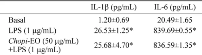
Inhibition of chopi-EO on the biosynthesis of
inflammatory mediators Additionally, chopi-EO did
not show the effect of the biosynthesis of inflammatory cytokines in LPS-induced RAW 264.7 cells (Table 3). E-selectin is constitutive, and was expressed by immunological inflammatory agent-modulation. Also, the expression of E-selectin protein is regulated by mitogen-activated protein kinases (MAPKs) and NF-κB pathways (32).
As mentioned above, the inhibitory effect of chopi-EO
on NO production was not due to cell viability or chemical NO scavenging effect. These results suggest that chopi-EO
may inhibit iNOS gene transcriptional level through it exerts its anti-inflammatory activity. Most of reports regarding the inhibition of NO production have elucidated only iNOS protein suppression without investigation of chemical NO scavenge. However the chemical quenching should be elucidated for inhibition of NO production in RAW 264.7 cells. And chopi-EO inhibited the cellular
adhesion through the suppression of E-selectin transcription level in HUVEC monolayers. Taken together, chopi-EO
may be useful as a functional food component or an alternative medicine for the relief and retardation of immunological inflammatory responses, and its action may occur through the reduction of inflammatory mediators.
Acknowledgments
This work was supported by Priority Research Centers Program through the National Research Foundation of Korea (NRF) funded by the Ministry of Education, Science and Technology (2009-0094018).
References
1. Marletta MA. Nitric oxide synthase structure and mechanism. J. Biol. Chem. 268: 12231-12234 (1993)
2. Palmer RM, Ashton DS, Moncada S. Vascular endothelial cells synthesize nitric oxide from L-arginine. Nature 333: 664-666 (1988)
3. Xie Q-W, Cho HJ, Calaycay J, Mumford RA, Swiderek KM, Lee TD, Ding A, Troso T, Nathan CF. Cloning and characterization of inducible nitric oxide synthase from mouse macrophages. Science 256: 225-228 (1992)
4. Hibbs JB, Taintor RR, Vavrin Z. Macrophage cytotoxicity: Role for
L-arginine deiminase and imino nitrogen oxidation in nitrite.
Science 235: 473-476 (1987)
5. Chung MS. Volatile compounds of Zanthoxylum piperitum A.P. DC.
Food Sci. Biotechnol. 14: 529-532 (2005)
6. Caldefie-Chezet F, Guerry M, Chalchat JC, Fusillier C, Vasson MP, Guillot J. Anti-inflammatory effects of Melaleuca alternifolia
essential oil on human polymorphonuclear neutrophils and monocytes. Free Radical Res. 38: 805-811 (2004)
7. Hart PH, Brand C, Carson CF, Riley TV, Prager RH, Finlay-Jones JJ. Terpinen-4-ol, the main component of the essential oil of
Melaleuca alternifolia (tea tree oil), suppresses inflammatory mediator production by activated human monocytes.Inflamm. Res. 49: 619-626 (2000)
8. Fiebich BL, Mueksch B, Boehringer M, Hull M. Interleukin-1β
induces cyclooxygenase-2 and prostaglandin E2 synthesis in human neuroblastoma cells: Involvement of p38 mitogen-activated protein kinase and nuclear factor-κB. J. Neurochem. 75: 2020-2028 (2000) 9. Silverman MD, Zamora DO, Pan Y, Texeira PV, Planck SR, Rosenbaum JT. Cell adhesion molecule expression in cultured human iris endothelial cells. Invest. Ophth. Vis. Sci. 42: 2861-2866 (2001)
10. Gómez NE, Witte L. A simple method to extract essential oils from tissue samples by using microwave radiation. J. Chem. Ecol. 27: 2351-2359 (2001)
11. Van den Dool H, Kratz PD. A generalization of the retention index system including linear temperature programmed gas-liquid partition chromatogram. J. Chromatogr. 11: 463-471 (1963)
12. Kondjoyan N, Berdague JL. A Compilation of Relative Retention for the Analysis of Aromatic Compounds. Laboratorie flaveur, de Recherches sur la Viande. Clemont-Ferrand, France. pp. 15-138 (1996)
13. Chattopadhyay S, Bhaumik S, Purkayastha M, Basu S, Chaudhuri AN, Gupta SD. Apoptosis and necrosis in developing brain cells due to arsenic toxicity and protection with antioxidants. Toxicol. Lett. 136: 65-76 (2002)
14. Babu BH, Shylesh BS, Padikkala J. Antioxidant and hepatoprotective effect of Acanthus ilicifolius. Fitoterapia 72: 272-277 (2001) 15. Wei T, Chen C, Hau J, Zhao B, Xin W, Mori A. The antioxidant
EPC-K1 attenuates NO-induced mitochondrial dysfunction, lipid peroxidation, and apoptosis in cerebellar granuele cells. Toxicology 134: 117-126 (1999)
16. Lee JH, Choi SI, Lee YS, Kim GH. Antioxidant and anti-inflammatory activities of Allium victorialis subsp. platyphyllum
extracts. Food Sci. Biotechnol. 16: 796-801 (2007)
17. Ahn KS, Noh EJ, Zhao HL, Jung SH, Kang SS, Kim YS. Inhibition of inducible nitric oxide synthase and cylooxygenase II by
Platycodon grandiflorum saponins via suppression of nuclear
factor-κB activation in RAW 264.7 cells. Life Sci. 76: 231-2328 (2005)
18. Sasaki M, Ohara-Nemoto Y, Tajika S, Kobayashi M, Yamaura C, Kimura S. Antigenic characterization of a novel Streptococcus anginosus antigen that induces nitric oxide synthesis by murine peritoneal exudates cells. J. Med. Microbiol. 50: 952-958 (2001) 19. Choi JS, Choi YJ, Park SH, Kang JS, Kang YH. Flavones migrate
tumor necrosis factor-α-induced adhesion molecule upregulation in
cultured human endothelial cells: Role of nuclear factor-κB. J. Nutr.
134: 1013-1019 (2004)
20. Rasmussen LM, Hansen PT, Nabipour MT, Olesen P, Kristiansen MT, Ledet T. Diverse effects of inhibition of 3-hydroxy-3-methylglutaryl-CoA reductase on the expression of VCAM-1 and E-selectin in endothelial cells. Biochem. J. 360: 363-370 (2001) 21. Kim JH, Lee KS, Oh WT, Kim KR. Flavor components of the fruit
peel and leaf oil from Zanthoxylum piperitum DC. Korean J. Food Sci. Technol. 21: 562-568 (1989)
22. Chung MS. Compositional changes in essential oil of Zanthoxylum piperitum A.P. DC during storage. Korean J. Food Culture 21: 433-438 (2006)
23. Babior BM. The respiratory burst oxidase. Adv. Enzymol. RAMB 65: 49-95 (1992)
24. Lu XG, Feng BA, Zhan LB, Yu ZH. D-Limonene induces apoptosis
of gastric cancer cells. Zhongguo Shi Yan Xue Ye Xue Za Zhi 25: 325-327 (2003)
25. Whysner J, Williams GM. D-Limonene mechanistic data and risk
assessment: Absolute species-specific cytotoxicity, enhanced cell proliferation, and tumor promotion. Pharmacol. Therapeut. 71: 127-136 (1996)
Table 3. Effect of chopi-EO on the biosynthesis of cytokines
IL-1β (pg/mL) IL-6 (pg/mL)
Basal 1.20±0.69 020.49±1.65*
LPS (1 µg/mL) 26.53±1.25* 839.69±0.55* Chopi-EO (50 µg/mL)
+LPS (1 µg/mL) 25.68±4.70* 836.59±1.35*
26. Belmont HM, Levartovsky D, Goel A, Amin A, Giorno R, Rediske J, Skovron ML, Abramson SB. Increased nitric oxide production accompanied by the up-regulation of inducible nitric oxide synthase in vascular endothelium from patients with systemic lupus erythematosus. Arthritis Rheum. 40: 1810-1816 (1997)
27. Amin AR, Attur M, Abramson SB. Nitric oxide synthase and cyclooxygenases: Distribution, regulation, and intervention in arthritis. Curr. Opin. Rheumatol. 11: 202-209 (1999)
28. Leikert JF, Räthel TR, Wohlfart P, Cheynier V, Vollmar AM, Dirsch VM. Red wine polyphenols enhance endothelial nitric oxide synthase expression and subsequent nitric oxide release from endothelial cells. Circulation 106: 1614-1617 (2002)
29. Cauin-Glaser T, Garcia-Cardena G, Sarrel P, Sessa WC, Bender JR. 17β-Estradiol regulation of human endothelial cell basal nitric oxide
release, independent of cytosolic Ca2+ mobilization. Circ. Res. 81:
885-892 (1997)
30. Mordan LJ, Burnett TS, Zhang LX. Inhibitors of endogenous nitric oxide formation block the promotion of neoplastic transformation in C3H10T1/2 fibroblasts. Carcinogenesis 14: 1555-1559 (1993) 31. Chan MM, Huang HI, Fenton MR, Fong D. In vivo inhibition of
nitric oxide synthase gene expression by curcumin, a cancer preventive natural product with anti-inflammatory properties. Biochem. Pharmacol. 55: 1955-1962 (1998)
32. Lee CW, Lin WN, Lin CC, Luo SF, Wang JS, Pouyssegur J, Yang CM. Transcriptional regulation of VCAM-1 expression by tumor necrosis factor-α in human tracheal smooth muscle cells:
Involvement of MAPKs, NF-κB, p300, and histone acetylation.J.