A THESIS
FOR THE DEGREE OF DOCTOR OF PHYLOSOPHY
MOLECULAR INSIGHT INTO THE IMMUNE SYSTEM OF
THE DISK ABALONE (Haliotis discus discus): cDNA
MICROARRAY, TRANSCRIPTIONAL PROFILING AND
FUNCTIONAL ASPECTS
Mahanama De Zoysa
Department of Aquatic Life Medicine
GRADUATE SCHOOL
JEJU NATIONAL UNIVERSITY
2010-02
MOLECULAR INSIGHT INTO THE IMMUNE SYSTEM OF
THE DISK ABALONE (Haliotis discus discus): cDNA
MICROARRAY, TRANSCRIPTIONAL PROFILING AND
FUNCTIONAL ASPECTS
Mahanama De Zoysa
(Supervised by Professor Jehee Lee)
A thesis submitted in partial fulfillment of the requirement for the degree of
DOCTOR OF PHILOSOPHY
2010-02
The thesis has been examined and approved by
Thesis Director, Gi-Young Kim, Professor of Marine Biomedical Sciences
Choon-Bok Song, Professor of Marine Biomedical Sciences
Moon-Soo Heo, Professor of Marine Biomedical Sciences
Joon Bum Jeong, Professor of Marine Biomedical Sciences
Jehee Lee, Professor of Marine Biomedical Sciences
2010-01-01
Department of Aquatic Life Medicine Date
GRADUATE SCHOOL
JEJU NATIONAL UNIVERSITY
REPUBLIC OF KOREA
CONTENTS
요약문 II
SUMMERY VI
LIST OF FIGURES X
LIST OF TABLES XII
ABBRIVIATIONS XIII
GENERAL INTRODUCTION 01
OUT LINE OF THE STUDY 08
CHAPTER 1: cDNA microarray analysis of bacteria and VHS virus challenged disk abalone Haliotis discus discus
09
ABSTRACT 10
1.1 INTRODUCTION 11
1.2 MATERIALS AND METHODS 13
1.3 RESULTS AND DISCUSSION 21
CHAPTER 2: Molecular and functional characterization of molluskan TNF-α and Fas ligand genes from disk abalone
56
2.1 INTRODUCTION 58
2.2 MATERIALS AND METHODS 59
2.3 RESULTS AND DISCUSSION 64
CHAPTER 3: Immune regulatory transcription factors from disk abalone: lipopolysaccharide-induced TNF-α factor (LITAF) and Rel family nuclear factor kappa B (Rel/NF-kB)
90
3.1 INTRODUCTION 92
3.2 MATERIALS AND METHODS 94
3.3 RESULTS AND DISCUSSION 95
CHAPTER 4: Antimicrobial peptides (AMPs) from disk abalone: defensin and “Abhisin” a histone H2A derived AMP
109
4.1 INTRODUCTION 111
4.2 MATERIALS AND METHODS 113
4.3 RESULTS AND DISCUSSION 116
CHAPTER 5: Disk abalone antioxidant enzymes and immune responses against bacteria and VHSV challenge
142
5.1 INTRODUCTION 144
5.2 MATERIALS AND METHODS 147
5.3 RESULTS AND DISCUSSION 148
II 요약문 전복의 초기 면역 시스템은 다른 무척추동물들에서 처럼 주된 방어체계로 사 용된다. 따라서, 분자적 수준에서 다양한 미생물의 공격에 대한 면역 응답 및 그 기작을 이해하는 것이 중요하다. 본 연구에서는, 까막전복으로부터 면역에 관련된 유전자들을 분자적 수준에서 이해하기 위해 cDNA microarray, 전사 profiling 및 각각의 단백질들에 대한 기능적 특성 분석을 수행함으로써 전복의 면역 응답 체계를 이해하고자 하였다.
본 연구에서는, 까막전복의 cDNA library로부터 4.2 K의 cDNA chip을 제작하였
고, cDNA microarray를 이용한 mRNA 차원에서의 전사 수준 분석은 까막전복에 혼합 박테리아 (Vibrio alginolyticus, Vibrio parahemolyticus, Listeria monocytogenes) 50 μL (5x10⁷ cells/mL)와 viral hemorrhagic septicemia virus (VHSV) 50 μL (1 X 10⁸
pfu/ml)를 각각 주입하여 면역 공격 실험을 통해 수행하였다. 면역 공격에 대한 전사 응답 반응은 박테리아와 VHSV의 공격 실험 후 24시간째에 아가미, 소화 관, 그리고 혈구에서 조사하였다. 박테리아가 공격된 전복에서는, 아가미에서 68개 (1.6%)와 소화관에서112개 (2.7%)의 전사 상의 변화를 확인함으로써 발현 수준의 의미있는 변화(≥2 or ≤2 배)를 관찰할 수 있었다. 본 연구의 결과에서는 박테리아 공격에 대한 전복의 초기 면역 반응과 관련하여, 전사인자들 또는 그들의 활성인자들 (KLF, NFIL-3, IK-B), 염증과 apoptosis와 관련된 단백질들 (AIF, TNF-α, archeron), cytokine들 (IFN-44-like, SOCS-2)과 항산화 효소들 (glutathione S transferase, TRx-2, TPx)이 활
성화 될 수 있다는 것을 보여주었다.
VHSV 공격 실험에서는 PBS를 대조구로 했을 때와 비교하여 아가미와 혈구에 서 280개(6.6%)의 의미있는 발현수준의 변화(≥1.5 or ≤1.5배)를 보였다. 아가미와 혈구에서 각각 88개와 65개 유전자의 발현이 증가하였다. 확인된 유전자들은 염증과 apoptosis 관련 유전자 (TNF super family, Fas ligand), IFN 조절 단백질 (IFN-44 like, IFN inducible GTPase), 전사 인자 (C-jun, NFIL-3), 해독 단백질 (glutathione peroxidase)과 같이 면역 기능에 따라 분류하였다. 전사가 조절된 유 전자들의 상당수는 기능이 알려져 있지 않거나 GenBank에서 유사한 유전자들을 찾을 수 없었지만, 전복에서의 면역시스템에 관련하는 새로운 유전자들일 가능 성을 보인다. TNF super-family에 속하는 단백질들은 염증 반응 조절, apoptosis 및 면역 조 절들에 관여하는 다양한 역할을 한다. 그들 사이에서, TNF-α 와 Fas ligand들은
다양한 기능적 면역 조절자처럼 여겨져 왔다. 까막전복으로부터 TNF-α 와 Fas l igand를 코딩하는 유전자들을 AbTNF-α 와 AbFas ligand로 각각 명칭하였으며, microarray 결과들에서 전사 수준이 증가함을 보였다. AbTNF-α 와 Fas ligand의 아미노산 서열들은 TNF family signature와 N-terminal transmembrane domain들을 가지고 있었다. 계통 분석결과, AbTNF-α 와 Fas ligand는 다른 무척추동물의 것과 가까운 관계를 보였다. qRT-PCR 결과, AbTNF-α 와 AbFas ligand는 전복의 혈구, 아가미, 외투막, 근육, 소화관, 간췌장에서 지속적으로 발현되는 것을 확인할 수 있었으며, 박테리아, VHSV와 LPS 공격 실험에 의한 AbTNF-α 와 AbFas ligand의 전사 수준은 아가미와 혈구에서 의미있는 증가(p<0.05)를 보였다. AbTNF-α 와 AbFas ligand 를 코딩하는 유전자는 발현 벡터에 재조합하여, Escherichia coli 에서 재조합 단백질의 과잉 생산을 유도하였고, pMAL protein fusion system을 이용하여 재조합된 단백질을 정제하였다. 재조합 AbTNF-α 와 AbFas ligand 단백질은 전복
의 혈구세포와 인간의 THP-1 세포에서 superoxide anion (O-2) 유도에 의해 생물
학적 활성을 나타내었다. 또한, 몇 가지 면역 유전자들은 (defensin, SOCS-2, NF-kB) 전복의 혈구세포에 재조합 AbTNF-α 와 AbFas ligand를 처리했을 때 발현 수준이 다르게 나타나는 것을 볼 수 있었다. 두 재조합 단백질의 생물학적 활 성을 통해, 박테리아, 바이러스, LPS의 공격 때와는 반대로 AbTNF-α와 AbFas ligand의 전사 수준이 증가함을 봤을 때, 우리는 미생물 주입했을 때 ROS 처럼
O-2 유도에 의해 전복의 TNF-α 와 Fas ligand가 응답할 수 있다는 것을 제안 할
수 있다.
LPS에 유도되는 TNF-α factor (LITAF)와 Rel family nuclear factor kappa B (Rel/NF-kB)는 염증 cytokine들과 apoptosis, 면역 과련 유전자들의 조절에 있어 두 가지 중요한 작용을 하는 전사 요소들이다. 최근 연구에서는 전복의 LITAF (AbLITAF)
와 Rel/NF-kB (AbRel/NF-kB) 상관 관계와 그들의 면역 응답에 대한 연구가 진행
되었다. AbLITAF의 염기서열 분석결과 147개의 아미노산으로 구성되고 두 개의
CXXC motifs (82CPHC85 and 134CPNC137)를 포함하는 LITAF (Zn+2) binding domain이 확
인되었다. 그리고, 계통분석 결과 AbLITAF가 LITAF family 단백질에 속하는 것을
확인할 수 있었다. AbRel/NF-kB는 무척추동물과 척추동물의 것과 유사한 Rel
homology domain (RHD), Rel protein signature (107FRYECR112), DNA binding motif
(103RGLRFFRYEC101), nuclear localization signal (NLS)과 transcription factor
immunoglobulin–like fold (TIG) 같은 다수의 domain을 포함하고 있었다. 조직 특
IV 투막을 제외한 모든 조직에서 더 높게 나타났다. 다른 연체동물들과 본 연구의 전복 TNF-α, LITAF, Rel/NF-kB 데이터로부터 종합된 결과들은, 연체동물 문 전체 에 걸쳐 일어날 수 있는 LITAF와 NF-kB의 독립적인 방향을 강하게 제시할 수 있다. 본 연구의 4장에서는 병원체 감염에 대한 전복의 초기 면역 반응에 중요한 구성 요소로 알려진 defensin과 histone H2A로 명명된 두 개의 항균펩타이드들을 암호화하는 유전자에 대해서 서술한다. 전복의 defensin (pro-defensin)은 66개의 아미노산을 암호화하는 198 bp의 염기서열로 구성된다. 무척추동물의 최근 연구 에서 3차 구조 상에 defensin family domain, 6개의 cysteine 잔기의 배열 그리 고 그들의 이황화 결합 (C1-C4, C2-C5, C3-C63), alpha helix 구조가 보여졌고, 우 리는 전복의 defensin이 다른 무척추동물 defensin의 새로운 member임을 계통적 관계를 통해 제시하였다.
자극을 주지 않은 전복에서, defensin의 전사 수준은 혈구세포, 아가미, 외투막, 근육, 소화관 그리고 간췌장을 포함하는 모든 실험된 조직들에서 지속적으로 발 현되어졌다. 또한, V. alginolyticus, V. parahemolyticus와 L. monocytogenes 같은 박테 리아 공격 실험 상에서도 전복 defensin의 전사 수준은 혈구세포, 아가미, 소화 관에서 의미있게 유도되어졌다. 4장의 두 번째 절에서는 전복으로부터 histone H2A cDNA 유전자를 클로닝하였고, 전복의 histone H2A의 N말단 서열로부터 “Abhisin”를 지칭하는 40개의 아미노산 항균펩타이드를 서열을 확인하였다. Abhisin는 net positive charge (+13), 높은 소수성 잔기들(27%)과2.82 Kcal/mol protein binding potential을 가지는 항균펩타이드들의 특징적인 점을 보여준다. 본 연구에서 합성된 abhisin 을 250 µg/mL 처리했을 때 박테리아 계통인 L.
monocytogenes, V. ichthyoenteri와 fungi 계통인 Pityrosporum ovale에 대해 성장을
저해하는 것으로 확인되었다. 하지만, 그람 음성균보다 그람 양성균에 대해 더
강한 활성을 나타내는 것으로 보여졌다. 부가적으로, P. ovale를 대상으로 abhisin
을 처리했을 때의 손상 정도를 주사 전자 현미경 (SEM)을 통해 관찰 하였다.
흥미로운 점은, THP-1 leukemia cancer cell에 abhisin을 50 µg/mL 처리했을 때 생 존 능력이 약 25% 감소하는 것을 확인할 수 있었고, 일반 vero cell에는 영향을
나타내지 않았다. 이 결과를 통해 abhisin이 일반 cell보다 cancer cell에 세포
독성을 가진다고 할 수 있다. 그리고, 전복에 박테리아 공격 실험을 수행했을
때 아가미와 소화관에서 Histone H2A 전사 수준이 의미있게 유도되어졌음을
qRT-PCR 결과를 통해 확인할 수 있었다. 종합적인 결과를 봤을 때 defensin과 histone H2A 전구체 또는 그것의 N-말단 아미노산 (abhisin)이 까막 전복에서 강
력한 항균펩타이드들이고, 그들은 감염들에 대한 보호체계를 증가시키는 면역 방어 반응에 포함된다고 할 수 있다. ROS는 침입된 병원체들에 대한 무척추동물의 가장 중요한 숙주 방어 요소 중 하나이다. 항산화 인자는 과도한 ROS에 의해 생성되는 산화적 스트레스에 대한 잠재적인 indicator이다. 따라서 ROS 수준의 주입 상태에 따른 항산화 효 소들의 전사 수준은 산화적 스트레스에 대한 indicator로 사용할 수 있다. 미생 물의 주입에 대해 관계하는 항산화 효소들의 변화를 이해하기 위해, 박테리아와 VHSV 공격 실험 후에 전복 항산화 효소들 [Mn-superoxide dismutase (Mn-SOD); CuZn-superoxide dismutase (CuZn-SOD); catalase; thioredoxin peroxidase (TPx); mitochondrial thioredoxin-2 (Mt-TRx-2); Selenium dependant glutathione peroxidases (SeGPx)]의 전사 수준을 분석하였다. 그 결과, Mn-SOD, SeGPx에서 면역 공격 실 험 동안 각각 다른 시점에 뚜렷한 변화를 확인할 수 있었다. 더 나아가, 그것은 전복에서 항산화 효소가 박테리아와 VHSV 공격에 대해 식균 작용 활성과 다른 초기 면역 방어 응답으로 유도될 수 있다는 것을 제시할 수 있다.
본 연구에서는 전복의 cDNA microarray 분석 결과를 가지고 ESTs에 결합하여 접근함으로써 박테리아와 VHSV 공격 실험 상에서 방어 작용에 관계되는 전복 의 유전자들을 탐색하였다. 이 접근 방식은 다른 병원체들에 대한 전복의 방어 체계에 관계된 유전자들의 확인을 위한 좋은 잠재력을 지닌다. 현재까지 연구에 서는 전복의 초기 면역 시스템에 대한 다른 숙주 방어 인자들 하에 다양한 유
전자 집단을 보여주었다. 전복의 면역에 관련된 유전자들과 그 단백질들은 염증
및 apoptosis 조절자, 전사 인자들인 NF-kB, LITAF, NFIL-3 C-Jun, cytokine 조절 인 자인 SOCS-2와 IFN-44, 항균펩타이드 같은 defensin과 histone H2A 그리고 항산 화 효소 등을 포함한다. 결론적으로, 본 연구는 척추동물과 무척추동물간에 면
역 기능에 관련된 유전자들의 진화적 관계를 규명하는 단초를 제공한다. 선택된
면역 관련 유전자들에 대해서는 이후에 전복의 면역 방어 기작에 대한 연구를
VI
Summary
The innate immune system is the main host defense in abalone like invertebrates. Hence, it is important to understand the immune responses and their mechanisms against different microbial challengers at molecular level. In this study, molecular analysis of immune genes in disk abalone, Haliotis discus discus was investigated using cDNA microarray, transcriptional profiling and functional properties of respective proteins.
An abalone 4.2 K cDNA microarray (cDNA chip) was manufactured using the abalone cDNA library clones (ESTs). The cDNA microarray analysis was performed with two immune challenge experiments by injecting 50 mL (5x10⁷cells/mL) of bacteria mixture
(Vibrio alginolyticus, V. parahemolyticus, and Listeria monocytogenes) and 50 μL (1 X 10⁸
pfu/ml) of viral hemorrhagic septicemia virus (VHSV). Transcriptional response of abalone gills, digestive tract, and hemocytes was investigated at 24 h post challenge of bacteria and VHSV. In bacteria challenged abalone, 68 (1.6%) and 112 (2.7%) transcripts were significantly (≥2 or ≤2 –fold) changed in their expression levels in gills and digestive tract, respectively. There were 46 tissue-specific transcripts that were up-regulated only in digestive tract. In contrast, only 13 transcripts showed gill-specific up-regulation. Our results showed that challenge of bacteria, may activate the transcription factors or their activators (KLF, NFIL-3 and Ik-B), inflammatory and apoptosis-related proteins (AIF, TNF-α, archeron), other cytokines (IFN-44-like, SOCS-2) and antioxidant enzymes (glutathione S transferase, TRx-2 and TPx) as a part of the innate immune responses of abalone.
Upon VHSV challenge, 280 (6.6%) transcripts were significantly (≥1.5 or ≤1.5 – fold) changed in their expression level of gills and hemocytes compared to respective PBS controls. Total of 88 and 65 genes were up-regulated in gills and hemocytes, respectively. Identified genes were grouped under different immune-functional categories such as inflammatory and apoptosis related genes (TNF super family, Fas ligand), IFN regulatory proteins (IFN-44 like, IFN inducible GTPase), transcription factors (C-jun, NFIL-3) and detoxification proteins (glutathione peroxidase). Considerable number of regulated transcripts were matched with either hypothetical (un-known) sequences or showed no GenBank match (no hit) suggesting that those immune responded transcripts may represent novel genes related to immune system in abalone.
Members of TNF super-family play diversified roles in mediating the inflammatory responses, apoptosis and immune regulation. Among them, TNF-α and Fas ligands are considered as multifunctional immune modulators. The gene encoding TNF-α and Fas ligand,
which showed up-regulation in microarray results were isolated from abalone cDNA library, denoted as the AbTNF-α and AbFas ligand, respectively. The AbTNF-α and Fas ligand amino acid sequences showed their characteristic TNF family signature and N-terminal transmembrane domains. Phylogenic analysis results showed that AbTNF-α and Fas ligand were closely related with their invertebrate counterparts. qRT-PCR results showed that AbTNF-α and AbFas ligand transcripts were constitutively expressed in abalone hemocytes, gills, mantle, muscle, digestive tract and hepatopancrease in a tissue-specific manner. Transcription level of AbTNF-α and AbFas ligand was significantly (p<0.05) up-regulated in gills and hemocytes by bacteria, VHSV and LPS challenge. The recombinant AbTNF-α and AbFas ligand proteins were over-expressed in Escherichia coli (E. coli) and purified using a pMAL protein fusion system. Recombinant AbTNF-α and AbFas ligand showed its biological activity by inducing O
-2 in abalone hemocytes and human THP-1 cells. Also, several immune
genes such as defensin, SOCS-2, NF-kB have shown marked differences at transcriptional level when abalone hemocytes treated with recombinant AbTNF-α and AbFas ligand. Correlating the transcriptional up-regulation of AbTNF-α and AbFas ligand against bacteria, virus and LPS challenge with the biological activity of two recombinant proteins, we could suggest that the abalone TNF-α and Fas ligand may response to microbial infection by inducing O
-2 like ROS.
LITAF and Rel/NF-kB are two important transcription factors which play major role in the regulating inflammatory cytokines, apoptosis and immune related genes. Present study describes the discovery of abalone LITAF (AbLITAF) and Rel/NF-kB (AbRel/NF-kB) homologues and their immune responses. Analysis of AbLITAF sequence shows that it shares characteristic LITAF (Zn+2) binding domain with two CXXC motifs (82CPHC85 and 134CPNC137) with putative peptide of 147 aa. Phylogenetic analysis results further confirms
that AbLITAF is a member of LITAF family proteins. AbRel/NF-kB shares numerous signature motifs such as RHD, Rel protein signature (107FRYECR112), DNA binding motif (103RGLRFFRYEC101), NLS and transcription factor TIG fold similar to their invertebrate
and vertebrate counterparts. Tissue specific analysis results showed that both AbLITAF and AbRel/NF-kB mRNA is expressed ubiquitously in all selected tissues in constitutive manner. However, constitutive expression of AbLITAF was higher than AbRel/NF-kB in all tissues except mantle. Upon immune challenged by bacteria and VHSV, AbLITAF showed the significant up-regulation in gills and hemocytes while AbRel/NF-kB transcription was not
VIII
TNF-α, LITAF, Rel/NF-kB from abalone provide strong evidence that LITAF and NF-kB are independent pathways likely to occur throughout the Phylum mollusc.
In the fourth chapter, it describes the gene-encoded two AMPs namely defensin and histone H2A derived AMP as important components of the abalone innate immune response against pathogen invasion. Abalone defensin (pro-defensin) consists of 198-bp coding sequence of putative 66 aa acids which includes the 48 aa mature peptide. The present of invertebrate defensin family domain, arrangement of six cysteine residues and their disulfide linage in C1-C4, C2-C5, and C3-C6, alpha helix in three dimensional structure and phylogenetic
relationship suggest that abalone defensin could be a new member of invertebrate defensin family closely related to arthropod defensins. In non-stimulated abalone, defensin transcripts were constitutively expressed in all examined tissues including hemocytes, gills, mantle, muscle, digestive tract and hepatopancreas. Also, abalone defensin transcripts were significantly induced in hemocytes, gills and digestive tract upon bacterial challenge containing V. alginolyticus, V. parahemolyticus and L. monocytogenes.
In the second section of chapter four, describes a 40-amino acid AMP designated as “Abhisin” identified from the N-terminus of the abalone histone H2A. Abhisin displays characteristic features of AMPs including net positive charge (+13), higher hydrophobic residues (27%) and 2.82 Kcal/mol protein binding potential. Our results showed that growth inhibition of L. monocytogenes, V. ichthyoenteri bacteria, and fungi (yeast) Pityrosporum
ovale by synthetic abhisin at 250 µg/mL. However, stronger activity was displayed against
the Gram positive than negative bacteria. Additionally, SEM observation results confirmed that P. ovale cells were damaged by abhisin treatment. Interestingly, abhisin treatment (50 µg/mL) decreased the viability of THP-1 leukemia cancer cells approximately by 25% but there was no effect on the normal vero cells, suggesting that abhisin has cytotoxicity against cancer cells than normal cells. qRT-PCR results revealed that histone H2A transcription was significantly induced after bacteria challenge in abalone gills and digestive tract. Our overall results suggest that defensin and precursor histone H2A or its N-terminal peptide (abhisin) are potent AMPs in disk abalone that could involve in immune defense reactions to increase protection against infections.
ROS are highly microbicidal and considered as one of the most important components of host defense of invertebrates against invading pathogens. Antioxidants are potential indicators of oxidative stress due to excessive ROS. Hence, transcriptional responses of antioxidant enzymes could be used as an indicator of oxidative stress (ROS
level) as well as infectious status. In order to understand the change of antioxidant enzymes in relation to microbial infection, abalone antioxidant enzymes (Mn-SOD, CuZn-SOD, catalase, TPx, Mt-TRx-2, and SeGPx) transcriptional responses were analyzed after bacteria and VHSV challenge. Results showed that several antioxidant enzymes such as Mn-SOD, catalase, SeGPx, were induced but the responses were shown marked differences at different time points during the immune challenge. Furthermore, it could be suggested that bacteria and VHSV challenge may induce the oxidative stress associated with the activation of phagocytosis and other innate immune defense responses in abalone.
In conclusion, the approach to combine the abalone ESTs with cDNA microarray analysis has been resulted for the screening of abalone genes involved in the defense reaction upon a bacteria and VHSV challenge. This approach has good potential for identifying genes as immune markers involved in abalone defenses against different pathogens. Present study clearly showed abalone innate immune system has a number of diversified gene families under different host defense components. Genes and their proteins involved in the disk abalone immunity include sets of TNF-α, Fas ligand, caspase like inflammatory and apoptosis regulators; NF-kB, LITAF, NFIL-3 C-Jun like transcription factors; SOCS-2 and IFN-44 like cytokine regulators; defensin and histone H2A like AMPs and classical set of enzymatic antioxidant genes. Final outcome of this study provides the linkage of evolutional relationship of immune functional genes between invertebrates and vertebrates with better insight into abalone immune defense as a highly complex and diversified system. Further functional studies on these selected immune response genes involved in the abalone host defense mechanisms have to be initiated in future.
X
LIST OF FIGURES
Figure 1: Schematic diagram of cDNA microarray principle for gene expression
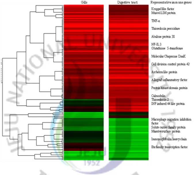
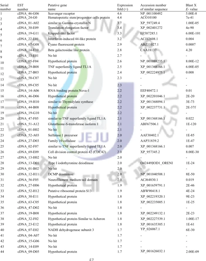
Figure 2: Gene expression profiles of significantly regulated transcripts in abalone gills and digestive tract at 24 h after bacterial challenge
Figure 3: Classification of significantly responded transcript numbers in abalone after bacterial challenge
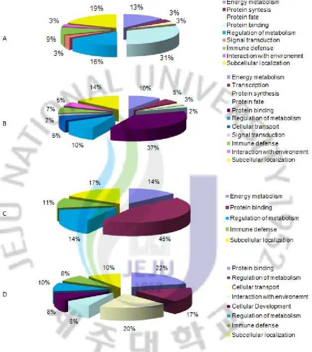
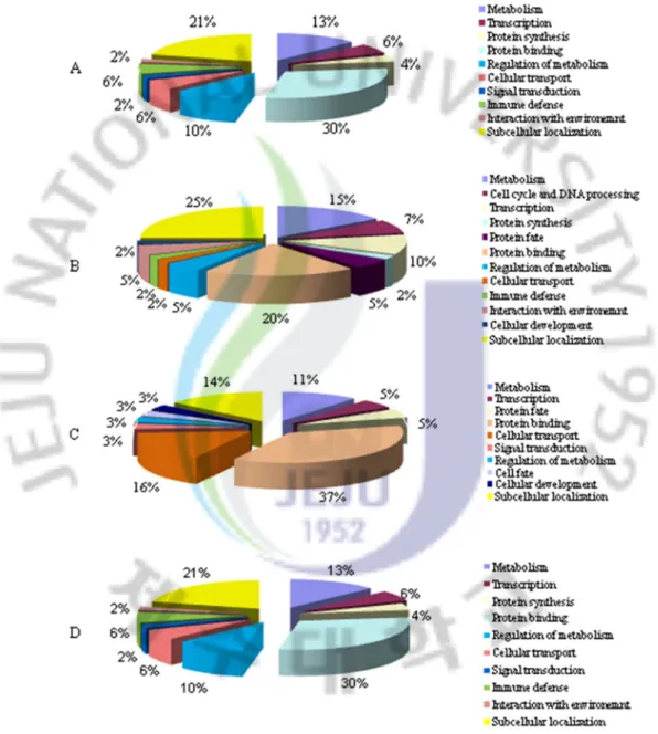
Figure 4: GO analysis of significantly regulated transcripts in abalone at 24 h after bacterial challenge
Figure 5: qRT-PCR analysis of potential (candidate) immune genes identified in the microarray analysis in abalone gills and digestive tract after bacterial challenge Figure 6: Gene expression profiles of significantly regulated transcripts in abalone gills and
hemocytes at 24 h after VHSV challenge
Figure 7: Classification of significantly responded transcript numbers in abalone after bacterial challenge
Figure 8: GO analysis of significantly regulated transcripts in abalone at 24 h after VHSV challenge
Figure 9: qRT-PCR analysis of potential (candidate) immune genes identified in the microarray analysis in disk abalone gills and hemocytes after VHSV challenge Figure 10: Schematic diagram of cloning of AbTNF-α and AbFas ligand coding sequences
into pMAL-c2X
Figure 11: The nucleotide and deduced amino acid sequences of the abalone TNF-α cDNA Figure 12: Phylogenetic analysis of disk abalone TNF-α with selected vertebrate and
invertebrate TNF superfamily ligands
Figure 13: Tissue expression profile and transcriptional responses of AbTNF-α after bacteria and VHSV challenge by qRT-PCR
Figure 14: Over expression and purification of recombinant TNF-ά fusion protein
Figure 15: Analysis of intracellular ROS generation (represented by O-2) by recombinant
abalone TNF-ά
Figure 16: Recombinant TNF-ά induced immune response genes in abalone hemocytes Figure 17: The complete nucleotide and deduced amino acid sequences of the abalone Fas
ligand cDNA
Figure 18: Phylogenetic analysis of disk abalone Fas ligand amino acid sequence with other known Fas ligand, TNF-α and Lymphotoxin-α sequences
Figure 19: Tissue expression analysis and transcriptional responses of AbFas ligand after bacteria and VHSV challenge by qRT-PCR
Figure 21: Analysis of intracellular ROS generation (represented by O-2) by recombinant
abalone Fas ligand
Figure 22: Recombinant Fas ligand induced immune response genes in abalone hemocytes Figure 23: The nucleotide and deduced amino acid sequences of abalone LITAF
Figure 24: Phylogenetic relationship of abalone LITAF
Figure 25: The nucleotide and deduced amino acid sequences of abalone Rel/NF-kB gene Figure 26: Alignments of AbRel/NF-kB with characteristic motifs of other Rel proteins Figure 27: Phylogenetic relationship of abalone Rel/NF-kB
Figure 28: Tissue expression profile of abalone LITAF and Rel/NF-kB
Figure 29: Transcriptional responses of AbLITAF and AbRel/NF-kB after bacteria and VHSV challenge by qRT-PCR
Figure 30: The nucleotide and deduced amino acid sequences of the abalone defensin cDNA Figure 31: Multiple sequence alignment of the abalone defensin mature peptide
Figure 32: The predicted three-dimensional defensin structure of abalone and mosquito
Anopheles gambiae
Figure 33: Phylogenetic relationship of abalone defensin
Figure 34: Tissue expression profile and transcriptional responses of abalone defensin after bacteria challenge by qRT-PCR
Figure 35: The nucleotide and deduced amino acid sequences of the abalone histone H2A cDNA
Figure 36: ClustalW multiple alignment of abhisin with known H2A derived AMPs Figure 37: Predicted α-helical secondary structure of abalone abhisin
Figure 38: Comparison of antimicrobial activities of synthetic abhisin against L.
monocytogenes, V. ichthyoenteri, and P. ovale
Figure 39: Scanning electron microscope image of P. ovale after treated with synthetic abhisin peptide
Figure 40: Transcriptional responses of abalone histone H2A after bacterial infection Figure 41: Effect of synthetic abhisin peptide on cell viability
Figure 42: Transcriptional analysis of abalone antioxidant enzymes in gills
Figure 43: Transcriptional responses of abalone antioxidant enzymes after bacteria and VHSV challenge by qRT-PCR
XII
LIST OF TABLES
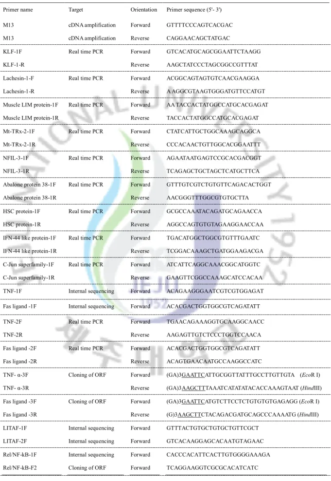
Table 1: Diseases in abalone aquaculture; a summery of recent reports Table 2: Description of the gene specific primers used in microarray analysis
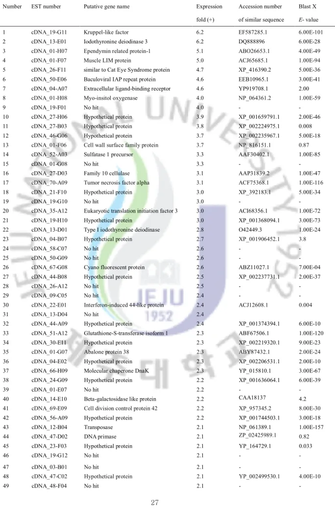
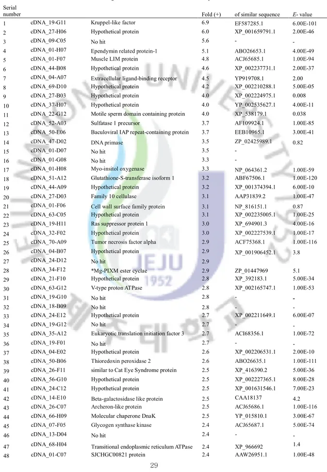
Table 3: List of genes significantly up-regulated in abalone gills after bacterial challenge Table 4: List of genes significantly up-regulated in abalone digestive tract after bacterial
challenge
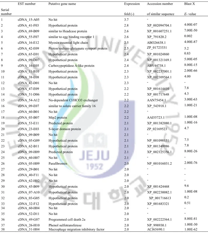
Table 5: List of genes significantly down-regulated in abalaone gills after bacterial challenge
Table 6: List of genes significantly down-regulated in abalone digestive tract after bacterial challenge
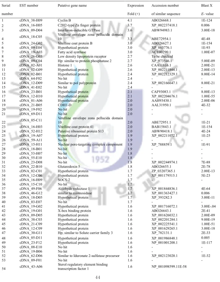
Table 7: List of genes significantly up-regulated in abalone gills after VHSV challenge Table 8: List of genes significantly up-regulated in abalone hemocytes after VHSV
challenge
Table 9: List of genes significantly down-regulated in abalaone gills after VHSV infection
Table 10: List of genes significantly down-regulated in abalone hemocytes after VHSV challenge
Table 11: Percentage of identity of the abalone Fas ligand to other Fas ligand, TNF-α and LT-α
Table 12: A comparison of abhisin with selected histone-derived AMPs
Table 13: Descriptions of the disk abalone antioxidant and immune response genes Table 14: Summary of the transcriptional responses of abalone antioxidant genes against
bacteria and VHSV challenge
ABBREVIATIONS
Ab abalone
AIF allograft inflammatory factor
α alpha
AMP antimicrobial peptide(s)
ANOVA analysis of variance
APD antimicrobial peptide database
AVG abalone viral ganglioneuritis
β beta
BLAST Basic Local Alignment Tool
bp base pair(s)
CIP calf intestinal phosphatase
oC centigrade
cDNA complementary deoxyribonucleic acid
Cu,Zn-SOD copper zinc super oxide dismutase
DEPC diethyl pyrocarbonate
DIZ disc inhibition zone
DMEM Dulbecco's modified eagle medium
DMSO dimethyl sulfoxide
DNA deoxyribonucleic acid
dNTP deoxynucleotide-triphosphate
DTT dithiothreitol
E.coli Escherichia coli
EDTA ethylene diamine tetra acetic acid
EST expressed sequence tag(s)
FBS fetal bovine serum
γ gama x g gravity G+ Gram positive G- Gram negative GO gene ontology GSH glutathione
GTPase guanosine triphosphatase
HE hydroethidium
XIV
IFN interferon
I-kB inhibitor of kappa B
IL interleukin
IPTG isopropyl-beta-D-thiogalactopyranoside
K kilo
Kb kilobase(s)
Kcal kilo calorie
KDa kilo dalton
KLF krüppell-like factor
L liter
LB Luria-Bertani
LITAF lipopolysaccharide-induced tumor necrosis factor alpha factor
LPS lipo polysaccharide
LT-α lymphotoxin alpha
MBP maltose binding protein
mg mili gram(s)
mL miilliliter(s)
MnSOD managanese super oxide dismutase
mol mole
MTT 3-(4,5-dimethylthiazol-2-yl)-2,5-diphenyltetrazolium bromide Mt-Trx-2 mitoocondrial thioredoxin 2
mRNA messenger RNA
Mx myxovirus myxovirus-resistance protein
MW molecular weight
μL microlitre(s)
NADPH nicotinamide adenine dinucleotide phosphate NCBI National Center for Biotechnology Information NFIL-3 nuclear factor interleukin-3
NF-kB nuclear factor kappa B
NK cells natural killer cells
NLS nuclear localization signal
nm nanometer(s)
NOX NADPH oxidase
O-2 superoxide anion
1O
2 singlet oxygen
OD optical density
ORF open reading frame
PBS phosphate buffered saline
PCR polymerase chain reaction
pfu plaque forming units
pI isoelectri point
PMN polymophonuclear leukocytes
PMT photo-multiplicator
PRD proline rich domain
PRR pattern-recognition receptors
proPO prophenoloxidase-activating system
PRx peroxiredoxin
qRT-PCR quantitative real time polimerase chain reaction Rel/NF-kB rel family nuclear factor kappa B
ROS reactive oxygen species
rpm revolutions per minutes
RT-PCR reverse transcription-polymerase chain reaction
SD standard deviation
SDS sodium dodecyl sulphate
SeGPx selenium dependent glutathione peroxidase
SEM scanning electron microscope
SOCS-2 supressor of cytokine signaling 2
TACE TNF alpha converting enzyme
Taq Thermus aqaticus
THP-1 human acute monocytic leukemia cell line
TIG transcription factor immunoglobulin
TNF tumor necrosis factor
TNF-α tumor necrosis factor-alpha
TPx thioredoxin peroxidase
TRxR thioredoxin reductase
TRx-2 thioredoxin 2
U unit
UTR untranslated region
1
GENERAL INTRODUCTION
Abalone biology and taxonomy
Abalone (from Spanish word `Abulón`) belongs to Phylum Mollusk one of the largest phyla in the animal kingdom. The phylum takes its name from Latin word “mollus” meaning of “soft”. A significant characteristic of mollusks is their soft body which is generally protected by a hard, calcium- containing shell except in some forms such as in slugs and octopuses which their shell has been lost in the course of evolution. Mollusks are a highly diverse group of animals that include three major classes namely Class bivalve (oysters, mussels, scallops, and clams), Class gastropod (abalone, snails, limpets, sea hares and slugs), and Class cephalopod (cuttlefish, squids, and octopuses). Abalone includes in class gastropod, the largest and most successful class of mollusks containing over 60,000-75,000 known living species and 15,000 fossil forms. All abalones belong to the Family Haliotidae and Genus Haliotis (http://simple.wikipedia.org/wiki/Gastropoda). There are over 70 species of halitosis species, which all of are marine. The most important species with regard to aquaculture in North America is the red abalone, Haliotis rufescens, while in Asia,
H. discuss hannai, H. discus discus and H. diversicolor supertexta are predominantly cultured
(McBride, 1998).
Abalone Aquaculture and diseases
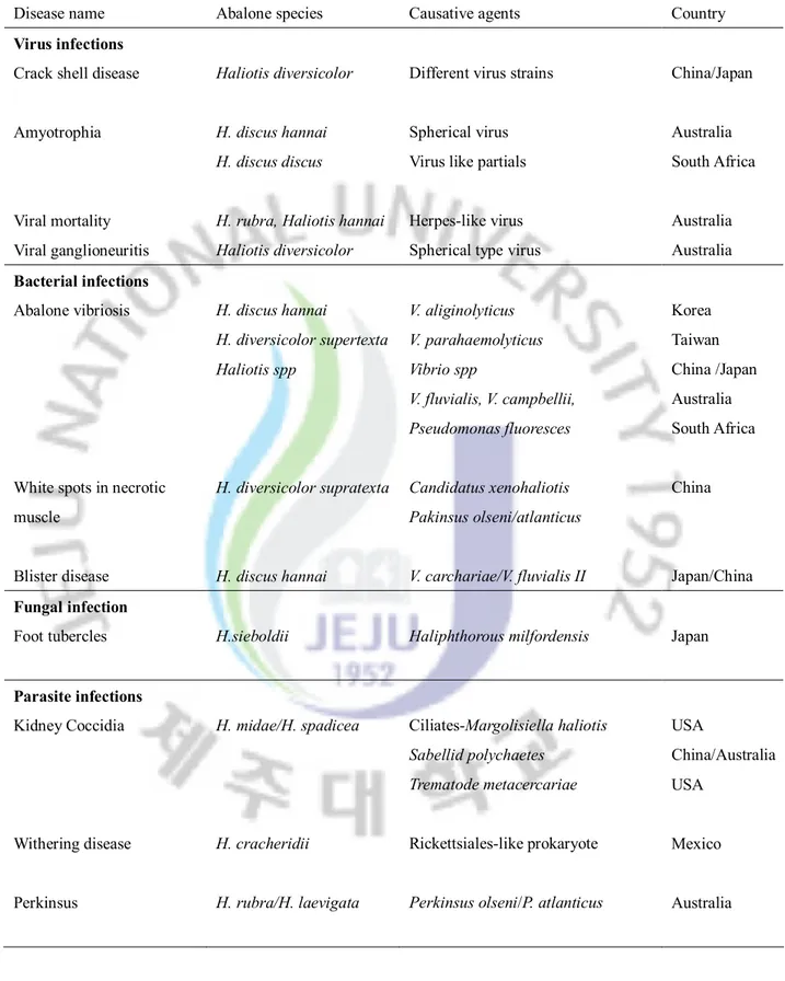
The emergence of new pathogens, particularly intracellular bacteria and viruses represent serious risks for the health of aquatic animals (Villena, 2003). A mass mortality has been reported from of marine mollusks including abalone due to pathogenic infections in different parts of the world resulting massive economic loss (Hooper at al., 2007). Recently reported abalone disease infections and causative agents are listed in table 1.
Major innate immune defense components in invertebrate mollusks
A correctly functioning immune system is vital to efficient aquaculture for both defense against invading pathogens and response to different stress (Villena, 2003). Innate immunity is the first line of defense against infection and invertebrates including abalone. Under the intensive conditions of aquaculture, some of the innate defenses are easily compromised and can allow pathogens more easily gain access to tissues and cells for infecting the host. The high immune specificity is usually considered as an exclusive property of vertebrate adaptive immunity which has been assumed that invertebrates lack of highly
specific immune defense (Loker at al., 2004).
To identify genetic diversity in genome scale and their phylogenetic as well as functional relationships “model organism concept” has been adapted from long time in different invertebrates including Drosophila (insect) and Caenorhabditis elegans (nematode). Genome sequencing projects of those two invertebrates have been already completed and genomic data are available to the public. Hence, it facilitates the unlimited genetic base to research community for understanding the immune defense mechanisms of those model species. From invertebrate model systems, we have learnt that the primary components of innate immunity fall into three categories that define the effectiveness of an immune response. First, the organism distinguishes between self and nonself; second, the organism mounts a defensive response that can kill or disable the invader; and finally, the organism recognizes and can eliminate its own damaged or diseased cells. These requirements lead to the three essential components of innate immunity: phagocytosis (cell-mediated); activation of humoral responses leading to opsonization, melanization, and coagulation (cell-free); and the production of humoral antimicrobial compounds (cell-free). Invertebrate immune defense against pathogens mainly occurs by major innate immunity systems or components and pathways which include; PRRs, hemolymph coagulation, AMP, respiratory burst activity, proPO, phagocytosis activity, lectin agglutinin system, antiviral, antifungal, antibacterial systems (Iwanaga et al., 2005).
The mechanisms underlying these host defense components depend on the presence of functional proteins in appropriate quantities, within a crucial time window. These proteins are encoded by genes whose transcription is tightly coordinated by complex programs of gene expression. As a summery, each of above individual immune defense systems consist functional genes under specific signaling pathways. In many occasions pathways are interrelated and work coordinately. Based on the several previous studies, researchers have described the important fact that the invertebrate’s immune defense mechanisms are heterogeneous, complex and poorly understood. In order to understand the entire immune defense mechanism in a whole organism, we need to know the regulatory mechanism and specific function of individual genes of each immune system. In general, it believes that molecular understanding of the genes and pathways regulating the mollusk immune system has lagged behind compared to that reported for other invertebrates such as insects and nematodes. Transcriptional analysis is one of the basic steps which could be applied to
3
Table 1: Diseases in abalone aquaculture; a summery of recent reports
Disease name Abalone species Causative agents Country
Virus infections Crack shell disease
Amyotrophia Viral mortality Viral ganglioneuritis Haliotis diversicolor H. discus hannai H. discus discus
H. rubra, Haliotis hannai Haliotis diversicolor
Different virus strains
Spherical virus Virus like partials
Herpes-like virus Spherical type virus
China/Japan Australia South Africa Australia Australia Bacterial infections Abalone vibriosis
White spots in necrotic muscle Blister disease H. discus hannai H. diversicolor supertexta Haliotis spp H. diversicolor supratexta H. discus hannai V. aliginolyticus V. parahaemolyticus Vibrio spp V. fluvialis, V. campbellii, Pseudomonas fluoresces Candidatus xenohaliotis Pakinsus olseni/atlanticus V. carchariae/V. fluvialis II Korea Taiwan China /Japan Australia South Africa China Japan/China Fungal infection
Foot tubercles H.sieboldii Haliphthorous milfordensis Japan
Parasite infections Kidney Coccidia Withering disease Perkinsus H. midae/H. spadicea H. cracheridii H. rubra/H. laevigata Ciliates-Margolisiella haliotis Sabellid polychaetes Trematode metacercariae Rickettsiales-like prokaryote
Perkinsus olseni/P. atlanticus
USA
China/Australia USA
Mexico
DNA microarray for global expression profiling
The recent increase in availability of gene expression technologies has the potential to dramatically expand our understanding of cellular immunology in molecular detail. By studying the gene expression of genes in various cellular contexts, it is possible to assign putative function to protein encoded by genes. Moreover, level of gene expression reflects the cellular activity and physiology. Analysis of gene expression at mRNA level (transcriptional) could be performed in several ways such as northern blot, RT-PCR etc. Alternatively, DNA microarray or DNA chip is a one of the latest high-throughput method used to analyze the large scale gene expression profiles of biologic samples which technology began in 1995 (Lamartine, 2006). Also, DNA microarray technology has given rise to the study of functional genomics. The entire set of genes of an organism can be microarrayed on an area as small as a fingernail and the expression levels of thousands of genes are simultaneously studied in a single experiment. DNA microarray technology allows comparisons of gene expression levels on a genomic scale in all kinds of combinations of samples derived from normal and diseased tissues, treated and nontreated time courses, and different stages of differentiation or development. Further computational analysis of microarray data allows the classification of known or unknown genes by their mRNA expression patterns. Global gene expression profiles in cells or tissues will provide us with a better understanding of the molecular basis of phenotype, pathology, or treatment.
Microarray manufacturing technology
The measurement of mRNA abundance is based on the capacity of every nucleic acid strand to recognize complementary sequences through base pairing. In 1995, Pat Brown et al., used glass support associated with fluorescence detection and robotically spotted 10,000 DNA probes onto a microscope slide and hybridized with a double-labeled mRNA population. This approach is now largely used in large-scale transcriptome analysis and called “cDNA microarray.” Its principle is described in fig. 1: A microarray is a solid substrate, such as a silicon wafer or glass slide, on which DNA or oligonucleotides from either host or pathogen could be attached. These nucleic acids are complementary to thousands of genes of both known and unknown function. There are two main types of microarray: spotted DNA and oligonucleotide arrays (Schena et al., 1995).
5
Spotted DNA arrays
The spotted DNA method entails attaching single-strand or disassociated double-strand DNA (from a cDNA library or genomic products amplified by PCR) to the slide. In most slides, duplicate spots are attached as controls, either next to or distant from the first spot, with up to 20 000 spots per slide. RNAs are extracted from two cell cultures from which expression level needs to be compared. Messenger RNAs are then transformed into cDNA by reverse transcription. At this stage, cDNA from the first culture is labeled with a red dye (Cy5), whereas cDNA from the second culture is labelled with a green dye (Cy3). Green-labeled and red-Green-labeled cDNAs are mixed together (called the target), put on the matrix of spotted single-strand DNA (called the probe) and incubated for one night. The fluorescent cDNA will then hybridize on the probe DNA spots. For slide scanning, a laser excites each spot and the fluorescent emission is gathered through a PMT coupled to a confocal microscope. Two images are obtained where green and red scales represent fluorescent intensities read. By superimposing these two images, one image composed of spots going from red (where only DNA from the first culture is fixed) to green (where only DNA from the second culture is fixed) passing through the yellow color (where DNAs from the two cultures are fixed in equal amounts) is obtained. Since the amount of fluorescent DNA fixed is proportional to the amount of mRNA present in each cell at the beginning, the red/green fluorescence ratio can be calculated. If this ratio is greater than 1 (red on the image), the gene expression is greater under the first experimental condition; if this ratio is smaller than 1 (green on the image), the gene expression is greater in the second condition (Schena et al., 1995).
Oligonucleotide arrays
Oligonucleotides can either be spotted as above or directly synthesized in situ on to the solid substrate, negating the need for attachment methods. Each oligonucleotide is usually about 50–70 nucleotides long rather than a whole gene. Over the slide there are several different oligonucleotide sequences from the same gene. This range produces more robust results because the same gene can be probed independently several times in the same experiment. A refinement of this method-Affymetrix microarrays (Affymetrix, CA, USA)-allows even greater specificity and may have up to 600 000 oligonucleotides per chip. For each approximately 20 nucleotide sequence there is a duplicate sequence with a single base
7
to a specific sequence and background hybridization to non-specific sequences and “true” quantification. Another advantage of oligonucleotidearrays is that they can be designed to include different alleles and splicing variants. The main disadvantage if there is hybridization with a single sample only is that more detailed data processing is needed to determine changes in the abundance of each transcript in two populations. The vast quantity of data generated from microarray experiment is only useful if it can be interpreted. The knowledge that a gene is expressed under a particular condition is not an answer in itself but only the first step in understanding. For example, whether expression of a gene is vital for host defense or involved in tissue injury requires further investigation. In addition, expression of a gene does not always translate into production of a protein. Although gene expression reveals nothing directly about translational and post-translational events, protein activation status alterations can and do frequently alter the expression of downstream target genes. These transcriptional consequences of protein activation can result in reproducible signatures of gene expression, specific to such an activation pathway. Microarrays therefore provide a stepping-off point for a wide range of further biological investigations (Bryant et al., 2004).
OUTLINE OF THE STUDY
Previous studies on pathogenic infections and immune challenge revealed that several genes involved in the immune defense response of disk abalone. These studies have been mainly conducted with a single or very few genes at a time. Since immune response reactions are highly complex and interrelated in the cellular level, DNA microarray based approaches allow researchers to interrogate host and pathogen genomes. It enables the investigation of entire transcriptional responses, biological pathways as well as previously unknown genes and their functions. No information is available reference to the immune response genes of abalone upon bacterial and viral challenge in a large scale gene expression using cDNA microarray technique.
Chapter one of this study has been focused on the construction of 4.2 K cDNA microarray using disk abalone cDNA library clones (ESTs) and analysis of transcriptional response in gills, digestive tract and hemocytes upon immune modulation (challenge) of abalone with bacteria (Vibrio alginolyticus, Vibrio parahemolyticus, and Listeria
monocytogenes) and VHSV. This chapter further describes the identification of novel immune
response genes from disk abalone and their functional categories under different host defense components of invertebrate immune defense systems including antimicrobial, inflammatory, apoptosis, antioxidant, protease inhibitory, cytokines regulatory genes and immune regulatory transcription factors. Based on the resulted microarray gene expression profiles, it was possible to select and grouped immune potential genes for further characterization which provides molecular insights of host immune defense systems against pathogens.
Chapter 2, 3, 4, and 5 have been focused to molecular characterization, transcriptional analysis (expression profiling) and functional aspects of genes and their respective proteins selected from microarray analysis and abalone cDNA library. Inflammatory and apoptosis related genes such as TNF-α, Fas ligand have been discussed in chapter 2. Also, two important transcription factors namely Rel/NF-kB and LITAF have been characterized in chapter 3. Chapter 4 describes the role of two AMP namely defensin and histone H2A derived AMP called “abhishin”. Additionally, immune responses of other genes such as antioxidant enzymes, cytokine regulators (SOCS-2), protease inhibitors have been discussed. Finally, the results presented in all chapters are summarized and discussed with suggesting (hypothesis) possible mechanisms, pathways and other potential immune relevant genes which may exist in abalone as host defense system.
9
CHAPTER 1
cDNA microarray analysis of bacteria and virus
(VHSV) challenged disk abalone
ABSTRACT
In this study, cDNA microarray technology has been used to analyze the transcriptional profiles of immune-challenged disk abalone using bacteria (Vibrio
alginolyticus, V. parahemolyticus, and Listeria monocytogenes) and VHSV to select potential
genes as immune-markers as well as to screen novel immune response genes. An abalone 4.2 K cDNA microarray was manufactured using the abalone ESTs, which has been constructed in our laboratory.
In bacteria challenged microarray, 68 (1.6%) and 112 (2.7%) transcripts changed their expression levels significantly (≥2 or ≤2 –fold) in gills and digestive tract tissues, respectively. There were 46 tissue-specific transcripts that were up-regulated only in digestive tract tissue. In contrast, only 13 transcripts showed gill-specific up-regulation. Verification of microarray results was done by selecting six candidate genes namely KLF, lachesin, muscle Lim protein, TRx-2, NFIL-3, abalone protein 38 using qRT-PCR. Our results indicate that post challenge of bacteria (at 24 h), may activate the transcription factors or their activators (KLF, inhibitor of NF-kB or Ik-B), inflammatory and apoptosis-related proteins (NFIL-3, AIF, TNF-α, archeron), cytokines (IFN-44-like, SOCS-2) and antioxidant enzymes (glutathione S transferase, TRx-2 and TPx) as a part of the innate immune responses in abalone.
Upon VHSV challenge, 280 (6.6%) transcripts were significantly (≥1.5 or ≤1.5 – fold) changed their expression level in gills and hemocytes against VHSV challenge compared to respective PBS controls. Total of 88 and 65 genes were up-regulated in gills and hemocytes, respectively. These genes included under various immune-functional categories such as KLF, inflammatory and apoptosis related genes (TNF super family members, Fas ligand), IFN regulatory proteins (IFN-44 like protein, IFN inducible GTPase) and detoxification proteins (glutathione peroxidase). In contrast to up-regulated responses, 25 and 102 genes were shown down-regulation in gills and hemocytes, respectively.
Considerably higher number of ESTs was matched to hypothetical (unknown) proteins while several ESTs that had no GenBank match (no hit). These significantly responded unknown and no hit transcripts may be novel genes involving immune defense role. Based on the microarray results, we selected TNF-α, Fas ligand, AIF, SOCS-2 and as potential immune-markers in abalone against bacterial and viral infection which required investigating further with different type of microorganisms. Additionally, the identification of immune-relevant genes and their expression profiles in this microarray will permit detailed
11 1.1. INTRODUCTION
Abalones are frequently infected by a wide range of pathogens including bacteria, viruses and parasites resulting huge economic losses (table 1). Bacterial infection is one of the serious threats to abalone aquaculture among the wide range of pathogens. It is generally known that bacterial pathogencity is associated with structural components of their cell wall, such as LPS or active secretions of substances that either damage host tissue or protect the bacteria from host defense (Paillard et al., 2004). Bacterial infections into abalone lead to major diseases such as abalone vibriosis (Elston et al., 1983), blister or white spot necrosis (Li et al., 1997), and brown ring disease (Shepheerd et al., 1997). Additionally, high mortality outbreaks have been reported in abalone Haliotis tuberculata by pathogenic V. carchariae (Nicolas et al., 2002) at elevated seawater temperature. It was reported that abalones are frequently infected by Vibrio species (Li et al., 1997).
Various types of viruses have been reported from mollusk species associated with specific types of infection or disease conditions (Renault and Novoa 2000). However, only few viral diseases were reported from abalone species. Nakatsugawa et al., (1990) identified the virus in a primary culture of hemocytes of Japanese black abalone Nordotic discus discus which virus caused to “amyotrophia” disease. Later, amyotrophia infection was identified in
H. discus hannai (Yu et al., 2007), H. discus discus, H. madaka species (Momoyama et al.,
1999). Also, a spherical enveloped virus related to disease called “crack shell” was identified in Haliotis discus hannai, (Wang Li 1997) and Haliotis diversicolor Reeve (Wang et al., 2004). Furthermore, this virus was mainly detected in the cytoplasm of hemocytes, connective tissues of other organs such as digestive gland and intestine. Fang et al., (2002) identified the DNA virus in the cytoplasm of the digestive gland, kidney, and intestine of H.
diversicolor supertexta. Abalone viral mortality is caused by several spherical viruses which
could be grouped mainly into four virus types (type I, II, III, IV). Type I is the least virulent while types II, III and IV are highly virulent, resulting in mass mortalities. A herpes-like virus which caused infectious disease named as abalone viral ganglioneuritis (AVG) was identified from H. diversicolor supertexta (Chang et al., 2005; Hooper et al., 2007). However, very little is known about the immune responses of abalone against virus challenge at a molecular level.
McGuire and his group (McGuire et al., 1998) emphasized that greater understanding of the complex interactions between host, pathogen and environmental factors could lead to the identification of the host defense strategies. Therefore, a better understanding of interrelated immune defense responses may help in developing strategies
and tools to reduce infectious diseases in abalone. To address this goal, a wide screening program of immune-response genes and their interactions in abalone is necessary. To date, most of the abalone immune defense systems have been studied at the molecular level based on the functions of individual genes “single gene research”, no studies have utilized the “whole genome approach”. To overcome the limitations, barriers and misinterpretations of “single gene” analysis approaches, scientists are increasingly concentrating on large-scale gene expression studies using genomic techniques associated with bioinformatics.
A DNA microarray is a high-throughput tool that allows the transcriptional analysis of large number of genes simultaneously to expand our understanding of cellular immunology in molecular level. There are several reports on microarray analysis to investigate transcriptional responses of pathogen-infected or immune-stimulated invertebrate animals such as
Drosophila (Pal et al., 2008; Johansson et al., 2005), the moth Plutella xylostella (Eum et al.,
2007), mosquito (Aguilar et al., 2005), and shrimp (Dhar et al., 2003). So far, only a few microarray studies have been conducted in mollusk species such as the oyster Crassostrea
gigas, and C. virginica (Jenny et al., 2007; Lang et al., 2009). However, there is no report on
large-scale gene expression study using cDNA microarray to identify immune response genes and their functional groups of disk abalone or other mollusks. Therefore, the construction of large-scale transcriptome profiling using a cDNA microarray against bacteria and virus challenge would provide much needed information for identification and characterization of different components of the abalone immune system.
The first chapter of the present study describes the application of a cDNA microarray technique for investigating immune-related genes, their transcriptional responses and possible interactions in abalone. For that, 4.2 K cDNA chip was constructed by adding 4188 transcripts from disk abalone ESTs, and described the microarray transcriptional profiling in gills and digestive tract tissues of bacterial (V. alginolyticus, V. parahemolyticus and L.
13 1.2. MATERIALS AND METHODS
1.2.1. Disk abalone cDNA library and 4.2 K microarray construction
An abalone cDNA library was constructed using mRNA isolated from whole tissues of disk abalone and cDNA library construction kit (Creator™ SMART™, Clontech, USA). The cDNA library was normalized using Trimmer-Direct cDNA normalization kit according to the manufacturer’s protocol (Evrogen, Russia). Then, the cDNA clones were ligated into the pDNR-LIB vector at SfiI restriction site. After obtaining the plasmids with different cDNA inserts, 6700 clones were sequenced from the 5' end with M13 forward vector primer utilizing a Big Dye Terminator sequencing kit and ABI 3700 sequencer (Macrogen, Korea). Finally, disk abalone normalized cDNA library ESTs were obtained and used to select cDNA sequences for microarray construction. The disk abalone microarray platform was produced by GenomicTree Inc (Daejon, Republic of Korea). Briefly, a set of cDNA clones representing 4188 genes were selected from abalone ESTs to construct the cDNA microarray. In order to construct the cDNA region of each clone, PCR amplification was carried out using M13 forward and reverse universal primers (Table 2) under the following conditions: initial denaturation at 94 oC for 5 min, followed by 30 cycles of 94 oC for 1 min, 55 oC for 1 min and
72 oC for 90 s, and a final extension step at 72 oC for 10 min. The PCR-amplified products
were examined by 1% agarose gel electrophoresis, purified using Sephadex G-50 columns, air-dried and resuspended in 50% DMSO solution. Purified DNA samples were spotted using an OmniGridTM Microarrayer (GeneMachines, San Carlos, CA) onto silanized glass slides (GAPS-IITM, Corning, Charlotte, NC). Each slide was crosslinked with 300 mJ of short wave ultraviolet (UV) irradiation (Stratalinker, Stratagene, La Jolla, CA) and stored in humidity and light-controlled conditions until use. Finally, a total of 4188 amplicons were spotted in a total of 16 sub arrays in a 17 x 16 raw-column arrangement.
1.2.2. Abalone culture
Healthy disk abalones (H. discus discus) with an average body weight of 50-60 g were obtained from the Youngsoo abalone farm (Jeju Island, Republic of Korea). Abalones were maintained in flat-bottomed fiberglass tanks (60 L) with aerated and sand-filtered seawater at 18–20 °C during the experiment. Animals were acclimatized for a one week prior to the experiment. They were fed daily with fresh seaweed (Undaria pinnatifida) diet. A maximum of 20 animals per tank were maintained during the experiment.
1.2.3. Isolation of abalone tissues and hemocytes
Healthy animals were selected to examine the tissue specific expression profile of selected genes. Abalones were carefully dissected on ice and tissues (gills, mantle, muscle, digestive tract and hepatopancreas) were collected and pooled from three abalones. The abalone hemolymph was withdrawn from the cephalic arterial sinus, accessed anteriorly at the angle between the foot and the head using a syringe fitted with a 22-gauge needle. Collected hemolymph was immediately transferred into micro tubes and kept on ice. Then, hemolymph was centrifuged at 1500 g for 10 min at 4 oC. The supernatant was removed and hemocytes were collected. All the tissue samples were snap-frozen in liquid nitrogen immediately after obtained from abalone and stored at -80 oC until used for RNA isolation.
1.2.4. Bacterial challenge for microarray
Abalones were immune-challenged using two Gram negative (G-) bacteria V.
alginolyticus (KCTC2472), V. parahemolyticus (KCTC2729); and, Gram positive (G+) L. monocytogenes (KCTC3710). All three bacteria were obtained from the Korean Collection
for Type Cultures. The three bacteria mixture was intramuscularly injected into the abalone. Briefly, V. alginolyticus and V. parahemolyticus were cultured in marine broth for 16-20 h at 25o C, while shaking at 200 rpm. L. monocytogenes was cultured in LB broth for 16 h at 30o C.
Then all three bacterial cultures (1.5 mL) were centrifuged at 7000 x g for 5 min at 4o C. The supernatant fluid was removed and the bacterial pellets were re-suspended in PBS and an equal volume of each bacterial media was mixed to make bacterial stock with a cell density of 1.0 at 600 nm absorbance. Three abalones (n=3) were injected intramuscularly with 50 mL/animal (5x10⁷ cells/mL) of bacterial stock. Three control abalones (n=3) were injected
with the same amount of sterile PBS and treated identically as the bacteria-injected group. Gills and digestive tissues were harvested at 24 h post challenge of bacteria and PBS control groups for microarray analysis.
1.2.5. Virus challenge for microarray
For the virus challenge experiment, abalones were intramuscularly injected with 50 mL VHSV solution (1 X 10⁸ pfu/mL per abalone). The control group was injected with same
volume of PBS. Abalone gills and hemocytes were isolated after 24 h of VHSV challenge for microarray analysis. To analysis the transcriptional up-regulation of immune relevant genes
15 1.2.6. RNA isolation
An equal amount (50 mg) of gills, digestive tract or hemocytes samples were obtained from immune-challenged and un-induced or PBS-injected abalones (n=3) separately. RNA was extracted from pooled tissue of three replicate tissue samples using the Tri Reagent™ Kit (Sigma, USA) according to manufacturer’s protocol. RNA quality was assessed by spectrophotometer using a Nanodrop ND-1000 (Nanodrop, USA) and run on a Bioanalyzer 2100 (Agilent, USA) for microarray analysis.
1.2.7. Microarray hybridization
RNA labeling, array hybridization and scanning were carried out by GenomicTree Inc (Daejon, Republic of Korea). Briefly, for bacteria-infected (test) and control RNAs, the synthesis of target cDNA probes and hybridization were performed using Agilent’s Low RNA Input Linear Amplification Kit PLUS (Agilent Technologies, USA) according to the manufacturer’s instructions. Briefly, 1 μg of each disk abalone total RNA was mixed with dT-promoter primer and M-MLV-reverse transcriptase, followed by incubation at 40 oC for 2 h. The reverse-transcribed samples were linear-amplified using T7 polymerase with 2 h incubation at 40 oC. During amplification, control cRNAs and test (from bacteria-infected
tissues) cRNAs were labeled with Cy3-CTP and Cy5-CTP, respectively. Both fluorescent-labeled control and test samples were combined and resuspended in 80 μL of hybridization buffer (3X SSC and 0.3% SDS, 50% formamide). After 3 min boiling at 95 oC and 3 min cooling in ice, the hybridization mixtures were directly introduced to disk abalone 4.2K cDNA microarray. For hybridization, the microarray slide was incubated in a humidified hybridization chamber at 42o C for 16 h with mild agitation to perform competitive hybridization reactions between labeled probes and targets on microarray. Washing of hybridized arrays was performed with gentle agitation to eliminate the nonspecific binding as follows: 5 min at 42 oC in 1´SSC and 0.2% SDS, 5 min at room temperature in 1´SSC and 0.2% SDS, and 2 min at room temperature in 0.1´SSC (twice). Finally, microarrays were spin-dried and stored in dark condition until scanned.
1.2.8. Microarray data analysis
The hybridization images were visualized with Axon GenePix 4000B scanner (Axon Instruments, CA) and analyzed by GenePix Pro 6.0 program (Axon Instruments, USA). The
average fluorescence intensity of each spot was calculated and local background was subtracted. All data normalization and selection of the fold-changed genes were performed using GeneSpring 7.3.1 (Agilent Technologies, USA). Intensity-dependent normalization (LOWESS) was performed, where the ratio was reduced to the residual of the Lowes fit of the intensity vs. ratio curve. The averages of normalized ratios were calculated by dividing the average of normalized test channel intensity by the average of normalized control channel intensity. Up-down regulated genes were analyzed using the algorithms BLASTX and BLASTN that are available at NCBI to identify similar (homology) sequences. When the expected value (E) was less than 1.E-05, an EST was considered as a significant match to a specific homology sequence. Then, the identified sequences were classified into functional groups using a GO web based application and manual adjustments.
1.2.9. Bacteria and VHSV challenge for microarray validation
To validate the microarray results, two independent time course experiments were conducted by applying same method of bacterial and VHSV challenge, as described for the microarray experiments. Briefly, abalones were challenged with bacteria mixture and VHSV. Tissue samples of gills, digestive tract (from bacterial challenged) and gills and hemocytes (VHSV challenged) were isolated at 3, 6, 12, 24, and 48 h. Selected tissues were collected from three individuals at every time point while control samples were collected from PBS-injected abalones.
1.2.10. cDNA synthesis
RNA isolation was done as previously described method and purified RNA was diluted up to 1 mg/mL concentration before synthesis of cDNA. A sample of 2.5 mg RNA was used to synthesize cDNA from each tissue using a Superscript III first-strand synthesis system for RT-PCR kit (Invitrogen, USA). Briefly, RNA was incubated with 1 mL of 50 mM oligo (dT)20 and 1 mL of 10 mM dNTP for 5 min at 65 °C. After incubation, 2 mL of 10´
cDNA synthesis buffer, 2 mL of dithiothreitol (DTT, 0.1 M), 1 mL of RNaseOUT™ (40 U/mL) and 1 mL of SuperScript III reverse transcriptase (15 U/mL) were added and incubated for 1 h at 50 °C. The reaction was terminated by adjusting the temperature to 85 °C for 5 min. Then, 1 mL of RNase H was added to each cDNA and incubated at 37 °C for 20 min. Finally, synthesized cDNA was diluted 10 fold (total 200 mL) before storing at -20 °C.