Vol. 46, No. 3, pp. 425 - 430, 2005
Advanced Hodgkin's disease is usually treated with six or more cycles of combination chemotherapy. Spontaneous re-gression of the cancer is very rarely reported in patients with Hodgkin's disease. We present an unusual case of a patient with Hodgkin's disease who experienced complete remission with a single cycle of chemotherapy, followed by pneumonia. The case was a 36-year-old man diagnosed with stage IVB mixed cellularity Hodgkin's disease in November 2000. After treatment with one cycle of COPP-ABV (cyclophosphamide, vincristine, procarbazine, prednisone, doxorubicin, bleomycin, and vinblastine) chemotherapy without bleomycin, the patient developed interstitial pneumonia and was cared in the inten-sive care unit (ICU) for two months. Follow-up chest compu-terized tomography (CT), performed during the course of ICU care, revealed markedly improved mediastinal lymphomatous lesions. Furthermore, follow-up whole body CT and 18-fluoro-deoxyglucose positron emission tomography showed complete disappearance of the lymphomatous lesions. Four years later, the patient is well and without relapse. This report is followed by a short review of the literature on spontaneous regression of Hodgkin's disease. To the best of our knowledge, this is the first case report of spontaneous remission of Hodgkin's disease in Korea.
Key Words: Spontaneous remission, Hodgkin's disease, in-complete chemotherapy
INTRODUCTION
Hodgkin's disease has been diagnosed in
appro-ximately 7 per 100,000 persons annually since the disorder was first described by Thomas Hodgkin in 1832.1 Like other malignant diseases, spontaneous regression of Hodgkin's lymphoma has been reported as a very rare phenomenon. Many possible etiologies of this phenomenon have been proposed, however, the most prevalent view is that immunologic factors in the host are responsible. Spontaneous regression of Hodgkin's disease occurs very rarely compared to non-Hodgkin's lymphoma. To date, only 15 cases have been reported in the literature. We report an unusual case of a patient with Hodgkin's disease who had complete remission during intensified care for interstitial pneumonia of an unidentified organism following one cycle of combination chemotherapy.
CASE REPORT
The 36-year-old male patient was admitted through the emergency room on November 30, 2000 with a 15-day history of intermittent spiking fevers and chills. He had a temperature of 38.1 , a pulse rate of 80/min, a respiratory rate of 16/ min, and a blood pressure of 130/80mmHg. There was no palpable peripheral lymph node enlarge-ment, hepatomegaly, or splenomegaly. Initial lab-oratory studies revealed a hemoglobin of 11.0g/ dL, a hematocrit 31.1%, a white blood cell count 4,960/ l (neutrophils, 75.5%; lymphocytes, 12%;μ monocytes, 7.3%; eosinophils, 0.6%; basophils, 0.5%), and a platelet count 328,000/ l. The C-μ
An Unusual Case of Spontaneous Remission of Hodgkin's
Disease after a Single Cycle of COPP-ABV Chemotherapy
Followed by Infectious Complications
Seungmin Bang1, June-Won Cheong1, Woo Ick Yang2, and Jee Sook Hahn1
Division of Hemato-oncology, Department of1Internal Medicine and2Pathology, Yonsei University College of Medicine, Seoul,
Korea.
Received December 30, 2004 Accepted February 28, 2005
Reprint address: requests to Dr. Jee Sook Hahn, Division of Hemato-oncology, Department of Internal Medicine, Yonsei University College of Medicine, 134 Shinchon-dong, Seodaemun-gu, Seoul 120-752, Korea. Tel: 82-2-2228-1935, Fax: 82-2-393-6884, E-mail: [email protected]
reactive protein was 11.9 mg/dL (normal < 0.8 mg/dL). He had an AST/ALT of 98/271 IU/L, total protein 6.38 g/dL, albumin 3.08 g/dL, total biliru-bin 0.5 mg/dL, alkaline phosphatase 299 IU/L (normal 38-115 IU/L), BUN 6.0 mg/dL, and cre-atinine 0.8 mg/dL. The LDH was 460 IU/L (nor-mal 225-455 IU/L) with an increased LDH5 iso-enzyme fraction and β2-microglobulin 3.6 mg/ dL. The tests for the IgM antibody to herpes simplex virus (HSV), varicella zoster virus (VZV) and Epstein-Barr virus (EBV) early antigen were nega-tive, and the cytomegalovirus (CMV) early anti-gen was not detected. Microbiologic studies in-cluding cultures of sputum, urine, stool and blood were all negative. A posteroanterior chest radio-graph was normal.
A neck computerized tomography (CT) scan showed multiple lymphadenopathy (0.5-1.2 cm in size) on the left level V, the left level II, and the right level II with well-defined, round contours without necrotic portions, strongly suggestive of benign reactive hyperplasia. A chest CT scan
showed multiple areas of lymphadenopathy in the mediastinum, high and lower right paratracheal, pretracheal, aortopulmonary window, prevascu-lar, subcarinal, paraesophageal, right and left cardiophrenic areas and the right supraclavicular fossa. The largest lymph node was measured at 3.5×2 cm in the right cardiophrenic area (Fig. 1A). The lungs were clear, without evidence of abnor-mal nodules (Fig. 1D). An abdominopelvic CT scan revealed hepatosplenomegaly with multiple low-density nodules on the liver and spleen as well as multiple enlarged lymph nodes in the aortocaval area, paraaortic area, and along the left iliac lymphatic chain (4 × 3 × 6 cm), and a conglo-merated lymph node (3 × 2 cm) on the left renal hilum. Whole body 18-fluorodeoxyglucose posi-tron emission tomography (18FDG-PET) showed massive areas of hot uptake in the mediastinum, intra-abdominal lymph nodes, liver, and spleen; it showed multiple areas of hot uptake near the entire vertebra, bilateral shoulder, rib, left femur, and pelvic bones, as well as an abnormal lesion
Fig. 1. Serial follow up images of chest CT scans. A. Enlargement of multiple lymph nodes at the paratracheal and aortopulmonary window was noted at the time of initial diagnosis. B. The enlarged lymph nodes, noted on the initial CT scan, were found to be markedly improved on the 21st day of hospitalization. C. A CT scan performed nine months after initial diagnosis showed no abnormalities. D. No abnormalities in the lung parenchyma were found at the time of diagnosis. E. A CT scan performed at 21th day of hospitalization showed ground glass opacities in the peripheral areas of both lung parenchyma. F. Although minimal honey combing and reticular patterns remained, the ground glass opacities, noted at D, were not found with CT nine months after initial diagnosis.
on the lungs (Fig. 2A). A whole body bone scan did not show any evidence of bone involvement. A laparoscopic biopsy of the left iliac lymph node and liver, performed on the 7th hospital day, showed mixed celluarity with high contents of epitheloid histiocytes and a few Reed-Sternberg giant cells, compatible with Hodgkin's disease (Fig. 3). Additionally, a bone marrow specimen showed no infiltration of lymphomatous cells.
The patient was diagnosed with mixed cellu-larity type, stage IVB Hodgkin's disease. On the 15th hospital day, he received combination che-motherapy with COPP-ABV but without bleo-mycin because of insufficient pulmonary function. One day after initiation of the chemotherapy, the patient complained of dyspnea on exertion and even at rest. A chest radiograph revealed slightly increased peripheral ground-glass opacities with
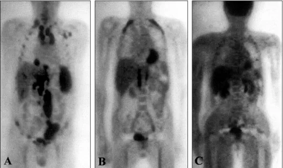
Fig. 2.Serial follow up images of 18FDG-PET scans. A. The initial18FDG-PET scan showed massive areas of hot uptake
in the mediastinum, intra-abdominal lymph nodes, spleen, possibly liver, near the entire vertebrae, bilateral shoulder, rib, and left femur. B. A follow-up18FDG-PET scan, performed 3 months after the initial chemotherapy, showed markedly
decreased hot uptakes in the liver, spleen, vertebral bodies and iliac lymph nodes. C. Nine months after initial chemotherapy, there was no definite abnormal uptake noted on the 18FDG-PET scan.
Fig. 3. Pathologic features. A. Left iliac lymph node biopsy. Microscopic examination of the specimen showed mixed cellularity type Hodgkin's disease with high contents of epitheloid histiocytes and a few Reed-Sternberg cells. (H-E stain, ×400). B. Liver biopsy. Reed-Sternberg cells with background reactive cells infiltrated and replaced the liver parenchyma (H-E stain, ×40).
peribronchial cuffing in both lower lung fields. He also had a positive serology for the candida anti-gen. Therefore, amphotericin B and acyclovir were administrated in addition to his previous anti-biotics regimen, composed of cefoperazone/sul-bactam and amikacin, for treatment of suspected interstitial pneumonia. Repeat microbiologic studies did not reveal any pathogens. Virologic studies including HSV IgM, VZV IgM, CMV IgM, CMV early antigen, and EBV IgM were all nega-tive. Two days following the initiation of chemo-therapy, the patient was transferred to the inten-sive care unit (ICU) for low PaO2(68mm Hg). His chest radiographs, consistent with interstitial pneumonia, continued to worsen in appearance despite the addition of trimethoprim/sulfome-thoxazole and a switch of acyclovir to ganciclovir. On the 21st hospital day, a chest CT scan, per-formed to evaluate the abnormal chest radio-graphic findings, showed ground glass opacities on the periphery of both lungs with smooth bronchovascular bundle thickening and interlobu-lar septal thickening; however, the eninterlobu-larged lymph nodes in mediastinum and right supracla-vicular fossa were markedly improved (Fig. 1B, 1E). The patient's dyspnea did not improve de-spite vigorous therapy and on the 30th hospital day the patient was started on mechanical ventila-tion in the ICU. One day after endotracheal intu-bation, trimethoprim/sulfomethoxazole was re-placed with pentamidine because of the former's bone marrow suppressive effect. During the period of ICU care and mechanical ventilation, the patient experienced left foot drop, with sensory loss due to left peroneal neuropathy, nephrogenic diabetes inspidus, and pancreatitis secondary to the pentamidine. However, with maximal conser-vative care, on the 74th hospital day he improved enough to be transferred to the general ward and no longer required mechanical ventilation. Whole body CT scans were repeated on the 90th hospital day to assess the lymphadenopathy, noted to be improved upon follow-up chest CT scan on the 20th hospital day, because the patient had refused further chemotherapy. Interestingly, follow-up CT scans of the neck, chest and abdomen-pelvis showed no definite evidence of lymphadenopathy in the neck, intrathoracic mediastinum, markedly decreased lymphadenopathy in the paraaortic
area and the left iliac chain, as well as improved hepatosplenomegaly with resolution of the previous low attenuating nodules (Fig. 1C). On 18FDG-PET images there was no evidence of lymphomatous involvement of the liver, spleen, iliac, inguinal area or spine, except for a residual lesion in the upper abdominal lymph node chain along the inferior vena cava and aorta (Fig. 2B). These findings were thought to be compatible with a partial response. Nine months following the initial chemotherapy, whole body CT (neck, chest, abdomen and pelvis) scans, 18FDG-PET, and a whole body bone scan, performed for restaging,
did not show any definite evidence of
lymphomatous lesions, and thus were compatible with complete remission status (Fig. 2C). On the 103rd hospital day, the patient's tracheotomy site was sealed. On the 167th hospital day, pulmonary function tests showed a vital capacity of 39.6%, FVC of 40.8%, FEV1 of 49.4%, FEV1/FVC of 122, and DLco of 58.3%. Chest radiography at this time showed diffuse and coarse reticular changes. The patient, with the support of his family, decided not to undergo further chemotherapy and he was discharged on the 198th day of hospitalization with slight dyspnea on exertion, but otherwise feeling well. Four years following hospitalization, the patient is living well, even climbing up high mountains, and is without recurrence of Hodgkin's disease.
DISCUSSION
Spontaneous regression of cancer is defined as the complete or partial disappearance of a malig-nant tumor in the absence of or inadequacy of therapy that is capable of inducing antineoplastic effects.2 This definition of spontaneous regression does not necessarily imply a spontaneous cure of the cancer, because it also applies to cases of in-complete or temporary regression of cancer. Al-though spontaneous regression of cancer is uncom-mon, 761 reports of individual cases and small case studies have been published between 1900 and 1987.3,4 Common clinical entities reported to have shown spontaneous regression include hypernephroma, malignant melanoma, neuroblas-toma, leukemia and non-Hodgkin's lymphoma.2
Spontaneous regression in malignant lymphoma has been reported primarily in patients with low-grade non-Hodgkin's lymphoma,5,6 but rare case reports have described regression in patients with intermediate or high-grade non- Hodgkin's lymphoma and Hodgkin's disease.7-9
Untreated Hodgkin's disease has a 5-year sur-vival rate of less than 5%.10 Spontaneous regres-sion of Hodgkin's lymphoma is a very rare event; a search of MEDLINE identified only 15 cases of spontaneous regression of the disease, with vari-ous follow-up periods ranging from several months to eight years.11-16 Of these patients, the subtypes of Hodgkin's disease were reported in only eight cases, with the mixed cellularity being the most frequent subtype (mixed cellularity, 4 cases; lymphocyte predominant, 3 cases; nodular sclerosis, 1 case). The case described here was also of the mixed cellularity subtype. Among the 15 reported cases of spontaneous regression, five cases occurred in children following measles in-fection; however, all five of these patients still required treatment with chemotherapy following the regression.14,15
The etiologies underlying spontaneous remis-sion remain unclear. The proposed mechanisms to explain this phenomenon have included the role of immunological factors, concomitant infections, hormonal factors, tumor necrosis, angiogenesis inhibition, apoptosis, elimination of carcinogens, surgical trauma on the primary tumor, or induc-tion of differentiainduc-tion.3,17
Among these possible etiologic factors, immu-nological mechanisms have been the most fre-quently used to explain spontaneous regression of Hodgkin's disease.11,16 Drobynski and Qazi re-viewed the indirect experimental evidence sup-porting the notion that host immunological mech-anisms are important in B cell lymphoma regres-sion.17 Ono et al. suggested that highly elevated natural killing activity might be one of the pos-sible mechanisms responpos-sible for spontaneous regression of malignant lymphoma.18 In this study, the patients with spontaneous regression had significantly higher natural killing activities prior to cancer regression than either controls or patients without regression in the absence of episodes suggesting viral infection.
Nonspecific stimulation of the immune system
may result in enhancement of host immune re-sponses against tumors. The temporal association found between concurrent infections and sponta-neous remissions of tumor suggests that infection may stimulate the immune system to induce tumor regression. Similar immunologic changes may also play a significant role in the patho-physiology of post-transplant lymphoproliferative disease (PTLD). In PTLD, immunosuppressive agents for prevention of graft rejection can induce disruption of T cell control of B cell growth, resulting in the proliferation of Ebstein-Barr virus infected B cells, which is followed by the develop-ment of hyperplasia or malignancy.19 This dis-ruption of the normal immune system can be recovered by tapering immunosuppressive agents. In addition, the restored immune system can identify the abnormal lymphomatous cells and clear them out.20
It was strongly suggested that the spontaneous regression observed in our patient was most likely due to enhanced endogenous immune regulation from a concomitant unidentified infectious disease such a viral pneumonia. The immune system in our patient may have been activated during the struggle with severe interstitial pneumonia, and this intensified cellular immunity may be respon-sible for the spontaneous regression of the tumor. In addition, various cytokines or toxins associated with infections may also play a role in mediating regression.21,22
The experience with this case could support the possible role of infections as an important etio-logic factor for spontaneous regression of Hodgkin's disease via nonspecific ostentation of host im-munity. However, it is impossible to exclude the possibility that the tumor cells may have been ex-tremely sensitive to the chemotherapeutic agents used. Therefore, the phenomenon of spontaneous regression should be studied further because a more precise understanding may make it possible to harness the host immune system to mediate tumor regression in neoplastic diseases such as Hodgkin's disease.
REFERENCES
Can-cer Res 1966;26:1189-200.
2. Papac RJ. Spontaneous regression of cancer. Cancer Treat Rev 1996;22:395-423.
3. Challis GB, Stam HJ. The spontaneous regression of cancer. A review of cases from 1900 to 1987. Acta Oncol 1990;5:545-9.
4. Gattiker HH, Wiltshaw E, Galton DA. Spontaneous regression in non-Hodgkin's lymphoma. Cancer 1980; 45:2627-32.
5. Horning SJ, Rosenberg SA. The natural history of ini-tially untreated low-grade non-Hodgkin's lymphomas. N Engl J Med 1984;311:1471-5.
6. Krikorian JG, Portlock CS, Cooney P, Rosenberg SA. Spontaneous regression of non-Hodgkin's lymphoma: a report of nine cases. Cancer 1980;46:2093-9.
7. Poppema S, Postma L, Brinker M, de Jong B. Spontane-ous regression of a small non-cleaved cell malignant lymphoma (non-Burkitt's lymphoblastic lymphoma). Morphologic, immunohistological, and immunoglo-bulin gene analysis. Cancer 1988;62:791-4.
8. Kumamoto M, Nakamine H, Hara T, Yokoya Y, Kawai J, Ito H, et al. Spontaneous complete regression of high grade non-Hodgkin's lymphoma. Morphologic, immu-nohistochemical, and gene amplification analyses. Can-cer 1994;74:3023-8.
9. Heibel H, Knodgen R, Bredenfeld H, Wickenhauser C, Scheer M, Zoller JE. Complete spontaneous remission of an aggressive non-Hodgkin's lymphoma with pri-mary manifestation in the oral cavity. Leuk Lymphoma 2004;45:171-4.
10. Weinshel EL, Peterson BA. Hodgkin's disease. CA Can-cer J Clin 1993;43:327-46.
11. Mangel J, Barth D, Berinstein NL, Imrie KR. Spontane-ous regression of Hodgkin's disease: two case reports and a review of the literature. Hematology 2003;8:191-6.
12. Williams MV. Spontaneous regression of cutaneous Hodgkin's disease. Br Med J 1980;280:903.
13. Szur L, Harrison CV, Levene GM, Samman PD. Pri-mary cutaneous Hodgkin's disease. Lancet 1970;1:1016-20.
14. Taqi AM, Abdurrahman MB, Yakubu AM, Fleming AF. Regression of Hodgkin's disease after measles. Lancet 1981;1:1112.
15. Zygiert Z. Hodgkin's disease: remissions after measles. Lancet 1971;1:593.
16. Parekh S, Koduri PR. Spontaneous regression of HIV-associated Hodgkin's disease. Am J Hematol 2003;72: 153-4.
17. Drobyski WR, Qazi R. Spontaneous regression in non-Hodgkin's lymphoma: clinical and pathogenetic con-siderations. Am J Hematol 1989;31:138-41.
18. Ono K, Kikuchi M, Funai N, Matsuzaki M, Shimamoto Y. Natural killing activity in patients with spontaneous regression of malignant lymphoma. J Clin Immunol 1996;16:334-9.
19. Loren AW, Porter DL, Stadtmauer EA, Tsai DE. Post-transplant lymphoproliferative disorder: a review. Bone Marrow Transplant 2003;31:145-55.
20. Ryu HJ, Hahn JS, Kim YS, Park K, Yang WI, Lee JD. Complete resolution of posttransplant lymphoproli-ferative disorder (diffuse large B-cell lymphoma) with reduction of immunosuppressive therapy. Yonsei Med J 2004;45:527-32.
21. Nauts H. The treatment of malignant tumors by bacterial toxins, as developed by the late William B. and Coley M.D. reviewed in the light of modern re-search. Cancer Res 1946;6:205-16
22. Balkwill FR, Naylor MS, Malik S. Tumour necrosis factor as an anticancer agent. Eur J Cancer 1990;26:641-4.