Control of Rapsyn Stability by the CUL-3-containing E3
Ligase Complex
*
□SReceived for publication, October 28, 2008, and in revised form, January 13, 2009 Published, JBC Papers in Press, January 20, 2009, DOI 10.1074/jbc.M808230200
Seunghee Nam‡1,2, Kyoengwoo Min‡1,3, Hyejin Hwang§, Hae-ock Lee‡, Jung Hwa Lee‡, Jongbok Yoon¶,
Hyunsook Lee‡, Sungsu Park§, and Junho Lee‡4
From the‡Research Center for Cellulomics, Institute of Molecular Biology and Genetics, School of Biological Sciences, Seoul National University, 151-742 Seoul, Korea, the§Division of Nano Sciences (BK21), Ewha Womans University, 120-750 Seoul, Korea, ¶Protein Network Research Center, Department of Biochemistry, Yonsei University, 134 Shinchon, 120-749 Seoul, Korea
Rapsyn is a postsynaptic protein required for clustering of nicotinic acetylcholine receptors (nAChRs) at the neuromuscu-lar junction. Here we report the mechanism for posttransla-tional control of rapsyn protein stability. We confirmed that C18H9.7-encoded RPY-1 is a rapsyn homolog in Caenorhabdi-tis elegans by showing that human rapsyn rescued rpy-1 mutant phenotypes in nematodes, as determined by levamisole assays and micropost array behavioral assays. We found that RPY-1 was degraded in the absence of functional UNC-29, a non-␣ subunit of the receptor, in an allele-specific manner, but not in the absence of other receptor subunits. The cytoplasmic loop of UNC-29 was found to be critical for RPY-1 stability. Through RNA interference screening, we found that UBC-1, UBC-12, NEDD-8, and RBX-1 were required for degradation of RPY-1. We identified cullin (CUL)-3 as a component of E3 ligase and KEL-8 as the substrate adaptor of RPY-1. Mammalian rapsyn was ubiquitinated by the CUL3/KLHL8-containing E3 ligase in vitro, and the knockdown of KLHL-8, a mammalian KEL-8 hom-olog, inhibited rapsyn ubiquitination in vivo, implying evolu-tionary conservation of the rapsyn stability control machinery. kel-8 suppression and rpy-1 overexpression in C. elegans pro-duced a phenotype similar to that of a loss-of-function mutation of rpy-1, suggesting that control of rapsyn abundance is impor-tant for proper function of the receptor. Our results suggest a link between the control of rapsyn abundance and congenital myasthenic syndromes.
Neuromuscular junctions, which are composed of a presyn-aptic terminal, basal lamina, and a postsynpresyn-aptic terminal, are highly specialized structures through which neuronal signals are transmitted to muscles (1). Acetylcholine (ACh)5is a major excitatory neurotransmitter at the neuromuscular junctions in various organisms, including nematodes and vertebrates (2– 4). Nicotinic acetylcholine receptors (nAChRs), which consist of two␣ and three non-␣ subunits, cluster to facilitate ACh sig-naling at neuromuscular junction postsynaptic terminals (5). During cholinergic neuromuscular synaptogenesis in mam-mals, nAChRs cluster at the synapse; this activity is induced by neural agrin (6 –9) and its postsynaptic receptor, muscle-spe-cific kinase (MUSK) (10 –13). Various cytoplasmic signaling proteins are involved in MUSK-mediated clustering of AChRs, including Abl (14), casein kinase 2 (15),-catenin (16), ade-nomatous polyposis coli (17), and rapsyn (12, 18, 19). Rapsyn is essential for AChR clustering. Previous research demonstrated that coexpression of rapsyn and AChR, but not expression of AChR alone, in heterogeneous cells resulted in AChR ing (18 –21). Rapsyn was also shown to stabilize AChR cluster-ing by bindcluster-ing to and inhibitcluster-ing calpain, which plays a role in AChR cluster disassembly together with cyclin-dependent kinase 5 (22–24). A genetic study using mutant mice lacking rapsyn confirmed that it is essential for viability; the rapsyn knock-out mice died within hours after birth due to severe neu-romuscular defects (12). In addition, it is well known that rapsyn is a causal gene for congenital myasthenic syndrome (CMS) (25, 26).
Control of rapsyn abundance has been known to be impor-tant for proper AChR clustering (27). Inhibition of AChR clus-tering by down-regulation or overexpression of rapsyn implies that tight regulation is necessary for homeostatic postsynaptic AChR clustering (28, 29). Such regulation can be accomplished by transcription factors, such as kaiso and ∂-catenin (30). The balance between positive and negative regulation of protein degradation also affects protein levels. The abundance of postsynaptic proteins, such as PSD-95, Shank, GKAP, AKAP79/150, and glutamate receptors is controlled by ubiq-uitination and proteasome-mediated turnover (31–35). Inter-estingly, a recent report suggested that the stability of rapsyn *This work was supported in part by Research Center for Cellulomics, Seoul
National University funding from the Korea Science and Engineering Foun-dation (KOSEF), South Korea (to J. Lee), Molecular and Cellular BioDiscov-ery Research Program Grant M10748000241-07N4800-24100 from the Ministry of Science and Technology, Korea (to J. Lee), KRF grant KRF-2005-C00097 (to J. Lee), and KOSEF Grant M10536090002-05N3609-00210 funded by the Korean government (MEST) (to S. P.). The costs of publica-tion of this article were defrayed in part by the payment of page charges. This article must therefore be hereby marked “advertisement” in accord-ance with 18 U.S.C. Section 1734 solely to indicate this fact.
□S The on-line version of this article (available at http://www.jbc.org) contains supplementalFigs. S1–S8, Tables S1–S3, and Movies 1–7.
1Both authors contributed equally to this work.
2Supported by a postdoctoral grant from KOSEF (2004), the BK21 program
(2006), and the Seoul R&BD Program (2006).
3Supported by a postdoctoral grant from the Korean Research Foundation
Grant 2005-037-C00027, BK21 program (2007), and the Seoul R&BD Pro-gram (2007).
4To whom correspondence should be addressed. Fax: 82-2-877-2661; E-mail:
5The abbreviations used are: Ach, acetylcholine; nAChR, nicotinic
acetylcho-line receptor; MUSK, muscle-specific kinase; CL, cytoplasmic loop; CMS, congenital myasthenic syndrome; RNAi, RNA interference; CUL-3, cullin 3; WT, wild type; GFP, green fluorescent protein; PI, proteasome inhibitor; HA, hemagglutinin; Ub, ubiquitin.
at Ewha Medical Library on March 22, 2017
http://www.jbc.org/
can also be regulated at the posttranslational level (36). How-ever, a detailed molecular mechanism for rapsyn regulation at the posttranslational level has not been elucidated.
In this report, we investigated rapsyn regulation at the post-translational level using Caenorhabditis elegans as a model or-ganism. Levamisole-sensitive nAChRs in the muscles of C.
el-egansare composed of LEV-1, UNC-29, UNC-38, and UNC-63, and resemble the mammalian nAChR in structure and function (2). Although no clear homolog of neural agrin or MUSK has been found (37, 38), rpy-1 is a putative homolog of rapsyn. We first show that rpy-1 of C. elegans is a structural and functional homolog of mammalian rapsyn. Through levamisole resistance assays and recently developed post-microassays, we show that
rpy-1is required for AChR-mediated muscle function. We then elucidate a posttranslational mechanism for rapsyn stability control. We show that a functional reporter of muscular C.
el-egansRPY-1 is degraded by the ubiquitin-proteasome pathway in the absence of a non-␣ nAChR subunit, UNC-29. Through RNAi screening, we identified factors that are required for deg-radation of rapsyn. In particular, we identified cullin (CUL)-3 as a component of the E3 ligase complex and KEL-8 as the adaptor between CUL-3 and rapsyn; both proteins were conserved in mammals. We then show that the CUL-3-containing E3 ligase complex degraded rapsyn in vitro, and that knockdown of KLHL8, the mammalian KEL-8 homolog, inhibited ubiquitina-tion of rapsyn in mammalian cells. Finally, we show that inhi-bition of RPY-1 degradation produced a phenotype that was similar to that seen with a loss-of-function mutation of the
rpy-1gene. We conclude with a discussion of the potential link between rapsyn abundance and CMS.
EXPERIMENTAL PROCEDURES
Nematode Experiments—The C. elegans Bristol strain N2 was used as the WT strain (39). The N2 and mutant animals were grown at 20 °C on NGM plates. The unc-29 (e193) and
rpy-1 (ok145) mutant strains were generous gifts from Cae-norhabditis Genetics Center (Minneapolis, MN). The rpy-1 (ok145) strain was backcrossed at least four times before being used in the experiments.
Assays for rpy-1 Mutant Phenotypes—The effect of levami-sole on muscle contraction and egg laying was examined. For the studies of muscle hypercontraction, adult animals were placed on 55-mm Petri dishes containing 50Mlevamisole. After 4 h, the animals were observed for hypercontraction, and photographs were taken using a stereomicroscope. To measure the sensitivity of egg laying to levamisole, levamisole-treated animals were examined for the numbers of eggs that they laid over 3 h in the presence and absence of levamisole.
Microfabrication of Post-microarrays—We used photoli-thography (40) to prepare masters containing arrays of posts of SU-8 photoresist (MicroChem, Newton, MA) with 300-m diameter, 100-m height, and a distance of 150 m between the posts (41). The actual masters that were used for molding of the agar (positives) were replicas (negatives) of these photo-lithographically prepared masters in Sylgard 184 polydimeth-ylsiloxane (Dow-Corning, Midland, MI). Bacto-agar (4%) in NGM buffer (50 mMNaCl, 1 mMCaCl2, 1 mMMgSO4, and 25 mMKH2PO4) was poured at 70 °C onto the
polydimethylsilox-ane mold in a Petri dish and left to cool at room temperature. Air bubbles trapped in the small features of the mold were released by scraping the surface of the agar with the edge of a razor blade. After 1 h, the agar was cut and lifted from the mold and placed structure-side up onto a Petri dish. Just before use, the agar grid structure was filled with NGM buffer. L4 and young adult worms were selected from growth plates with a platinum worm pick, washed in NGM buffer, and placed into the fluid-filled agar microstructure. Worms were confined to the structure by the surface tension of the liquid layer. NGM buffer was added to the structure to offset evaporation.
Data Collection from Post-microarray Experiments—Movies of worms moving in the microstructures were taken using an SMZ 1500 stereomicroscope (Nikon Instruments, Japan) with a CCD camera (DS-Fi1; Nikon Instruments, Japan). The motion of the worms was analyzed using ImageJ (National Health Insti-tutes, Bethesda, MD).
Construction of the Reporter Genes and rpy-1 Derivatives— For examination of rpy-1 expression patterns, we constructed a reporter gene that encoded the full-length C. elegans rpy-1 gene fused to GFP and Discosoma sp. red reporter genes. To ensure avoidance of mosaicism, which is often caused by extrachromo-somal arrays, we observed GFP patterns in at least five inde-pendent transgenic lines. To examine the role of the RING domain of RPY-1, we created a construct that contained a mutated RING domain in which two His histidine residues were changed to glutamines by an overlap PCR method using the indicated primer sets. PCR was first performed using primer sets 43-5/43-7 and 43-6/43-8; a second round of PCR was performed with the 43-5 and 43-8 primers and the initial PCR products as template. This mutant rpy-1 gene was then subcloned into the pPD95.75 (pJL525) plasmid. The presence of the mutations in pJL525 was confirmed by sequencing. To examine whether the human RAPSN gene could rescue the
rpy-1(ok145) mutation, we designed constructs in which the cDNAs of the representative RAPSN isoform (NM_032645) were fused to the GFP reporter gene under the control of a muscle-specific promoter, myo-3. As a positive control, we designed a construct that contained the C. elegans rpy-1 cDNA fused to the GFP reporter gene. Each cDNA was amplified by PCR using the primer sets 43-132/43-133 (for C. elegans rpy-1 cDNA) and hRAPSN-1/hRAPSN-2 (for human RAPSN cDNA) and subcloned into the Pmyo-3::gfp vector (modified pPD114.108). The constructs were confirmed by sequencing. The sequences of the primers are listed in supplementalTable S3. All reporter vectors were a gift from Dr. Andrew Fire (Stan-ford University, CA).
The Construction of unc-29 Reporter Genes and Chimeric Derivatives—To examine the relationship between RPY-1 and UNC-29, we constructed the full-length C. elegans unc-29 gene by PCR with primer sets unc-29-9/unc-29-10. To test the role of the cytoplasmic loop region of UNC-29, we created a con-struct with this region deleted (unc-29⌬CL), as well as con-structs with deletions of transmembrane domain 1, 2 (unc-29 ⌬M1, 2), by PCR using the primer sets unc-29-29/unc-29-37, unc-29-38/unc-29-33, unc-29-9/unc-29-13, and unc-29-14/ unc-29-10, respectively. We made unc-29 chimeric constructs in which each cytoplasmic loop of UNC-38, UNC-63, and
at Ewha Medical Library on March 22, 2017
http://www.jbc.org/
LEV-1 was independently fused to the unc-29⌬CL construct. These constructs were confirmed by sequencing.
Germ Line Transformation—Germ line transformations were carried out using standard protocols (42). The coinjection marker used was the dominant rol-6 DNA pRF4 and sur-5 gene. At least three independent lines were established and used for experiments.
Proteasome Inhibitor Treatment and Analysis of RPY-1 in C. elegans—To test whether RPY-1 is degraded by the proteasome pathway, we treated the animals with a commonly used protea-some inhibitor mixture (PI) containing MG132 and lactacystin (Cayman Chemical, Ann Arbor, MI). The PI experiments were performed as previously described, with some modifications (43– 45). Briefly, the wild-type and unc-29 mutant animals con-taining the rpy-1::gfp reporter construct were grown in liquid culture (wormbook.org/chapters/www_strainmaintain), puri-fied by sucrose flotation, and collected into a 15-ml tube. PI was added at a final concentration of 50M, after which the animals were incubated for 6 h at 20 °C on a Nutator mixer before being washed with M9. To extract protein from PI-treated animals, collected animals were frozen at⫺80 °C and then ground in liquid nitrogen and resuspended in lysis buffer (50 mMHEPES/KOH (pH 7.6), 1 mMEDTA, 150 mMNaCl, 0.1% Nonidet P-40, 10% glycerol, 1 mMdithiothreitol, 1 mMPMSF, protease inhibitor mixture (Cal-biochem, Darmstadt, Germany), 10 mM N-ethylmaleimide (Sigma)). This lysis buffer was mixed as described (wormbook. org/chapters/www_intromethodscellbiology), with some mod-ifications. The extract was centrifuged at 18,000⫻ g for 1 h and pre-cleared with protein G-agarose (Upstate, Charlottesville, VA) for 1–2 h at 4 °C and then incubated with anti-GFP tag-agarose (MBL, Nagoya, Japan) overnight at 4 °C. After washing with the wash buffer (50 mMHEPES/KOH, pH 7.6, 1 mMEDTA, 150 mMNaCl, 0.1% Nonidet P-40, 1 mMdithiothreitol, 1 mM PMSF, protease inhibitor mixture), bound proteins were eluted in the reducing SDS-PAGE sample buffer. Samples were then centrifuged briefly and the supernatant loaded onto a 6% SDS-PAGE gel. The proteins were transferred onto a nitrocellulose transfer membrane. The blots were first incubated with pri-mary antibodies for 1 h and then with the secondary antibodies (horseradish peroxidase-conjugated anti-rabbit antibodies, diluted 1:5000; Amersham Biosciences) for 40 min in TBST. The RPY-1::GFP proteins were detected using an anti-RPY-1 antibody. An anti-actin antibody was used as a loading control.
RNAi Experiments—To identify genes responsible for rapsyn degradation by the proteasome pathway, we performed RNAi experiments. We investigated all available UBC genes by using the previously described feeding method (46). We used an RNAi library purchased from MRC (Cambridge, UK). In addi-tion, we created the ubc-3, cul-2, and cul-3 constructs for RNAi because these genes were not covered by the library. The cul-2, cul-3, and ubc-3 cDNAs were amplified by reverse tran-scriptase-PCR using primer sets cul-2-1/cul-2-2, cul-3-1/cul-3-2, and ubc-3-1/ubc-cul-3-1/cul-3-2, respectively, and subcloned into an L4440 feeding vector (pPD129.36). These constructs were con-firmed by sequencing. We transformed the obtained constructs into HT115 (DE3) bacterial cells and performed RNAi feeding. For genes whose RNAi-mediated knockdown caused embry-onic lethality, we fed L1 larvae with RNAi bacteria and observed
the phenotypes when they reached adulthood. The sequences of the primers are listed in supplemental Table S3.
Yeast Two-hybrid Assay—To determine whether rapsyn, KEL-8, and CUL-3 interact with one another, we performed yeast two-hybrid assays. The bait plasmids, cul-1::pAS2-1, cul-3::pAS2-1, WT rpy-1::pAS2-1, and mRING rpy-1:: pAS2-1, were constructed by subcloning cDNAs amplified by PCR using the primer sets cul-1-1/cul-1-2, cul-3-3/cul-3-2, and 43-114/43-18, respectively, into the pAS2-1 vector. We designed a prey plasmid (kel-8::pACTII) by subcloning kel-8 cDNA, which was amplified by reverse transcriptase-PCR using the primer set kel-8-10/kel-11, into the pACTII vector. These constructs were confirmed by sequencing. Each construct was transformed into yeast strain cells (Clontech, Mountain View, CA), which were then cultured on Trp⫺Leu⫺ or Trp⫺ Leu⫺His⫺plates. The sequences of the primers are listed in supplemental Table S3.
Antibody Production—The rpy-1 cDNA fragment encoding amino acids 453–597 of the RPY-1 protein was amplified by PCR using primer set 43-152/153 and then inserted into pRSETA using BamHI and HindIII (Invitrogen). This recombi-nant His6::RPY-1 protein was expressed in BL21 Escherichia
coliand purified according to the QIAexpressionist protocol (Qiagen). Antibody was produced by PEPTRON Inc. (Daejeon, Korea).
Cell Culture Experiments—We purchased human rapsyn cDNA from Invitrogen. Full-length cDNAs of KLHL8 (KIAA1328) and KEAP1 (KIAA0132) were generously pro-vided by the Kazusa DNA Research Institute. To construct plas-mids for the expression of the N-terminal FLAG-, MYC-, or HA-tagged proteins, cDNAs were amplified by PCR with the appropriate primers and ligated into the pcDNA3.1(⫹) vector (Invitrogen). For expression of C-terminal FLAG-tagged pro-teins, the PCR products, which were generated with appropri-ate primers, were ligappropri-ated into the p3XFLAG-CMV-14 vector (Sigma). The constructs for the expression of CUL3 and RBX1 were previously described (47). All protein processing was per-formed in accordance with standard protocols.
ts20 Cell Experiments—ts20 cells were transfected with MYC::RPY-1 at 34 °C. 24 h later, cells were split and maintained at either 34 or 40 °C for an additional 12 h, and then treated with cycloheximide (20g/ml). Cells were harvested at the indicated time points and Western blot analysis was performed for anti-MYC and anti-actin monoclonal antibody. The relative levels of MYC::RPY-1 and actin were obtained by densitometry using Multi Gause version 3.0 (Fujifilm). MYC::RPY-1/actin levels were arbitrarily set as 1 for the 0-h samples for each experimen-tal group.
In Vitro Ubiquitination—The constructs expressing HA-CUL3, HA-RBX1, and MYC-KLHL8 (or MYC-KEAP1) were co-transfected into a HeLa cell line. MYC-KLHL8 is a chimeric gene that contains the BTB domain of KEAP1 and the Kelch domains of KLHL8. This was done because the full-length KLHL8 protein was difficult to obtain as a soluble protein. The HA-CUL3/RBX1/MYC-KLHL8 complex (E3) was purified using HA-conjugated agarose beads (Sigma). Recombinant FLAG-RAPSYN was expressed in SF21 cells and purified by Newgex Inc. (Seoul, Korea). The Ub ligation reaction mixtures
at Ewha Medical Library on March 22, 2017
http://www.jbc.org/
were incubated at 37 °C for 1 h and proteins were analyzed by Western blotting.
Short Hairpin RNA Experiments—The short hairpin RNA expression lentiviral vectors used for targeting of the KLHL8 gene and a control vector were purchased from Macrogen Inc. (Seoul, Korea). 3T3 cells were infected with 35 ml of 10-fold concentrated virus for 8 h and then incubated for a further 48 h. Stable lines expressing short hairpin-red fluorescent protein were selected and used for further experiments.
RESULTS
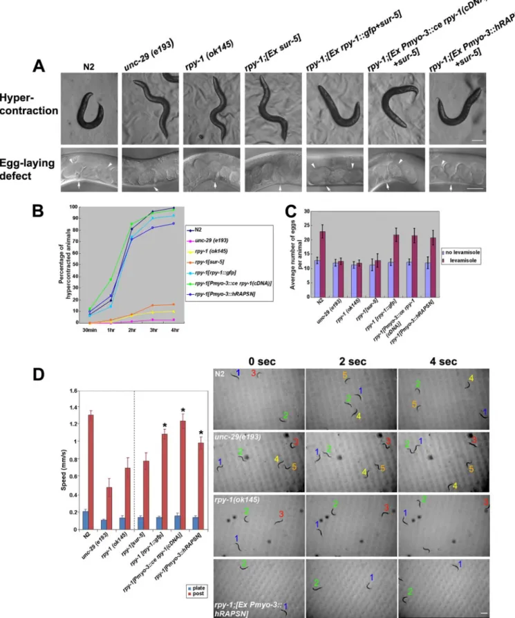
RPY-1 Is a Homolog of Rapsyn in C. elegans—The C. elegans RPY-1 protein, a putative rapsyn homolog encoded by C18H9.7, contains eight TPR domains and a RING domain. These domains are conserved in rapsyn proteins of other spe-cies (supplemental Fig. S1, A and C). We first examined the phenotype of a deletion mutation in the rpy-1 gene. The exam-ined mutation was rpy-1 (ok145), which contains a deletion of exons 4 through 10 that results in a severely truncated protein (supplemental Fig. S1B). Similar to animals that carry muta-tions in nAChR genes, rpy-1 (ok145) mutant animals were resistant to levamisole, an agonist of nAChRs (48, 49); they did not hypercontract or show increased egg laying after treatment (Fig. 1, A–C). Although rpy-1 mutant animals clearly showed levamisole resistance typical of nAChR mutations, they did not show significant defective motility on media plates in the absence of levamisole treatment (Fig. 1D). For increased sensi-tivity, we applied the post-microarray assay, which is a recently developed sensitive behavioral assay (41). With this method, we were able to detect a significant motility defect in rpy-1 (ok145) mutant animals (Fig. 1D). We then tested whether a GFP reporter gene harboring the rpy-1 gene (pJL524) could rescue rpy-1mutation phenotypic characteristics. Muscle hypercon-traction and increased egg laying caused by this mutation were rescued by injection of the rpy-1::gfp construct at a concentra-tion of 75g/ml (Fig. 1, A–C; supplemental Table S1). The motility defect detected by the post-microassay was also res-cued (Fig. 1D, supplemental Movies S1–S7). We then found that the expression of rpy-1 in muscles using the myo-3 pro-moter rescued the rpy-1 mutant phenotypes (Fig. 1, A–C; sup-plemental Table S1). Furthermore, we found that expression of the human rapsyn gene in the muscles of the nematode rpy-1 mutant rescued levamisole resistance (Fig. 1, A–C, supplemen-tal Table S2) and motility defects (Fig. 1D, supplemensupplemen-tal Movie S7). Although it is formally possible that RPY-1 and mamma-lian rapsyn may have distinct functions, the most probable interpretation of the data presented above is that RPY-1 is the functional homolog of rapsyn in the nematode.
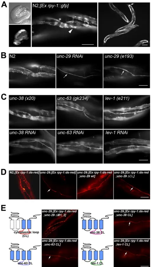
RPY-1 Is Expressed in Muscles and Neurons—Next, we exam-ined the expression pattern of rpy-1 using a reporter gene con-struct that contained the putative promoter region and full-length coding region of rpy-1. RPY-1 was first detected at 260 –270 min in embryonic development (data not shown) and persisted throughout development (Fig. 2A). In adult animals, RPY-1 was expressed in body-wall muscles, head muscles, and sex-specific muscles of hermaphrodites. The rpy-1::gfp reporter gene was also expressed in neurons (Fig. 2A).
Muscular RPY-1 Is Destabilized in the Absence of UNC-29, but Not Other nAChR Subunits—Knowing that rpy-1 is a rapsyn homolog, we started to investigate the unsolved ques-tion of whether and how RPY-1 stability is controlled. Proteins that are physically associated with rapsyn in vivo are good can-didates for effectors of RPY-1 protein stability control. To eval-uate whether RPY-1 protein abundance is dependent on the presence of its binding partner, nAChR, we examined whether the steady-state levels of RPY-1::GFP were influenced by the absence of functional nAChR subunits. We examined rpy-1::gfp expression in animals whose muscle nAChR subunit genes (unc-29, unc-38, unc-63, and lev-1) were individually knocked down by RNAi. We found that the steady-state level of RPY-1::GFP in muscle cells was reduced by RNAi targeting unc-29but not by RNAi against the other nAChR subunits (Fig. 2, B and C). We obtained identical results in experiments in which we examined the steady-state levels of RPY-1 in refer-ence mutant strains for each subunit (Fig. 2, B and C). The transcript level of rpy-1 in the unc-29 (e193) mutant worms was not reduced but rather increased probably due to compensa-tory effects (supplemental Fig. S2), suggesting that destabiliza-tion of RPY-1 is not due to the decrease in the rpy-1 transcrip-tion in muscles. Taken together, these results indicate that UNC-29, but not the other nAChR subunits, is required for stable maintenance of RPY-1 proteins.
Allele-specific Requirement of UNC-29 for Rapsyn Stability— We expressed the rpy-1::gfp reporter gene (pJL524) in various unc-29 mutant backgrounds to further evaluate the role of UNC-29 in control of RPY-1 stability. We examined the e193, e522, and x30 alleles (supplemental Fig. S3). The unc-29 (e193) mutation is a missense mutation of proline (Pro) to serine (Ser) at an evolutionarily conserved residue within the M2 trans-membrane domain, which forms the inner channel gate of the receptor (50 –52). The unc-29 (e522) mutation is a missense mutation of glycine (Gly) to glutamic acid (Glu) within the M4 transmembrane domain of the receptor, and the unc-29 (x30) mutation is a missense mutation of glutamic acid (Glu) to lysine (Lys) within the extracellular domain of the receptor. Although the expression of RPY-1::GFP was significantly reduced in the muscles of unc-29 (e193) mutant animals (Fig. 2B), RPY-1::GFP was not degraded in the unc-29 (e522) or unc-29 (x30) mutant backgrounds (supplemental Fig. S4). These results suggest that stable expression of RPY-1 in muscles requires a specific assem-blage of functional UNC-29 proteins and that specific amino acids located within the extracellular transmembrane domains or the M4 transmembrane domain may not be critical for the regulation of rapsyn stability.
The Cytoplasmic Loop of UNC-29 Is Crucial for RPY-1 Stability—Each subunit of nAChRs contains long cytoplasmic loops (CLs) between the third and fourth transmembrane domains, raising the possibility that the CL domain of UNC-29 is the RPY-1 binding domain. We found that expression of a unc-29construct that lacks the CL domain was unable to stabi-lize RPY-1 in unc-29 mutant animals (Fig. 2D). When the first two transmembrane domains of UNC-29 were deleted, leaving the CL domain intact, RPY-1 stability was restored (Fig. 2E), strongly suggesting that the CL domain is responsible for bind-ing to RPY-1. To closely examine the specific interactions
at Ewha Medical Library on March 22, 2017
http://www.jbc.org/
FIGURE 1. Analysis of rpy-1 mutant phenotype in C. elegans. A, the RPY-1::GFP reporter was functional in vivo. The rpy-1 mutant phenotypes were rescued by the expression of the rpy-1::gfp reporter or the human rapsyn gene under the control of both the rpy-1 promoter and muscle-specific promoter (Pmyo-3) in the levamisole assay. The upper panels show muscle hypercontraction, and the bottom panels show enhanced egg laying. The scale bars represent 100 (top row) and 50m (bottom row), respectively. Anterior is on the left and dorsal at the top. The arrows indicate the locations of the vulvae, and the arrowheads indicate two-cell stage eggs. B and C, the graphs summarize the results of the levamisole assay. B represents hypercontraction and C represents the egg-laying defect observed in A. D, the rescue of the rpy-1 mutant phenotypes by the rpy-1 expression using the rpy-1 promoter and the myo-3 promoter as analyzed by the micropost assay. The left graph represents post-array assay results and the right panels show the movements of the strains. *, p⬍ 0.001. The numbers indicate individual worms. The scale bar represents 500m.
at Ewha Medical Library on March 22, 2017
http://www.jbc.org/
FIGURE 2. The RPY-1 protein was unstable in the absence of UNC-29. A, the RPY-1 protein is expressed in muscles and neurons. The left panels show a 3-fold stage embryo; the middle and right panels are adults. The scale bars indicate 50 (middle image) and 100m (right panel). B, expression patterns of the RPY-1::GFP in N2, unc-29 RNAi, and mutant backgrounds. The arrows indicate the ventral nerve cord (VC). The scale bar represents 50m. C, GFP expression patterns of the RPY-1 protein in other nAChR subunit genes, unc-38, unc-63, and lev-1, RNAi, and mutant backgrounds, as indicated. The scale bar represents 50m. D, RPY-1 protein stability was dependent on the presence of the UNC-29 protein but not on the other nAChR subunits, UNC-38, UNC-63, or LEV-1 proteins. The arrows indicate the VC. The scale bar represents 50m. E, specific requirement of the cytoplasmic loop domain of UNC-29 for RPY-1 stability. The upper left panels show the effect of the deletion of two transmembrane domains of UNC-29 on the stability of RPY-1. The other panels show the effects of chimeric UNC-29 proteins, each of which contained the CL domain of UNC-38, UNC-63, or LEV-1, on the stability of RPY-1. The arrows indicate the VC. The scale bar represents 50m.
at Ewha Medical Library on March 22, 2017
http://www.jbc.org/
between the RPY-1 and UNC-29 CL domains, we constructed chimeric UNC-29 constructs, each of which contained the CL domain of UNC-38, UNC-63, or LEV-1. None of these con-structs restored RPY-1 stability in the unc-29 mutant back-ground (Fig. 2E), indicating that the CL domain of UNC-29 is indeed critical for RPY-1 stabilization.
RPY-1 Is Degraded by the Ubiquitin-proteasome Pathway— Next, we examined whether the ubiquitin-proteasome pathway is involved in the degradation of RPY-1. We found that unc-29 (e193) mutant animals containing the RPY-1::GFP transgene showed higher RPY-1 GFP expression in muscles after treat-ment with PI (PI mixture: MG132 and lactacystin) without increasing the transcript level of the gene (Fig. 3A, Fig. S2). Consistent with this finding, polyubiquitinated RPY-1 proteins were visible by Western blot analysis of anti-GFP immunopre-cipitates of PI-treated animals (Fig. 3B). To evaluate the post-translational control of RPY-1 under normal conditions, we examined the degradation of RPY-1 in wild-type animals injected with 25g/ml RPY-1 GFP transgene, which was one-third of the concentration used in the rescue experiment described above. The expression of the RPY-1::GFP was sub-stantially lower in these transgenic animals than in those injected with the concentration of 75g/ml of the transgene (supplemental Fig. S8). The transgenic animals were as sensi-tive to levamisole as nontransgenic WT animals (data not shown). We found that RPY-1::GFP was highly stabilized after PI treatment (supplemental Fig. S5). These results indicate that RPY-1 is ubiquitinated and degraded by the proteasome path-way not only in the absence of a functional UNC-29 protein but also in the wild-type background. We then examined whether nematode RPY-1 proteins are ubiquitinated in mammalian cells by expressing nematode RPY-1 in 293T cells. Fig. 3C shows that addition of Ub and MG132 greatly enhanced the ubiquitination of RPY-1 in these cells. In addition, we found that hamster ts20 cells containing a temperature-sensitive E1 mutation (53) could not degrade RPY-1 at a nonpermissive temperature (Fig. 3D). RPY-1 was efficiently degraded at the permissive temperature with a half-life of about 4 h and was not degraded after 4 h at the nonpermissive temperature (Fig. 3D). Of note, the overall level of MYC::RPY-1 expression was higher for the cells maintained at the nonpermissive temperature com-pared with that at the permissive temperature.These results suggest that rapsyn and its degradation machinery are evolu-tionarily conserved. Indeed, as shown below, all proteins
FIGURE 3. RPY-1 is degraded by the ubiquitin-proteasome pathway. A, the RPY-1::GFP protein was stabilized in vivo by treatment with a protea-some inhibitor mixture (PI). The RPY-1::GFP protein is in the unc-29 back-ground. The arrows indicate the ventral nerve cord. The scale bar represents
100m. B, PI treatment blocked RPY-1 protein degradation in the unc-29 mutant animals. A small amount of the RPY-1::GFP protein was present with-out PI treatment, probably due to its persistent expression in neurons (lanes 4 and 5). When treated with the PI, polyubiquitinated RPY-1::GFP proteins were visible (lane 6). The RPY-1::GFP protein was detected by Western blot analysis with anti-RPY-1 antibody. Western blot analysis using an anti-actin antibody was used as a loading control. C, RPY-1 was ubiquitinated in cultured cells. 293T cells were cotransfected with MYC-tagged WT RPY-1 and HA-tagged Ub. After 48 h, the cells were treated with 20MMG132 for 6 h (lane 5) or left untreated. RPY-1-Ub complexes were analyzed by immunoprecipitation (IP) with an anti-MYC antibody (9E10) followed by immunoblotting (IB) using monoclonal anti-HA antibodies. The same blot was reprobed with 9E10 to normalize RPY-1 immunoprecipitation. D, RPY-1 degradation depends on the ubiquitin pathway. The upper panels show representative results. The bottom graphs summarize the results of ts20 cell experiments. Each bar represents average value⫾ S.E. from 3 independent experiments. Actin was used as a loading control. CHX, cycloheximide; DMSO, dimethyl sulfoxide.
at Ewha Medical Library on March 22, 2017
http://www.jbc.org/
involved in rapsyn degradation have been well conserved throughout evolution.
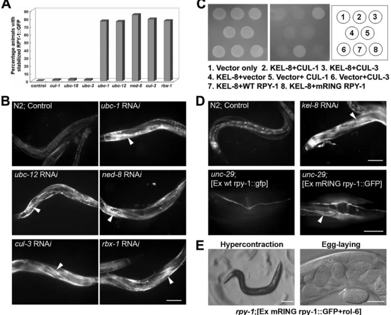
Identification of ubc-1, ubc-12, nedd-8, rbx-1, and cul-3 as Factors Required for Rapsyn Degradation—To identify protein factors responsible for rapsyn degradation, we performed RNAi experiments in which we knocked down each gene that had been previously linked to ubiquitination. Effects on RPY-1 deg-radation in wild-type animals were studied by analysis of ani-mals following injection of the transgene (25g/ml). We then knocked down most of the E2 enzyme genes in C. elegans and found that knockdown of ubc-1 and ubc-12 resulted in up-reg-ulation of RPY-1 (Fig. 4, A and B; supplemental Fig. S6). The
involvement of UBC-12, an E2 enzyme for neddylation, raised the possibility of the involvement of a cullin-E3 complex, because cullin complexes are E3-Ub ligase complexes that undergo neddylation (54, 55). We subsequently found that RNAi-mediated suppression of ned-8, rbx-1, and cul-3, but not other cullin genes, stabilized RPY-1 (Fig. 4, A and B, supple-mental Fig. S6). These RNAi results strongly suggest that the CUL-3-containing protein complex is the E3 ligase required for RPY-1 ubiquitination.
Identification of KEL-8 as a Substrate Adaptor That Binds Rapsyn—To identify the adaptor protein that links E3 ligase and RPY-1, we searched for proteins that had previously been
FIGURE 4. The identification of factors involved in RPY-1 degradation. A and B, identification of UBC-1, UBC-12, NED-8, RBX-1, and CUL-3. The graph (A) summarizes the results of RNAi assays with genes involved in the degradation of RPY-1. The y axis represents the percentage of animals with stabilized RPY-1::GFP in the muscles after each RNAi experiment. B, the panels show representative effects of RNAi on RPY-1 stability in the wild-type background. The arrowheads indicate stable RPY-1::GFP expression in the muscles. The scale bar represents 100m. C and D, KEL-8 is the substrate adaptor for RPY-1 ubiquiti-nation. C, the physical interaction of KEL-8 with rapsyn and CUL-3 as determined by yeast two-hybrid assays. KEL-8 interacts with CUL-3 and RPY-1. KEL-8 did not interact with RPY-1[mRING, H395, 398Q-RING]::GFP, which carries two His to Gln mutations in the RING domain. D, the degradation of RPY-1 required KEL-8 and the RPY-1 RING domain. The upper panels show the expression of RPY-1::GFP in wild-type animals with or without kel-8 RNAi. The arrowhead indicates the expression of RPY-1 GFP in the muscles. The scale bar represents 100m. The bottom panels show unc-29 (e193) animals carrying either a wild-type rpy-1 transgene (left panel) or the rpy-1[mRING]::gfp reporter (right panel). The arrowhead indicates the muscles. The scale bar represents 25m. E, the RING domain was required for the function of RPY-1. The left panel shows that muscle hypercontraction was absent after levamisole treatment; the right panel shows that enhanced egg laying was absent after levamisole treatment. The scale bars represent 100 (left panel) and 25m (right panel).
at Ewha Medical Library on March 22, 2017
http://www.jbc.org/
shown to physically interact with RPY-1. We found that KEL-8, encoded by W02G9.2, was a strong candidate as it was previ-ously identified as an interaction partner of RPY-1 in a yeast two-hybrid screening assay (56) and shown to contain a BTB/ POZ domain and six Kelch motifs. BTB/POZ domain proteins are known to directly interact with CUL3 and several proteins containing both BTB/POZ domain and six Kelch motif are sub-strate adapters for CUL3-containing E3 ubiquitin ligase com-plex (57– 62). We confirmed that KEL-8 interacted with the N-terminal domain of CUL-3 as well as with the full-length RPY-1 protein in yeast two-hybrid assays (Fig. 4C). Consistent with KEL-8 acting as an adaptor between the CUL-3 complex and RPY-1, we found that kel-8 RNAi stabilized RPY-1::GFP (Fig. 4D). We also found that an RPY-1 protein with a mutant RING domain failed to interact with KEL-8, indicating that the RING domain is required for the interaction (Fig. 4C). How-ever, the RING domain itself was not sufficient for binding to KEL-8 (data not shown). In addition, we found that RPY-1 pro-teins with a defective RING domain were stabilized, even in the
unc-29 (e193) mutant background (Fig. 4D), but that the
rpy-1::gfp reporter gene with the defective RING domain did not rescue the rpy-1 mutant phenotype (Fig. 4E).
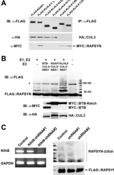
Proteins Involved in Rapsyn Degradation Are Conserved in Mammals—We noticed that all components required for RPY-1 degradation in the nematode are conserved in mam-mals. For example, KEL-8 is homologous to KLHL8 in humans (supplemental Fig. S7). To examine the evolutionary conserva-tion of the physical interacconserva-tion between the KEL-8 adaptor and rapsyn, we tested whether KLHL8 could bind to mamma-lian rapsyn. We found that KLHL8 was able to bind to human rapsyn and CUL3 by co-immunoprecipitation and Western blot analysis (Fig. 5A, lane 2). As a negative control, KEAP1, a protein that contains BTB and Kelch domains (59, 60), did not bind to rapsyn (Fig. 5A, lane 1). We then mapped the internal domain of KLHL8 for its interaction with CUL3 or rapsyn. For mapping, we generated four additional constructs that encode FLAG-tagged KLHL8 fragments (described in the figure leg-end) and co-expressed them with HA-tagged CUL3 or Myc-tagged rapsyn. We found that KLHL8 interacted with CUL3 through its BTB domain and bound to rapsyn via its Kelch domain (Fig. 5A, lanes 3– 6). Next, we performed in vitro ubiq-uitination assays using recombinant human rapsyn, yeast Uba1 (E1), human UbcH5A (E2), Ub, and the immunoprecipitated KLHL8/CUL3-containing E3 ligase complex (Fig. 5B). The KEAP1/CUL3 complex was used as a negative control. We found that rapsyn was polyubiquitinated by KLHL8-containing E3 ligase, but not by KEAP1-containing E3 ligase, clearly indi-cating that rapsyn is a direct substrate of KLHL8-containing E3 ligase in mammals. We next examined the effect of KLHL-8 depletion on the ubiquitination of rapsyn by performing RNAi experiments in mammalian cells. We found that knockdown of KLHL8 in 3T3 cells reduced the level of rapsyn ubiquitination (Fig. 5C), again indicating that the maintenance mechanism for rapsyn stability is conserved in mammals.
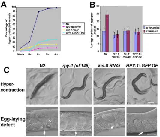
RPY-1 Accumulation by kel-8 Suppression or rpy-1 Overex-pression Resembles the rpy-1 Mutation—To examine the phys-iological consequences of RPY-1 accumulation following KEL-8 depletion, we examined the phenotypes of kel-8
knock-down animals in an otherwise wild-type background. Interest-ingly, these animals exhibited phenotypes similar to those of rpy-1 or unc-29 mutants, as they showed resistance to the AChR agonist levamisole as manifested by muscle hypercon-traction and acceleration of egg laying (Fig. 6, supplemental Table S2). These results indicate that accumulation of excess RPY-1 interferes with either the proper functioning of rapsyn-receptor complexes or their transport to the plasma membrane. Finally, we examined whether transgenic RPY-1 overexpres-sion in WT animals confers resistance to levamisole by
estab-FIGURE 5. The in vitro ubiquitination of mammalian rapsyn by
CUL3-con-taining E3 ligase and the effect of KLHL8 knockdown on the ubiquitina-tion of rapsyn. A, the physical interacubiquitina-tion between KLHL8, CUL3, and rapsyn.
293T cells transiently expressing epitope-tagged KEAP1, KLHL8 (KLHL8 F.L.), the BTB domain of KLHL8 (KLHL8 N332 (1–332 amino acids), and KLHL8 N178 (1–178 amino acids)), and the Kelch domain of KLHL8 (KLHL8 C462 (159 – 620 amino acids) or KLHL8 C309 (312– 620 amino acids)) were used. The BTB domain alone can interact with CUL3 (lanes 3 and 4, middle panel); the Kelch domain alone can interact with rapsyn (lanes 5 and 6, bottom panel). B, the in vitro ubiquitination of rapsyn by CUL3-containing E3 ligase. Purified recom-binant human rapsyn was incubated with immunoprecipitated E3 ligase complexes containing the BTB domain of KLHL8 alone (lane 2), full-length KEAP1 (lane 3), or full-length KLHL8 (lane 4) in the presence of purified E1, E2, and Ub proteins. The reaction mixture in lane 1 did not contain the E3 ligase complex, and lane 5 contained the purified rapsyn protein alone. The asterisk indicates a nonspecific product. C, the knockdown of KLHL8 inhibited the ubiquitination of rapsyn in 3T3 cells. IB, immunoblot; GAPDH, glyceraldehyde-3-phosphate dehydrogenase.
at Ewha Medical Library on March 22, 2017
http://www.jbc.org/
lishing a transgenic line injected with the RPY-1::GFP reporter at a concentration of 200g/ml, which was an 8-fold higher concentration than that used in the previous experiment. Quantitative PCR experiments showed that different concen-trations of transgenes gave rise to different gene doses of the transgenes (supplemental Fig. S8). We observed levamisole resistance in animals injected with the RPY-1::GFP reporter at a concentration of 200g/ml (Fig. 6, supplemental Table S2).
DISCUSSION
In this study, we elucidated the mechanism by which the abundance of rapsyn protein in muscles is regulated. We first proved that rpy-1, a putative nematode rapsyn homolog, is a functional homolog of rapsyn. It is interesting to note that agrin and MUSK, two key regulators of receptor clustering in mam-mals, are apparently not conserved in the nematode (37, 38). A nematode-specific protein such as LEV-10 is required for nAChR clustering in C. elegans, indicating that nonconserved proteins may play roles that are similar to those of the agrin-MUSK pathway in mammals (63). It is thus conceivable that rapsyn is the most ancient partner of AChRs. We also showed that RPY-1 stability is dependent on the presence of a non-␣ subunit of the receptor UNC-29, without which RPY-1 is degraded by the ubiquitin-proteasome pathway. We showed that the CUL-3-containing E3 ligase and KEL-8 are required for degradataion of RPY-1. We also showed that RPY-1 degradation
occurs under physiological condi-tions; failure of this degradation may cause phenotypes similar to those caused by a loss-of-function muta-tion of rpy-1. The evolumuta-tionary con-servation of the mechanism of rapsyn abundance control suggests that the failure to regulate rapsyn abundance in muscles may cause symptoms of myasthenic syn-dromes in humans.
Our results show that most RPY-1 proteins in muscle cells were significantly destabilized in the absence of UNC-29 but not in the absence of other nAChR subunits. This finding raises the possibility that the UNC-29-like non-␣ sub-unit is specifically involved in func-tional interactions with RAPSYN for the maintenance of stability in mammals. Furthermore, a specific unc-29mutant allele, e193, but not other alleles, destabilized RPY-1. Although it is still not clear whether UNC-29 directly interacts with RPY-1, the allele specificity of UNC-29 for RPY-1 degradation provides a genetic argument for a physical interaction between UNC-29 and RPY-1. It is possible that the unc-29 (e193) mutation caused a conformational change in UNC-29 or that the Pro to Ser mutation prevented proper membrane positioning of the receptors, thereby preventing the interaction with RPY-1. Mutations in the extracellular domain or in the M4 domain did not affect RPY-1 stability, suggesting that, although they caused a functional defect in the receptors, these mutations did not affect the interaction with RPY-1. Further examination of the regions within UNC-29 and the other nAChR subunits revealed that the cytoplasmic loop of UNC-29 was required the stable maintenance of RPY-1.
We found most players involved in rapsyn abundance con-trol (CUL-3, RBX-1, KEL-8, Nedd8, and UBC-12) to be compo-nents of E3 ligase. Although recent studies have suggested that RING domains often act as E3-Ub ligases for a variety of pro-teins (64 – 69), it is unlikely that rapsyn alone acts as the major E3 ligase for its own ubiquitination, because suppression of CUL-3 and other factors in the complex resulted in rapsyn sta-bilization, even in the presence of the intact RING domain. A similar case was reported for the polycomb protein Ring1B, where self-ubiquitination was not required for Ring1B degra-dation; degradation occurred through exogenous E3 ligases (70). It is possible that the RING domain of RPY-1 can be pro-tected from KEL-8-mediated ubiquitination if it is masked by physical association with the receptors.
Schaefer and Rongo (71) reported that KEL-8 is involved in the degradation of glutamate receptors in neurons. However,
FIGURE 6. Suppression of kel-8 and overexpression of RPY-1 cause phenotypes similar to that of the rpy-1
mutation. A and B, muscle hypercontraction (A) and enhanced egg laying (B) in the kel-8 RNAi and
RPY-1::GFP-overexpressing animals. N2 and rpy-1 (ok145) animals were used as controls. C shows representa-tive images of the phenotypes from B and C. The upper panels show muscle hypercontraction, and the bottom panels show enhanced egg laying after levamisole treatment. The arrows indicate the locations of the vulvae. The arrowhead indicates a two-cell stage egg. The scale bars represent 100 (top row) and 25m (bottom row).
at Ewha Medical Library on March 22, 2017
http://www.jbc.org/
KEL-8 did not directly interact with the glutamate receptors as the substrate adaptor. The direct target of KEL-8 was not iden-tified in that study. From our results, it is conceivable that KEL-8 interacts with a yet-to-be identified protein that is asso-ciated with glutamate receptors. This hypothesis could be tested by identifying proteins that physically interact with KEL-8. In contrast to rapsyn (RPY-1), which shows tissue-spe-cific expression, KLHL8 (KEL-8) is expressed in most tissues of mammals and C. elegans, implying that rapsyn is not the only substrate of the KLHL8/CUL3 complex (data not shown). It is also interesting that neuronal RPY-1 was not degraded in the
unc-29(e193) mutant animals. UNC-29 is a muscle-specific nAChR subunit, and the nematode contains many nAChR sub-units whose exact expression patterns are not completely known. It is possible that RPY-1 interacts with, and plays a role in clustering, other nAChR subunits in neurons.
Our data show that different concentrations of the rpy-1 transgene caused different effects; the transgene was able to rescue the rpy-1 mutant phenotype at a moderate tion, but it mimicked the rpy-1 mutation at a higher concentra-tion. Our interpretation is that highly excessive amounts of rapsyn inhibited receptor clustering. Although the presence of rapsyn at the postsynaptic membrane is absolutely required for AChR clustering in vivo (12, 18, 19, 72), there have been con-flicting reports about the effect of rapsyn abundance on AChR clustering and function. In some studies, expression of exoge-nous rapsyn stabilized AChR clustering, and in other cases, excessive expression of rapsyn inhibited the clustering of recep-tors (28, 29, 73). The latter cases are consistent with our results. Because the turnover rate of rapsyn is faster than that of AChR (74, 75), it is conceivable that moderate overexpression of rapsyn might increase the opportunity for rapsyn proteins to interact with the receptors, thus stabilizing more receptors. However, if rapsyn expression is too high, self-aggregation may inhibit clustering of the receptors by interfering with endoge-nous clustering. Collectively, these data strongly suggest that rapsyn abundance must be tightly regulated for proper muscle function.
It is known that multiple pathways of ubiquitin conjugation are activated in atrophying muscle (76). For example, the mus-cle-specific ubiquitin ligases MAFbx and MuRF1 are overex-pressed during muscular atrophy (77). These E3 ligases are responsible for degradation of structural proteins such as titin and transcription factors such as MyoD within muscles (78). Our finding that CUL-3-containing E3 ligase is involved in the degradation of rapsyn proteins within muscles raises the possi-bility that deregulation of the CUL-3 ligase complex in muscles could be associated with muscular disorders. Our data suggest that any defects leading to rapsyn depletion or excessive rapsyn accumulation could cause muscle defects similar to those seen in CMS in humans. CMS is caused by genetic defects, such as mutations in AChR, acetylcholinesterase, choline acetyltrans-ferase, the muscle sodium channel SCN4A, MuSK, and rapsyn genes; however, many patients do not have defects in any of these genes (25, 79 – 82). It is thus possible that factors that affect the abundance of rapsyn could be causal factors in CMS. In this respect, it might be worthwhile to examine these
patients for a new causal relationship between the CUL-3-E3 ligase complex and congenital myasthenic syndromes.
Acknowledgments—The nematode strains used in this study were provided by CGC (Minneapolis, MN). We thank A. Fire for the nem-atode expression vectors.
REFERENCES
1. Burden, S. J. (1998) Genes Dev. 12, 133–148
2. Rand, J. B. (2007) Worm Book, doi/10.1895/wormbook.1.131.1 3. Littleton, J. T., and Ganetzky, B. (2000) Neuron 26, 35– 43 4. Hubbard, J. I. (1973) Physiol. Rev. 53, 674 –723
5. Colledge, M., and Froehner, S. C. (1998) Curr. Opin. Neurobiol. 8, 357–363
6. Gautam, M., Noakes, P. G., Moscoso, L., Rupp, F., Scheller, R. H., Merlie, J. P., and Sanes, J. R. (1996) Cell 85, 525–535
7. Meier, T., Perez, G. M., and Wallace, B. G. (1995) J. Cell Biol. 131, 441– 451 8. Meier, T., Hauser, D. M., Chiquet, M., Landmann, L., Ruegg, M. A., and
Brenner, H. R. (1997) J. Neurosci. 17, 6534 – 6544
9. Ferns, M., Deiner, M., and Hall, Z. (1996) J. Cell Biol. 132, 937–944 10. Glass, D. J., Bowen, D. C., Stitt, T. N., Radziejewski, C., Bruno, J., Ryan,
T. E., Gies, D. R., Shah, S., Mattsson, K., Burden, S. J., DiStefano, P. S., Valenzuela, D. M., DeChiara, T. M., and Yancopoulos, G. D. (1996) Cell
85,513–523
11. Fuhrer, C., Sugiyama, J. E., Taylor, R. G., and Hall, Z. W. (1997) EMBO J.
16,4951– 4960
12. Gautam, M., Noakes, P. G., Mudd, J., Nichol, M., Chu, G. C., Sanes, J. R., and Merlie, J. P. (1995) Nature 377, 232–236
13. Apel, E. D., Roberds, S. L., Campbell, K. P., and Merlie, J. P. (1995) Neuron
15,115–126
14. Finn, A. J., Feng, G., and Pendergast, A. M. (2003) Nat. Neurosci. 6, 717–723
15. Cheusova, T., Khan, M. A., Schubert, S. W., Gavin, A. C., Buchou, T., Jacob, G., Sticht, H., Allende, J., Boldyreff, B., Brenner, H. R., and Hashe-molhosseini, S. (2006) Genes Dev. 20, 1800 –1816
16. Zhang, B., Luo, S., Dong, X. P., Zhang, X., Liu, C., Luo, Z., Xiong, W. C., and Mei, L. (2007) J. Neurosci. 27, 3968 –3973
17. Wang, J., Jing, Z., Zhang, L., Zhou, G., Braun, J., Yao, Y., and Wang, Z. Z. (2003) Nat. Neurosci. 6, 1017–1018
18. Froehner, S. C., Luetje, C. W., Scotland, P. B., and Patrick, J. (1990) Neuron
5,403– 410
19. Phillips, W. D., Kopta, C., Blount, P., Gardner, P. D., Steinbach, J. H., and Merlie, J. P. (1991) Science 251, 568 –570
20. Yu, X. M., and Hall, Z. W. (1994) J. Neurosci. 14, 785–795
21. Ramarao, M. K., and Cohen, J. B. (1998) Proc. Natl. Acad. Sci. U. S. A. 95, 4007– 4012
22. Lin, W., Dominguez, B., Yang, J., Aryal, P., Brandon, E. P., Gage, F. H., and Lee, K. F. (2005) Neuron 46, 569 –579
23. Misgeld, T., Kummer, T. T., Lichtman, J. W., and Sanes, J. R. (2005) Proc. Natl. Acad. Sci. U. S. A. 102,11088 –11093
24. Chen, F., Qian, L., Yang, Z. H., Huang, Y., Ngo, S. T., Ruan, N. J., Wang, J., Schneider, C., Noakes, P. G., Ding, Y. Q., Mei, L., and Luo, Z. G. (2007) Neuron 55,247–260
25. Ohno, K., Engel, A. G., Shen, X. M., Selcen, D., Brengman, J., Harper, C. M., Tsujino, A., and Milone, M. (2002) Am. J. Hum. Genet. 70, 875– 885 26. Dunne, V., and Maselli, R. A. (2003) J. Hum. Genet. 48, 204 –207 27. LaRochelle, W. J., and Froehner, S. C. (1986) J. Biol. Chem. 261,
5270 –5274
28. Yoshihara, C. M., and Hall, Z. W. (1993) J. Cell Biol. 122, 169 –179 29. Han, H., Noakes, P. G., and Phillips, W. D. (1999) J. Neurocytol. 28,
763–775
30. Rodova, M., Kelly, K. F., VanSaun, M., Daniel, J. M., and Werle, M. J. (2004) Mol. Cell. Biol. 24,7188 –7196
31. Colledge, M., Snyder, E. M., Crozier, R. A., Soderling, J. A., Jin, Y., Lange-berg, L. K., Lu, H., Bear, M. F., and Scott, J. D. (2003) Neuron 40, 595– 607 32. Ehlers, M. D. (2003) Nat. Neurosci. 6, 231–242
at Ewha Medical Library on March 22, 2017
http://www.jbc.org/
33. Burbea, M., Dreier, L., Dittman, J. S., Grunwald, M. E., and Kaplan, J. M. (2002) Neuron 35, 107–120
34. Juo, P., and Kaplan, J. M. (2004) Curr. Biol. 14, 2057–2062
35. van Roessel, P., Elliott, D. A., Robinson, I. M., Prokop, A., and Brand, A. H. (2004) Cell 119, 707–718
36. Brockhausen, J., Cole, R. N., Gervasio, O. L., Ngo, S. T., Noakes, P. G., and Phillips, W. D. (2008) Dev. Neurobiol. 68, 1153–1169
37. Hrus, A., Lau, G., Hutter, H., Schenk, S., Ferralli, J., Brown-Luedi, M., Chiquet-Ehrismann, R., and Canevascini, S. (2007) PLoS ONE 2, e731 38. Forrester, W. C., Dell, M., Perens, E., and Garriga, G. (1999) Nature 400,
881– 885
39. Brenner, S. (1974) Genetics 77, 71–94
40. Xia, Y., and Whitesides, G. M. (1998) Angew. Chem. Int. Ed. 37, 550 –575 41. Park, S., Hwang, H., Nam, S. W., Martinez, F., Austin, R. H., and Ryu, W. S.
(2008) PLoS ONE 3, e2550
42. Mello, C. C., Kramer, J. M., Stinchcomb, D., and Ambros, V. (1991) EMBO J. 10,3959 –3970
43. Bush, K. T., Goldberg, A. L., and Nigam, S. K. (1997) J. Biol. Chem. 272, 9086 –9092
44. Courbard, J. R., Fiore, F., Adelaide, J., Borg, J. P., Birnbaum, D., and Ollen-dorff, V. (2002) J. Biol. Chem. 277, 45267– 45275
45. Rocca, A., Lamaze, C., Subtil, A., and Dautry-Varsat, A. (2001) Mol. Biol. Cell 12,1293–1301
46. Jones, D., Crowe, E., Stevens, T. A., and Candido, E. P. (2002) Genome Biol.
3,RESEARCH0002
47. Min, K. W., Hwang, J. W., Lee, J. S., Park, Y., Tamura, T. A., and Yoon, J. B. (2003) J. Biol. Chem. 278, 15905–15910
48. Lewis, J. A., Wu, C. H., Berg, H., and Levine, J. H. (1980) Genetics 95, 905–928
49. Lewis, J. A., Wu, C. H., Levine, J. H., and Berg, H. (1980) Neuroscience 5, 967–989
50. Sine, S. M., and Engel, A. G. (2006) Nature 440, 448 – 455 51. Lee, W. Y., and Sine, S. M. (2005) Nature 438, 243–247
52. Lummis, S. C., Beene, D. L., Lee, L. W., Lester, H. A., Broadhurst, R. W., and Dougherty, D. A. (2005) Nature 438, 248 –252
53. Kulka, R. G., Raboy, B., Schuster, R., Parag, H. A., Diamond, G., Ciech-anover, A., and Marcus, M. (1988) J. Biol. Chem. 263, 15726 –15731 54. Parry, G., and Estelle, M. (2004) Semin. Cell Dev. Biol. 15, 221–229 55. Pan, Z. Q., Kentsis, A., Dias, D. C., Yamoah, K., and Wu, K. (2004)
Onco-gene 23,1985–1997
56. Li, S., Armstrong, C. M., Bertin, N., Ge, H., Milstein, S., Boxem, M., Vidalain, P. O., Han, J. D., Chesneau, A., Hao, T., Goldberg, D. S., Li, N., Martinez, M., Rual, J. F., Lamesch, P., Xu, L., Tewari, M., Wong, S. L., Zhang, L. V., Berriz, G. F., Jacotot, L., Vaglio, P., Reboul, J., Hirozane-Kishikawa, T., Li, Q., Gabel, H. W., Elewa, A., Baumgartner, B., Rose, D. J., Yu, H., Bosak, S., Sequerra, R., Fraser, A., Mango, S. E., Saxton, W. M., Strome, S., Van Den Heuvel, S., Piano, F., Vandenhaute, J., Sardet, C., Gerstein, M., Doucette-Stamm, L., Gunsalus, K. C., Harper, J. W., Cusick,
M. E., Roth, F. P., Hill, D. E., and Vidal, M. (2004) Science 303, 540 –543 57. van den Heuvel, S. (2004) Curr. Biol. 14, R59 –R61
58. Pintard, L., Willems, A., and Peter, M. (2004) EMBO J. 23, 1681–1687 59. Kobayashi, A., Kang, M. I., Okawa, H., Ohtsuji, M., Zenke, Y., Chiba, T.,
Igarashi, K., and Yamamoto, M. (2004) Mol. Cell. Biol. 24, 7130 –7139 60. Cullinan, S. B., Gordan, J. D., Jin, J., Harper, J. W., and Diehl, J. A. (2004)
Mol. Cell. Biol. 24,8477– 8486
61. Sumara, I., Quadroni, M., Frei, C., Olma, M. H., Sumara, G., Ricci, R., and Peter, M. (2007) Dev. Cell 12, 887–900
62. Angers, S., Thorpe, C. J., Biechele, T. L., Goldenberg, S. J., Zheng, N., MacCoss, M. J., and Moon, R. T. (2006) Nat. Cell Biol. 8, 348 –357 63. Gally, C., Eimer, S., Richmond, J. E., and Bessereau, J. L. (2004) Nature 431,
578 –582
64. Joazeiro, C. A., Wing, S. S., Huang, H., Leverson, J. D., Hunter, T., and Liu, Y. C. (1999) Science 286, 309 –312
65. Freemont, P. S. (2000) Curr. Biol. 10, R84 –R87
66. Tyers, M., and Jorgensen, P. (2000) Curr. Opin. Genet. Dev. 10, 54 – 64 67. Chen, A., Wu, K., Fuchs, S. Y., Tan, P., Gomez, C., and Pan, Z. Q. (2000)
J. Biol. Chem. 275,15432–15439
68. Leverson, J. D., Joazeiro, C. A., Page, A. M., Huang, H., Hieter, P., and Hunter, T. (2000) Mol. Biol. Cell 11, 2315–2325
69. Seol, J. H., Feldman, R. M., Zachariae, W., Shevchenko, A., Correll, C. C., Lyapina, S., Chi, Y., Galova, M., Claypool, J., Sandmeyer, S., Nasmyth, K., and Deshaies, R. J. (1999) Genes Dev. 13, 1614 –1626
70. Ben-Saadon, R., Zaaroor, D., Ziv, T., and Ciechanover, A. (2006) Mol. Cell
24,701–711
71. Schaefer, H., and Rongo, C. (2006) Mol. Biol. Cell 17, 1250 –1260 72. Noakes, P. G., Phillips, W. D., Hanley, T. A., Sanes, J. R., and Merlie, J. P.
(1993) Dev. Biol. 155, 275–280
73. Han, H., Yang, S. H., and Phillips, W. D. (2000) J. Neurosci. Res. 60, 155–163
74. Frail, D. E., Musil, L. S., Buonanno, A., and Merlie, J. P. (1989) Neuron 2, 1077–1086
75. Bruneau, E., and Akaaboune, M. (2007) J. Biol. Chem. 282, 9932–9940 76. Lecker, S. H. (2003) Curr. Opin. Clin. Nutr. Metab. Care 6, 271–275 77. Bodine, S. C., Latres, E., Baumhueter, S., Lai, V. K., Nunez, L., Clarke, B. A.,
Poueymirou, W. T., Panaro, F. J., Na, E., Dharmarajan, K., Pan, Z. Q., Valenzuela, D. M., DeChiara, T. M., Stitt, T. N., Yancopoulos, G. D., and Glass, D. J. (2001) Science 294, 1704 –1708
78. Tintignac, L. A., Lagirand, J., Batonnet, S., Sirri, V., Leibovitch, M. P., and Leibovitch, S. A. (2005) J. Biol. Chem. 280, 2847–2856
79. Hantai, D., Richard, P., Koenig, J., and Eymard, B. (2004) Curr. Opin. Neurol. 17,539 –551
80. Lecker, S. H., Jagoe, R. T., Gilbert, A., Gomes, M., Baracos, V., Bailey, J., Price, S. R., Mitch, W. E., and Goldberg, A. L. (2004) FASEB J. 18, 39 –51 81. Engel, A. G., Ohno, K., and Sine, S. M. (2003) Muscle Nerve 27, 4 –25 82. McConville, J., and Vincent, A. (2002) Curr. Opin. Pharmacol. 2, 296 –301
at Ewha Medical Library on March 22, 2017
http://www.jbc.org/
Jongbok Yoon, Hyunsook Lee, Sungsu Park and Junho Lee
doi: 10.1074/jbc.M808230200 originally published online January 20, 2009 2009, 284:8195-8206.
J. Biol. Chem.
10.1074/jbc.M808230200
Access the most updated version of this article at doi: Alerts:
When a correction for this article is posted
•
When this article is cited
•
to choose from all of JBC's e-mail alerts
Click here
Supplemental material:
http://www.jbc.org/content/suppl/2009/01/21/M808230200.DC1 http://www.jbc.org/content/284/12/8195.full.html#ref-list-1This article cites 80 references, 35 of which can be accessed free at
at Ewha Medical Library on March 22, 2017
http://www.jbc.org/