저작자표시-비영리-변경금지 2.0 대한민국 이용자는 아래의 조건을 따르는 경우에 한하여 자유롭게
l 이 저작물을 복제, 배포, 전송, 전시, 공연 및 방송할 수 있습니다. 다음과 같은 조건을 따라야 합니다:
l 귀하는, 이 저작물의 재이용이나 배포의 경우, 이 저작물에 적용된 이용허락조건 을 명확하게 나타내어야 합니다.
l 저작권자로부터 별도의 허가를 받으면 이러한 조건들은 적용되지 않습니다.
저작권법에 따른 이용자의 권리는 위의 내용에 의하여 영향을 받지 않습니다. 이것은 이용허락규약(Legal Code)을 이해하기 쉽게 요약한 것입니다.
Disclaimer
저작자표시. 귀하는 원저작자를 표시하여야 합니다.
비영리. 귀하는 이 저작물을 영리 목적으로 이용할 수 없습니다.
변경금지. 귀하는 이 저작물을 개작, 변형 또는 가공할 수 없습니다.
2019년 2월 박사학위 논문
Inhibitory Effects of
12, 14-Labdantrien-18-oic Acid, an Active Component Isolated from
Pinus densiflora Needle,
on Adrenal Catecholamine Secretion
조선대학교 대학원
의 학 과
김 남 철
[UCI]I804:24011-200000267200
Inhibitory Effects of
12, 14-Labdantrien-18-oic Acid, an Active Component Isolated from
Pinus densiflora Needle,
on Adrenal Catecholamine Secretion
솔잎에서 분리한 활성성분인 12, 14-Labdantrien-18-oic Acid 의 부신 카테콜아민분비에 대한 억제효과
2019년 2월 25일
조선대학교 대학원
의 학 과
김 남 철
Inhibitory Effects of
12, 14-Labdantrien-18-oic Acid, an Active Component Isolated from
Pinus densiflora Needle,
on Adrenal Catecholamine Secretion
지도교수 김 상 훈
이 논문을 의학 박사학위신청 논문으로 제출함
2018 년 10 월
조선대학교 대학원
의 학 과
김 남 철
김남철의 박사학위논문을 인준함
위원장 조선대학교 교수 박상학 ( 인 ) 위 원 조선대학교 교수 임동윤 (인)
위 원 전남대학교 교수 김재민 ( 인 )
위 원 조선대학교 교수 김승곤 ( 인 )
위 원 조선대학교 교수 김상훈 (인)
2018 년 12 월
조선대학교 대학원
CONTENTS
KOREAN ABSTRACT
---iI. INTRODUCTION
---1II. MATERIALS AND METHODS
---3Experimental procedure ---3
Isolation of adrenal glands ---3
Perfusion of adrenal gland ---4
Drug administration ---4
Collection of perfusate ---5
Measurement of catecholamines ---5
Measurement of NO release ---6
Statistical analysis ---6
Drugs and their sources ---6
III. RESULTS
---8Identification of chemical structure of LDTEA from methylene chloride fraction of Pinus densiflora needle, and comparison of some fractions on ACh-produced CA secretion from the perfused rat adrenal medullae---
----8Effects of LDTEA on the CA secretion produced by angiotensin II, ACh, DMPP, and McN-A-343 from the perfused adrenal medullae
---12Effects of LDTEA on the CA secretion produced by high K
+, Bay-K-8644, thapsigargin, and veratridine from the perfused adrenal medullae---
---17Influence of LDTEA plus L-NAME on CA secretion produced by ACh, DMPP,
McN-A-343, angiotensin II, High K
+, Bay-K-8644, thapsigargin, and veratridine
in the perfused adrenal medullae ---
---22Influence of LDTEA on the released level of nitric oxide in the perfused adrenal
medullae
---32IV. DISCUSSION
---34V. SUMMARY
---40REFERENCES
---42CONTENTS OF FIGURES
Fig. 1. Schematic drawing of the preparation used to study the CA secretion in the isolated perfused rat adrenal gland ---3
Fig. 2.Schematic diagram of fractionation from Pinus densifloraneedle ---9
Fig. 3. Identification of chemical structure by NMR of LDTEA from methylene chloride fraction ofPinus densiflora needle ---10
Fig. 4. Comparative effects of four fractions obtained from pine needle extract on acetylcholine-produced CA release in the perfused adrenal medullae ---11
Fig. 5. Dose-dependent effects of LDTEA on the secretory responses of catecholamines (CA) produced by acetylcholine (ACh) in the perfused adrenal medullae ---13
Fig. 6. Time-course effects of LDTEA on DMPP-produced CA secretion in the perfused adrenal medullae ---14
Fig. 7. Time-course effects of LDTEA on McN-A-343-produced CA release in the perfused adrenal medullae ---15
Fig. 8.Time-course effects of LDTEA on angiotensin II-produced CA secretion in the perfused rat adrenal medullae ---16
Fig. 9.Time-course effects of LDTEA on the high K+-produced CA secretion in the perfused adrenal medullae---18
Fig. 10.Time-course effects of LDTEA on Bay-K-8644-produced CA secretion in the perfused adrenal medullae ---19
Fig. 11.Time-course effects of LDTEA on thapsigargin-produced CA secretion in the perfused adrenal medullae ---20
Fig. 12.Time-course effects of LDTEA on veratridine-produced CA secretion in the perfused adrenal medullae ---21
Fig. 13. Effects of LDTEA plus L-NAME on acetylcholine-produced CA secretion in the perfused adrenal medullae ---23
Fig. 14. Effects of LDTEA plus L-NAME on DMPP-produced CA secretion in the perfused adrenal medullae ---24
Fig. 15. Effects of LDTEA plus L-NAME on McN-A-343-produced CA secretion in the perfused adrenal medullae ---25
Fig. 16. Effects of LDTEA plus L-NAME on angiotensin II-produced CA secretion in the perfused adrenal medullae ---26
Fig. 17.Influence of LDTEA plus L-NAME on high potassium-produced CA secretion in the perfused adrenal medullae ---28
Fig. 18. Effects of LDTEA plus L-NAME on Bay-K-8644-produced CA secretion in the perfused adrenal medullae ---29
Fig. 19. Effects of LDTEA plus L-NAME on thapsigargin-produced CA secretion in the perfused adrenal medullae ---30
Fig. 20. Effects of LDTEA plus L-NAME on veratridine-produced CA secretion in the perfused adrenal medullae ---31
Fig. 21. Effects of LDTEA on nitric oxide (NO) release in the perfused rat adrenal medullae ---33
Fig. 22. Schematic diagram of possible action site of LDTEA in the rat adrenal medullae ---39
< 국문초록 >
솔잎에서 분리한 활성성분인 12, 14-Labdantrien-18-oic Acid 의 부신 카테콜아민분비에 대한 억제효과
김 남 철
지도교수 : 김 상 훈 조선대학교 대학원 의학과
이전에 솔잎(Pinus densiflora needle)의 자가발효추출물이 흰쥐 관류부신수질에서
catecholamines (CA) 분비를 억제하며 (Shin 등, 2010), 또한 phenylephrine 과 high K+ 에 의한 흰쥐 적출 대동맥편의 수축반응을 억제한다고 알려졌다 (Cheong 및 Lim, 2010). 솔잎 자가발효액의 CH2Cl2추출물에서 분리한 Fraction 4-5가 acetylcholine (ACh)의 CA유리에 대 한 강력한 억제를 나타낸다고 하였다 (Hong 및 Lim, 2012). 솔잎 자가발효액의 ethylacetate 추출물에서 분리한 Fraction 4-5 역시 흰쥐 적출 관류신수질에서 콜린수용체 흥분 및 직접막탈분극에 의한 CA분비를 억제한다고 보고되었다 (Choi 등, 2013). 따라서 본 연구의 목적은 솔잎에서 분리한 가장 강력한 주요성분인 12, 14-labdantrien-18-oic
acid (LDTEA)가 흰쥐에서 분리 적출한 부신수질의 관류모델에서 CA유리작용에 미치는
효과를 탐색하여, 그 작용의 본태를 규명하고자 본 연구를 수행하여 얻어진 실험결과 는 다음과 같다.
LDTEA (70~600 µM)를 부신수질 내로 90분간 관류시 acetylcholine (5.32 mM, ACh)의 CA 유리작용을 비교적 용량 및 시간 의존적으로 억제시켰다. 또한 LDTEA (200 µM)를 관류하는 동안 DMPP (선택성 니코틴수용체 작용제), angiotesin II (Ang II, 100 ηM) 및 McN-A-343 (100 µM, 선택성 무스카린 M1수용체 작용제)의 CA 유리반응에 대한 시간
의존적인 억제작용을 나타내었다. 그러나, LDTEA 자체는 기초 CA 유리효과에 영향을 미치지 아니하였다. 또한, LDTEA (200 µM)를 과류하는 동안, 직접적인 새포막탈분극제인 고칼륨 (56 mM), 선택성 전압의존적 나트륨통로 활성화제, veratridine (50 µM), 전압의존적 L-형 dihyropyridine계 칼슘통로 활성화제, Bay-K-8644 (10 µM) 및 세포질 내 내형질세망막에서 Ca2+-ATPase 억제제, thapsigargin (10 µM)의 CA 분비반응이 역시 시간 의존적으로 감약되었다. 흥미롭게도, LDTEA (200 µM)와 L-NAME (30 µM, NO synthase 억제제)를 90분간 동시 관류하는 동안 ACh, DMPP, McN-A-343, Ang II, high K+, Bay-K-8644, veratridine 및 thapsigargin의 CA 분비효과가
LDTEA (200 µM)단독관류 시의 억제효과와 비교하여 상응하는 거의 대조치 수준으로
회복되었다. 또한 실제로 NO 유리량이 LDTEA (200 µM) 관류 후에 기초 유리량과 비교하여 현저하게 상승하였다.
이와 같은 연구결과를 종합하여 보면, LDTEA는 적출 흰쥐 부신의 관류모델에서 아세틸콜린수용체(니코틴 및 무스카린 수용체) 및 안지오텐신II 수용체 활성화에 의한 CA 분비작용에 대하여 현저한 억제작용을 나타내었다. 이러한 LDTEA의 억제작용은 흰쥐 부신수질에서 NO synthase 활성화에 의한 산화질소 생성증가로 인해 부신수질 크롬친화세포 내로 나트륨 및 칼슘통로를 통한 이들의 유입억제와 세포 내 칼슘저장고에서 칼슘유리에 대한 억제작용에 기인되며, 이는 니코틴수용체 및 안지오텐신 수용체 차단과 관련이 있는 것으로 생각된다. 이와 같은 결과를 바탕으로
LDTEA는 부신수질세포에서 CA유리를 억제하여, 결국 순환계의 CA농도를
저하시킴으로서 고혈압 및 협심증과 같은 심혈관계 질환 치료나 예방에 임상적으로 유용할 것으로 사료된다.
I. INTRODUCTION
Previously, it has been found that self-fermented pine needle (Pinus densiflora) extract inhibits secretion of catecholamines (CA) from the perfused rat adrenal medulla (Shin et al., 2010), and also the contractile responses of isolated rat aortic strips induced by phenylephrine and high K+ (Cheong & Lim, 2010). Fraction 4-5 among several fractions obtained from methylene chloride (CH2Cl2) extract of self-fermented pine needle showed the most potent inhibitory effects on the acetylcholine (ACh)-evoked CA release (Hong & Lim, 2012). Fraction 4-5 isolated from ethylacetate extract of self-fermented pine needle inhibits the CA secretion from the isolated perfused rat adrenal medulla evoked by stimulation of cholinergic receptors as well as by direct membrane-depolarization (Choi et al., 2013).
In general, it has also been found that the Pinus densiflora Sieb. et Zucc. (Pinaceae), red pine naturally grows or is planted in mountain districts of Japan, Korea, and China. Red pine needles have been traditionally used as a nutritive tonic agent Korean folk drug are frequently employed to brew a tea in Korea. Pine needle extract has been shown to cause several actions, such as an antioxidant activity in rats fed highly oxidized fat (Lee, 2003), cytotoxic effects on several cancer cell lines (Chung et al., 2002), inhibition of the pacemaker currents of interstitial cells of Cajal (ICC) by activating ATP-sensitive K+channels via the production of PGs (Cheong et al., 2005), and nitrite scavenging activities (Park et al., 2002). Hsu and his co-workers (2005) have shown that pine needle (Pinus morrisonicolaHay.) scavenges superoxide and suppresses the increase of leukemia cell U937. For the antioxidant activity of several fractions from Pinus densiflora, the following rank order of potency was obtained: ethyl acetate> n-butanol > water
> dichloromethane fraction (Jung et al., 2003). It has been reported that the pine bark extract was able to stimulate in vitro the production of nitric oxide, thus counteracting the vasoconstriction by adrenaline or noradrenaline in isolated aortic rings from rats (Fitzpatrick et al., 1998). Pine pollen powder, called ‘natural micro-nutrient storeroom’, is rich in many kinds of body-demanding amino acid, minerals, vitamin, enzyme, and flavonoids (Wang et al., 2005). It has been shown that pollen lipids of a pine species produce a marked reduction of platelet activating factor reactivity (Siafaka-Kapadai et al., 1986). Lee et al. (2009) proposed that the
pollen of pine tree is potentially an antioxidant, and beneficial for inflammatory conditions via down regulation of IL-1β-evoked JNK and matrix metalloproteinases (MMPs).
Thus, the present study was designed to examine the influence of 12, 14-labdantrien-18-oic acid, one of the active components isolated from ethyl acetate extract of Pinus densiflora needle on CA secretion in the perfused model of the adrenal medulla isolated from the rat, and also to verify its mechanism of action.
. MATERIALS AND METHODS
Ⅱ
Experimental procedure
Mature Sprague-Dowley (male) rats (DAMOOL SCIENCE, International Customer Service, Seoul, R.O.K.), weighing 180 to 300 grams, were employed in this study. The experimental animals individually were housed in separate cages. Food (Cheil Animal Chow, Korea) and tap water were freely allowed for about 7 to 10 days to adapt to circumstances. The rat, on the day of experiment, was given intraperitoneally with thiopental sodium (50 mg/kg) for anesthesia, and fixed in supine position on operating platform.
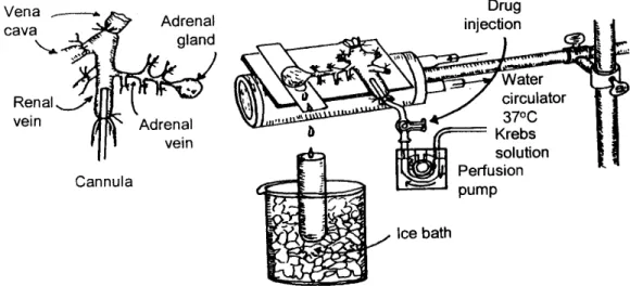
Fig. 1. Schematic drawing of the preparation used to study the CA secretion in the isolated perfused rat adrenal gland.
Isolation of adrenal glands: The adrenal medulla was isolated by some modification of previous method (Wakade, 1981). Through the midline incision, the abdomen was opened, and the left adrenal gland and its surrounding area were exposed by the three-hook retractors. The portions of the stomach, intestine and the liver were not subduced, but shunt to the right side and mantled with saline-soaked gauge pads, and urine in the bladder was removed in order to secure enough
working space for tying the blood vessels and cannulations. A cannula, employed for perfusion of the adrenal gland, was inserted into the distal end of the renal vein after the ligation of all branches of adrenal vein (if any), aorta and vena cava. Prior to ligation of vessels and cannulations, heparin (400 IU/mL) was given intravenously into vena cava to protect the blood coagulation. To make a small slit into just opposite side of adrenal vein, the adrenal cortex was cut down. The gland was initiated to perfuse, checking up there is no leakage, and the perfusion fluid flowed out only from the slit of the adrenal cortex. Then the adrenal gland, along with the cannula and the ligated blood vessels, was charily isolated from the rat and placed on the platform of a leucite chamber. The chamber was steadily circulated with water heated at 37 ±1℃ (Fig. 1).
Perfusion of adrenal gland
The perfusion of the adrenal medullae was done by means of peristaltic pump (Isco, St. Lincoln, NE, U.S.A.) with a rate of 0.31 mL/min. The perfusion was conducted with Krebs-bicarbonate solution including the following composition (mM): KH2PO4, 1.2; KCl, 4.7; CaCl2, 2.5; NaCl, 118.4; NaHCO3, 25; MgCl2, 1.18; glucose, 11.7. The perfusion solution was bubbled steadily with 95 % O2+ 5 % CO2and the pH of the Krebs-bicarbonate solution was persistently adjusted to 7.4 ~ 7.5. Ascorbic acid (100 mg/mL) and disodium EDTA (10 mg/mL) to protect oxidation of catecholamines were added into the Krebs-bicarbonate solution.
Drug administration
Angiotensin II (100 nM) and DMPP (100 μM) for 2 minutes and/or a single injection of and KCl (56 mM) and ACh (5.32 mM) in a volume of 50 μL were given into perfusion stream through a three-way stopcock, respectively. McN-A-343 (100 μM), veratridine (50 μM), thapsigargin (10 μM), and Bay-K-8644 (10 μM) were also administered by infusion for 4 min, respectively.
It was found that, in the preliminary studies, following the injection or perfusion of these drugs, the secretory responses to ACh, angiotensin II, KCl, McN-A-343, Bay-K-8644, veratridine and thapsigargin was recovered to pre-injection level in about 4 min, but the response to DMPP in 8 min.
Collection of perfusate
In general, the collection of perfusate prior to administration of various secretagogues, was done for 4 min to determine the basal CA release (background sample). Immediately after the background sample was collected, the perfusates were steadily collected in another tube as quick as the perfusion solution containing the stimulatory secretagogue got to the adrenal medulla. The perfusate of the stimulated sample was collected for 4 or 8 min. The amounts liberated in the background sample were subtracted from that liberated from the stimulated sample to get the net CA release, which is depicted in all of the figures.
Prior to examine the effect of LDTEA on the basal and produced CA release, the infusion of adrenal medulla was made with normal Krebs-bicarbonate solution for 90 min, and then the collection of perfusate was done for a certain period (background sample). Then the solution was changed with the one containing the stimulatory secretagogue or together with LDTEA, and the perfusates was collected for the same period as that for the background sample. The perfusate from the adrenal medulla was collected in chilled tubes.
Measurement of catecholamines
The content of CA, including epinephrine, norepinephrine and dopamine, in perfusate was directly assayed by the fluorometric method of Anton and Sayre (1962) without the intermediate purification process with alumina for the reasons described earlier (Wakade, 1981) using fluorospectrophotometer (Kontron Co., Milano, Italy).
The perfusate in volume of 0.2 mL was employed for the measurement reaction. The CA content in the perfusate of stimulated medulla by secretagogues used in the present work was fully enough to get readings several folds greater than the reading of unstimulated samples (control). The CA level in the perfusate was depicted in terms of norepinephrine (base) equivalents.
Measurement of NO release
The NO-selective microelectrode (ami700, Innovative Instruments Inc., Tampa
,
FL, USA) and an amplifier (inNo meter, Innovative Instruments Inc., Tampa,
FL, USA) were utilized for measurement of NO liberated from the perfused adrenal medulla. NO liberated from adrenal medulla was assayed as the integrated signal detected by the microelectrode after perfusion of LDTEA into rat adrenal medulla, as previously described (McVeigh et al., 2002). The value of electrode was calibrated by the established standard concentrations of NO in 0.5% (wt/vol) KI in 0.1 Mol/L H2SO4from NaNO2standards. NO liberation was measured as the current level detected at the electrode after infusion of LDTEA into adrenal medulla. The net NO release was expressed as picomoles in the figure.Statistical analysis
The difference between the control group and the drug-treated group was statistically analyzed by the Student's t-test. A P-value of less than 0.05 was regarded statistically to elicit significant changes unless specifically described in the text. Values depicted in the text refer to means and the standard errors of the mean (S.E.M.). The experimental data were statistically assayed by computer program (Tallarida and Murray, 1987).
Drugs and their sources
The following drugs were used: LDTEA (a gift from Professor HS Cheong, College of Natural Science, Chosun University, Gwangju, Korea), 3-(m-chloro-phenyl-carbamoyl-oxy)-2 -butynyltrimethyl ammonium chloride [McN-A-343] (RBI, U.S.A.), thapsigargin, norepinephrine bitartrate, acetylcholine chloride, veratridine hydrochloride, 1.1-dimethyl-4-phenyl piperazinium iodide (DMPP), potassium chloride (KCl), Sodium bicarbonate, calcium chloride, Nω-nitro-L-arginine methyl ester hydrochloride (L-NAME), angiotensin II, sodium chloride, potassium phosphate, glucose, ascorbic acid, methyl-1,4-dihydro-2,6-dimethyl-3-nitro -4-(2-trifluoro-methyl-phenyl)-pyridine-5-carboxylate [Bay–K-8644], disodium EDTA, magnesium chloride (Sigma Chemical Co., U.S.A.). Drugs were dissolved in distilled water (stock)
and then added to the normal Krebs-bicarbonate solution, Exceptively, Bay-K-8644 was dissolved in 99.5 % (stock) ethanol, and then diluted appropriately with the perfusion solution (final concentration of ethanol was less than 0.1 %). Concentrations of all reagents employed in the present study are expressed in terms of their molar base.
III. RESULTS
Identification of chemical structure of LDTEA from methylene chloride fraction of Pinus densiflora needle, and comparison of some fractions on ACh-produced CA secretion from the perfused rat adrenal medullae
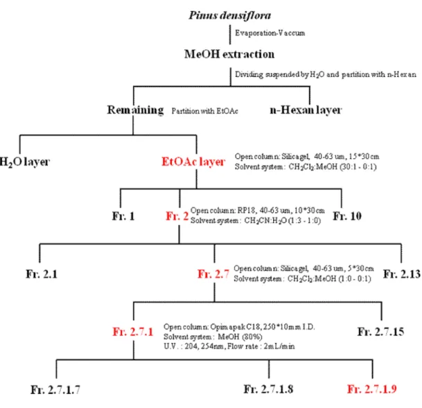
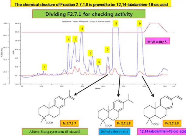
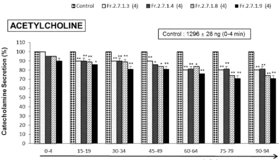
As shown Fig. 2 and 3, Fr. 2.7.1.9 (12, 14-Labdantrien-18-oic Acid, LDTEA), Fr. 2.7.1.8 (dehydroabietic acid), Fr. 2.7.1.4, and Fr. 2.7.1.3 were finally identified as the main components of several fractions extracted from Pinus densiflora needle through chemical analysis by nuclear magnetic resonance (NMR). Based on these components, effects of these fractions on adrenal CA secretion were compared each other. After the infusion of oxygenated Krebs-bicarbonate solution for 60 min, the basal CA release in the isolated perfused rat adrenal medullae was 23±2 ng for 2 min (n=12). Since it has been reported that self-fermented pine needle (Pinus densiflora) extract inhibits the CA secretion from the perfused rat adrenal medulla (Shin et al., 2010),it was tried initially to examine the effects of Fr. 2.7.1.9, Fr. 2.7.1.8, Fr. 2.7.1.4, and Fr. 2.7.1.3 themselves on CA secretion in the perfused adrenal medullae. However, in the present study, they did not influence the basal CA secretion from the perfused adrenal medullae (data not shown). Therefore, it was decided to compare effects of these four fractions on ACh-evoked CA secretory responses. ACh was administered at 15 min-intervals. These 4 fractions were loaded for 90 minutes after the check of the control secretion of the CA. When ACh (5.32 mM) in a volume of 50 μL was injected into the perfused medulla, the released CA amount was 1296±28 ng for 4 min. However, in the presence of each 4 fraction (60 μg/ml) for 90 min, ACh-produced CA release was markedly reduced in relatively time-dependent manner. For the ACh (5.32 mM)-produced CA secretion, the rank order of inhibitory potency was as follows: Fr.2-7-1-9 (71% of the control)> Fr.2-7-1-8 (74%)> Fr.2-7-1-3 (80%)> Fr.2-7-1-4(81%). As shown in Fig. 4, Fr. 2.7.1.9 (LDTEA) of 4 fractions produced the most powerful inhibitory effect on ACh-evoked CA secretory responses.
Therefore, the present study was tried to examine the effect of LDTEA aline on adrenal CA secretion.
Fig. 2.Schematic diagram of fractionation from Pinus densifloraneedle.
Fig, 3. Identification of chemical structure by NMR of LDTEA from methylene chloride fraction of Pinus densiflora needle. 9 Fractions were newly isolated from Fr. 2.7.1 by 80% methanol for NMR. Peak 8 and 9 of these fractions are main chemical components of Fr. 2.7.1. The wavelength of blue wave is 254 nm, and the wavelength of red wave is 204 nm.
Fig. 4. Comparative effects of four fractions obtained from pine needle extract on acetylcholine-produced CA release in the perfused adrenal medullae. The CA secretion by a single injection of ACh (5.32 mM) in a volume of 50µL was produced at 15 min intervals during perfusion of 200 μM of Fr. 2.7.1.9, Fr. 2.7.1.8, Fr. 2.7.1.4, and Fr. 2.7.1.3 for 90 min, respectively. The numbers in parentheses express the number of adrenal medullae.
Vertical bars on the columns express the standard error of the mean (S.E.M.). Ordinate:
the released CA amounts in the adrenal medullae (% of control). Abscissa: collecting time of perfusate (min). Statistical sinificance was obtained by comparing the corresponding control level with the same concentration (60 μg/ml)-treated group of each fraction. The perfusate after ACh injection was collected for 4 minutes. *: p<0.05, **: p<0.01.
Effects of LDTEA on the CA secretion produced by angiotensin II, ACh, DMPP,
and McN-A-343 from the perfused adrenal medullae
As shown in Fig, 4, since Fr. 2.7.1.9 (LDTEA) of four fractions revealed the most powerful inhibitory effect on Ach-evoked CA secretion, it was initially attempted to determine the effect of LDTEA itself on CA secretion from the perfused adrenal medullae. However, in the present study, LDTEA itself did not influence the basal CA secretion in the perfused adrenal medullae (data not shown). Therefore, it was decided to investigate effects of LDTEA on the CA secretory responses produced by activation of angiotensin II receptors as well as cholinergic receptors. Secretagogues used in this study were perfused or injected at 15 to 20 min-intervals. LDTEA was loaded for 90 minutes after the corroboration of the control secretion.
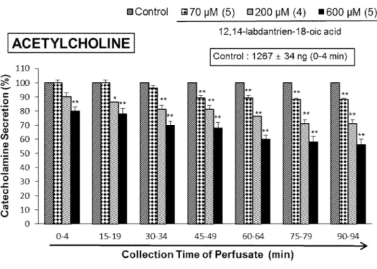
When ACh (5.32 mM) in a volume of 50 μL was administered into the perfused adrenal medullae, the released CA amount was 1267±34 ng for 4 min. But, in the presence of LDTEA in the range of 70 ~ 600 μM for 90 min, ACh-produced CA release was markedly reduced in relatively concentration- and time-dependent manner. As shown in Fig. 5, under the existence of LDTEA, the CA secretory responses were depressed maximally to 56% of the corresponding control secretion (100%).
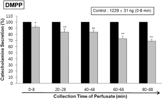
DMPP (100 μM), a selective neuronal nicotinic receptor agonist in autonomic sympathetic ganglia, produced a rapid and sharp increase in CA release (1229±31 ng for 0-8 min). However, as shown in Fig. 6, DMPP-produced CA secretion in the presence of LDTEA (200 μM) for 90 min was greatly reduced to 69% of the control secretion.
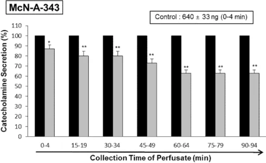
McN-A-343 (100 µM), a selective muscarinic M1-receptor agonist (Hammer and Giachetti, 1982), when perfused into an adrenal gland for 4 min, also increased the CA secretion (640±33 ng for 0-4 min). However, under the existence of LDTEA (200 µM), McN-A-343-produced CA release was greatly decreased to 63% of the corresponding control secretion as shown in Fig. 7.
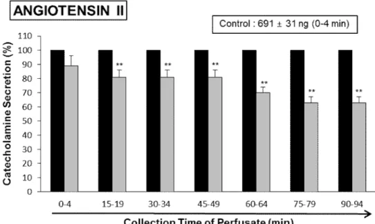
Since it has been found that Ang II increases epinephrine release from the adrenal medulla via the AT1 receptors (Hano et al., 1994), it was attempted to examine the effect of LDTEA on Ang II-produced CA secretion. Ang II (100 nM) greatly elevated the CA secretion (691±31 ng for 0-4 min), however in the presence of LDTEA (200 µM), Ang II-produced CA secretion was markedly inhibited to 63% of the corresponding control secretion (Fig. 8).
Fig. 5. Dose-dependent effects of LDTEA on the secretory responses of catecholamines (CA) produced by acetylcholine (ACh) in the perfused adrenal medullae. The CA secretion by a single injection of ACh (5.32 mM) in a volume of 50 µL was produced at 15 min intervals during infusion of 70, 200 and 600 μM of LDTEA for 90 min as denoted by the arrow marks, respectively. Statistical significance was obtained by comparing the corresponding control with each concentration-perfused group of LDTEA. The perfusate after ACh-injection was collected for 4 minutes. Other legends are the same as in Fig. 4.
*: p<0.05, **: p<0.01
Fig. 6. Time-course effects of LDTEA on DMPP-produced CA secretion in the perfused adrenal medullae. The CA release by infusion of DMPP (100 μM) for 2 min was performed at 20 min interval during perfusion of 200 μM LDTEA for 90 min. The perfusate after perfusion of DMPP was collected for 8 minutes. Other legends are the same as in Fig. 4.
*: p<0.05, **: p<0.01.
Fig. 7.Time-course effects of LDTEA on McN-A-343-produced CA release in the perfused adrenal medullae. The CA release by infusion of McN-A-343 (100 μM) for 4 min was performed at 15 min interval during perfusion of 200 μM of LDTEA for 90 min. The perfusate after perfusion of McN-A-343 was collected for 4 minutes. Other legends are the same as in Fig. 4. *: p<0.05, **: p<0.01.
Fig. 8. Time-course effects of LDTEA on angiotensin II-produced CA secretion in the perfused rat adrenal medulla. Angiotensin II (100 nM) was infused into an adrenal medulla for 1 min at 15 min intervals during perfusion of LDTEA (200 μM) for 90 min. The perfusate after perfusion of angiotensin II was collected for 4 minutes. Other legends are the same as in Fig. 4. **: p<0.01.
Effects of LDTEA on the CA secretion produced by high K
+, Bay-K-8644,
thapsigargin, and veratridine from the perfused adrenal medullae
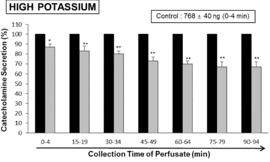
Also, high KCl, a depolarizing agent, markedly enhanced the CA secretion (768±40 ng for 0-4 min). High K+ (56 mM)-produced CA secretion in the presence of LDTEA (200 μM) for 90 min was maximally reduced to 68% of the corresponding control at 75~94 min periods, as in Fig. 9.
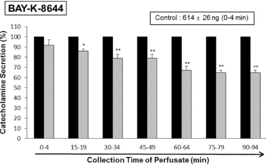
Since Bay-K-8644 is known to be a calcium channel activator, which enhances basal Ca2+uptake (Garcia et al., 1984) and CA release (Lim et al., 1992), it was of interest to determine the effects of LDTEA on Bay-K-8644-produced CA secretion from the perfused rat adrenal glands. Bay-K-8644 (10 μM)-produced CA secretion during perfusion with LDTEA (200 μM) was decreased to 65% of the control except the early 15 min period compared with the corresponding control level (614±26 ng for 0-4 min) from 5 rat adrenal medullas, as shown in Fig. 10.
The sarco–endoplasmic reticulum Ca2+-ATPase (SERCA) inhibitor thapsigargin is widely used to induce store-operated Ca2+entry (SOCE) in intact cells (Bird et al., 2008). Thapsigargin is also known to bind with subnanomolar affinity to the SERCA pump, thus protecting the Ca2+
sequestration from the cytoplasm into endopasmic reticulum (ER). Consequently, Ca2+is extruded from ER into the cytoplasm and is then rousted from the cell by the plasma membrane Ca2+pumps.
The inhibitory action of LDTEA on thapsigargin-produced CA release is shown as in Fig. 11. In 6 rat adrenal medullae, during the perfusion of LDTEA (200 µM) for 90 min, thapsigargin (10-5 M)-produced CA release was also reduced to 66% of the control level (619±21 ng for 0-4 min), however, it was not influenced only for the first period (0-4 min).
It has been found that veratridine-produced Na+influx mediated through voltage-dependent Na+ channels increased Ca2+influx via activation of voltage-dependent Ca2+channels and produced the exocytotic CA secretion in cultured bovine adrenal medullary cells (Wada et al., 1985a). As shown in Fig. 12, veratridine (50 µM) sharply increased the CA release (819±31 ng for 0-4 min). In 5 rat adrenal medullae, LDTEA (200 µM) also attenuated veratridine-produced CA secretion to 66% of the corresponding control release.
Fig. 9. Time-course effects of LDTEA on the high K+-produced CA secretion in the perfused adrenal medullae. The CA secretion by a single injection of K+ (56 mM) in a volume of 0.05 mL was produced at 15 min intervals during loading with 200 μM LDTEA for 90 min as indicated by the arrow marks. The perfusate after perfusion of high K+was collected for 4 minutes. Other legends are the same as in Fig. 4. *: p<0.05, **: p<0.01.
Fig. 10. Time-course effects of LDTEA on Bay-K-8644-produced CA secretion in the perfused adrenal medullae. Bay-K-8644 (10 μM) was given into an adrenal vein for 4 min at 15 min intervals during loading with LDTEA (200 μM) for 90 min. The perfusate after perfusion of Bay-K-8644- was collected for 4 minutes. Other legends are the same as in Fig. 4. *: p<0.05, **: p<0.01.
Fig. 11. Time-course effects of LDTEA on thapsigargin-produced CA secretion in the perfused adrenal medullae. Thapsigargin (10 μM) was infused into an adrenal medulla for 4 min at 15 min intervals during perfusion of LDTEA (200 μM) for 90 min. The perfusate after perfusion of thapsigargin was collected for 4 minutes. Other legends are the same as in Fig. 4. **: p<0.01. ns: Not statistically significant.
Fig. 12. Time-course effects of LDTEA on veratridine-produced CA secretion in the perfused adrenal medullae. Veratridine (50 μM) was infused into an adrenal medulla for 4 min at 15 min intervals during infusion of LDTEA (200 μM) for 90 min. The perfusate after perfusion of veratridine was collected for 4 minutes. Other legends are the same as in Fig.
4. *: p<0.05, **: p<0.01.
Influence of LDTEA plus L-NAME on CA secretion produced by ACh, DMPP,
McN-A-343, angiotensin II, high K
+, Bay-K-8644, thapsigargin, and veratridine in the perfused adrenal medullae
It was shown that, in the present work, LDTEA significantly suppressed the CA releasing response produced by activation of angiotensin II receptors as well as cholinergic receptors in the perfused adrenal medullae. Thus, in order to investigate the association between NO release and LDTEA-produced inhibitory effect on the CA release in the adrenal medullae, the impact of L-NAME on LDTEA-produced inhibition of CA release produced by ACh, DMPP, McN-A-343, Ang II, high K+, Bay-K-8644, thapsigargin and veratridine was examined.
In the present study, during the simultaneous infusion of L-NAME (30 μM) and LDTEA (200 μM) for 90 min in 10 rat adrenal medullae, ACh (5.32 mM)-produced CA release was largely recovered to 100~92% of the corresponding control level (1338±36 ng for 0-4 min) as compared with that of LDTEA (200 μM)-treated alone, as shown in Fig. 13.
Also, during the simultaneous infusion of L-NAME (30 μM) and LDTEA (200 μM) for 90 min, DMPP- and McN-A-343-produced CA secretory responses were also largely recovered to the control levels (100~92%) as compared with that of the LDTEA-treatment alone (Fig. 14 and 15).
Moreover, in 8 rat adrenal medullae, during the simultaneous presence of L-NAME (30 μM) and LDTEA (200 μM), the Ang II (100 nM)-produced CA secretory response was restored to 100~86% of the corresponding control release (650±26 ng for 0-4 min), as compared with the inhibitory effect of LDTEA-treatment alone, as shown in Fig. 16.
Fig. 13. Effects of LDTEA plus L-NAME on acetylcholine-produced CA secretion in the perfused adrenal medullae. The CA secretion evoked by a single injection of ACh (5.32 mM) in a volume of 50 μL was produced at 15 min intervals during simultaneous loading with LDTEA (200 μM) plus L-NAME (30μM) for 90 min. Statistical significance was obtained by comparing the corresponding control with only LDTEA-treated group or group treated with LDTEA+L-NAME. The perfusate after acetylcholine-injection was collected for 4 minutes. Other legends are the same as in Fig. 4. *: p<0.05, **: p<0.01.
Fig. 14.Effects of LDTEA plus L-NAME on DMPP-produced CA secretion in the perfused adrenal medullae. The CA release evoked by infusion of DMPP (100 μM) for 2 min was performed at 20 min intervals during simultaneous perfusion of LDTEA (200 μM) plus L-NAME (30μM) for 90 min. The perfusate after DMPP-perfusion was collected for 8 minutes. Other legends are the same as in Fig. 4. *: p<0.05, **: p<0.01.
Fig. 15. Effects of LDTEA plus L-NAME on McN-A-343-produced CA secretion in the perfused adrenal medullae. The CA secretion evoked by infusion of McN-A-343 (100 μM) for 4 min was performed at 15 min intervals during infusion of LDTEA (200 μM) plus L-NAME (30μM) for 90 min. The perfusate after perfusion of McN-A-343 was collected for 8 minutes. Other legends are the same as in Fig. 4. *: p<0.05, **: p<0.01.
Fig. 16. Effects of LDTEA plus L-NAME on angiotensin II-produced CA secretion in the perfused adrenal medullae. The CA secretion evoked by infusion of angiotensin II (100 nM) for 1 min was performed at 15 min intervals during infusion of LDTEA (200 μM) plus L-NAME (30μM) for 90 min. The perfusate after angiotensin II-perfusion was collected for 4 minutes. Other legends are the same as in Fig. 4. *: p<0.05, **: p<0.01.
Under the coexistence of LDTEA (200 μM) and L-NAME (30 μM) for 90 min in 6 adrenal medullae, high K+ (56 mM)-produced CA secretion was largely recovered to 100~88% of the control level (736±35 ng for 0-4 min), in which there was a great difference as compared with the inhibitory responses of LDTEA-treatment alone (Fig. 17).
During the simultaneous perfusion of LDTEA (200 μM) and L-NAME (30 μM) for 90 min, the CA secretion produced by Bay-K-8644 (10 μM) and thapsigargin (10 μM) was restored mostly to 100~88% (Bay-K-8644) and 100~85% (thapsigargin) of their control secretion (627±27 ng/0-4 min for Bay-K-8644; 598±25 ng/0-4 min for thapsigargin), respectively, in comparison to the inhibitory effect of LDTEA-treatment alone, as shown in Fig. 18 and 19.
Under the coexistence of LDTEA and L-NAME, there was also a almost full recovery (100~91%) of the control level (832±26 ng for 0-4 min) in veratridine (50 μM)-produced CA release as compared with that of the inhibitory effect of LDTEA-treatment alone (Fig. 20).
Fig. 17. Influence of LDTEA plus L-NAME on high potassium-produced CA secretion in the perfused adrenal medullae. The CA secretion evoked by a single injection of high potassium (56 mM) in a volume of 50 μL was produced at 15 intervals during simultaneous infusion of LDTEA (200 μM) plus L-NAME (30μM) for 90 min. Statistical significance was obtained by comparing the corresponding control level with group of LDTEA-treated alone or group treated with LDTEA+L-NAME. The perfusate after high potassium-perfusion were collected for 4 minutes. Other legends are the same as in Fig.
4. *: p<0.05, **: p<0.01.
Fig. 18. Effects of LDTEA plus L-NAME on Bay-K-8644-produced CA secretion in the perfused adrenal medullae. The CA secretion evoked by infusion of Bay-K-8644 (10 μM) for 4 min was performed at 15 min intervals during simultaneous infusion of LDTEA (200 μM) plus L-NAME (30μM) for 90 min. The perfusate after perfusion of Bay-K-8644 was collected for 4 minutes. Other legends are the same as in Fig. 4. *: p<0.05, **: p<0.01.
Fig. 19. Effects of LDTEA plus L-NAME on thapsigargin-produced CA secretion in the perfused adrenal medullae. The CA secretion by perfusion of thapsigargin (10 μM) for 4 min was performed at 15 min intervals during simultaneous infusion of LDTEA (200 μM) plus L-NAME (30μM) for 90 min. The perfusate after perfusion of thapsigargin was collected for 4 minutes. Other legends are the same as in Fig. 4. *: p<0.05, **: p<0.01.
Fig. 20. Effects of LDTEA plus L-NAME on veratridine-produced CA secretion in the perfused adrenal medullae. The CA secretion evoked by perfusion of veratridine (50 μM) for 4 min was performed at 15 min intervals during simultaneous infusion of LDTEA (200 μM) plus L-NAME (30μM) for 90 min. The perfusate after perfusion of veratridine was collected for 4 minutes. Other legends are the same as in Fig. 4. *: p<0.05, **: p<0.01.
Influence of LDTEA on the released level of nitric oxide in the perfused adrenal medullae
As depicted in Fig. 13~20, the CA secretory responses produced by ACh, DMPP, McN-A-343, Ang II, high K+, Bay-K-8644, thapsigargin and veratridine were recovered largely to their control levels under the coexistence of ginsenoside LDTEA and L-NAME. Therefore, it was decided directly to determine the level of NO released from rat adrenal medullae during the perfusion of LDTEA.
In 8 adrenal medullae, the basal release of NO in the adrenal medulla prior to perfusion of LDTEA was 13±2 picomoles. However, 8 min after loading with LDTEA (200 μM) it was greatly elevated to 25±3 picomoles, which was 192% of the basal release, as shown in Fig. 21.
Fig. 21. Effects of LDTEA on nitric oxide (NO) release in the perfused rat adrenal medullae. Following the infusion of LDTEA (200 μM) at a rate of 0.31 mL/min, perfusate sample was collected for 8 min. Ordinate: the released NO amounts in the adrenal medulla (% of control). Abscissa: Treatment (before and after LDTEA). . Statistical significance was calculated by comparing the control group with LDTEA-treated group. **:
p<0.01.
IV. DISCUSSION
The present experimental results are the first evidence showing that LDTEA significantly reduces the CA secretory effects produced by activation of acetylcholinergic nicotinic receptors as well as angiotensinergic receptors from the perfused model of the rat adrenal medullae. This LDTEA-produced inhibition of CA secretion seems to be mediated by suppressing influx of both sodium and calcium ions through their channels into the adrenal chromaffin cells and also by diminishing calcium release in the cytoplasmic calcium pool at least through the rise of NO formation via activation of neuronal nitric oxide synthase, which is relevant to the blocking action of angiotensin II receptors and neuronal cholinergic receptors.
In the present work, under the coexistence of LDTEA and L-NAME (an NO synthase inhibitor), the CA secretory effects produced by ACh, DMPP, McN-A-343, Ang II, high K+, Bay-K-8644, thapsigargin and veratridine were largely recovered to their control level as compared with their inhibitory effects of LDTEA-treatment alone. This result is well consistent with the report that, in a series of studies, ginsenoside-Rb2 depresses the CA secretion in the perfused rat adrenal medulla through increase in nitric oxide production due to activation of nNOS (Lim et al., 2014). Moreover, in this study, after loading of LDTEA into adrenal medulla, production of NO was significantly increased as depicted in Fig. 21. Taking account of these findings, in the present work, it appears that LDTEA suppresses the CA secretory response produced by cholinergic secretagogues or Ang II through elevated NO production in adrenomedullary chromaffin cells, since during the simultaneous perfusion of LDTEA and L-NAME (an inhibitor of NO synthase) for 90 min, the CA secretory responses produced by ACh, DMPP, McN-A-343, Ang II, high K+, Bay-K-8644, thapsigargin and veratridine were recovered mostly to the control level compared to that of LDTEA-treatment alone, and also substantially, LDTEA markedly increased NO release from the rat adrenal medullae.
In the support of this finding, it has been reported that the NOS inhibitor, L-NAME enhances K+-stimulated CA secretion in cultured bovine chromaffin cells (Torres et al., 1994) and also that sodium nitroprusside (SNP) inhibits ACh-produced CA secretion in bovine chromaffin cells (Uchiyama et al., 1994). Results of these studies indicate that NO may play an inhibitory role in the
regulation of the CA secretion. Moreover, the presence of endothelial cells has been reported to reduce the K+-produced or the nicotinic receptor agonist DMPP-produced CA secretion in cultured bovine chromaffin cells (Torres et al., 1994), suggesting that not only nNOS but also eNOS may play roles in modulating adrenal CA secretion. Based on the previous studies, the present results strongly imply that LDTEA can activate neuronal NO synthase (nNOS) in the adrenal medullary chromaffin cells, causing inhibition of the CA release via the rise of NO formation, besides the direct inhibitory action on the CA release. In supporting of this finding, among the ginsenosides of the protopanaxatriol and protopanaxadiol groups, ginsenoside Rg3 is known to be the most potent vasodilator (Kim et al., 1999; Kim et al., 2003). Previously, it has been found that Rg3 inhibits calcium-produced vascular contraction (Kim et al., 1999) as well as phenylephrine-produced vasocontraction as a consequence of NO production (Kim et al., 2003).
In contrast, it has been reported that L-NAME inhibits ACh-produced CA secretion in bovine chromaffin cells (Uchiyama et al., 1994), and also that the NO donor sodium nitroprusside (SNP) enhances nicotine-produced CA secretion in cultured bovine chromaffin cells (O'Sullivan and Burgoyne, 1990). These findings indicate that NO may enhance cholinergic agonist-produced CA secretion. On the other hand, a few in vivo studies have suggested that NO does not play a role in regulation of adrenal CA secretion (Breslow et al., 1992; 1993).
Commonly, the adrenal medulla has been employed as a good model system to investigate many cellular effects including noradrenergic nerve cells as well as neurons. During neurogenic acivation of the adrenal medulla, ACh is released from splanchnic nerve endings and stimulates cholinergic receptors on the chromaffin cell membrane (Viveros, 1975). This stimulation starts a series of activities as stimulus-secretion coupling, resulting in the exocytosis of CA and other constituents from synaptic vesicles into the extracellular cleft. Mostly, two mechanisms are associated with the release of adrenomedullary hormones. Upon stimulation of splanchnic nerves, the nerve terminals liberate ACh, and then this ACh activates cholinergic nicotinic receptors and produces the CA secretion. Based on this fact, the present findings demonstrated that LDTEA suppresses the CA secretion produced by activation of acetylcholinergic (nicotinic and muscarinic) receptors as well as Ang II receptors in the adrenal medulla. These experimental results suggest that this
LDTEA-produced inhibitory effect on the CA secretion can contribute at least to the antihypertensive mechanism. ACh, the physiological presynaptic transmitter at the adrenal medulla, which is released by depolarizing splanchnic nerve terminals and then stimulates nicotinic receptors, secrets the CA, and induces dopamine β-hydroxylase by calcium-dependent secretory process (Dixon et al., 1975; Viveros et al., 1968). In view of this finding, the present results demonstrate that LDTEA can attenuate CA secretion produced by cholinergic nicotinic stimulation in the splanchnic nerve terminal via the antagonism of neuronal cholinergic nicotinic (Nn) receptors.
The CA secretion from the adrenomedullary chromaffin cells evoked by splanchnic stimulation or cholinergic nicotinic agonist is mediated by activation of Nn receptors located on the adrenal chromaffin cells. The exocytotic CA release from the chromaffin cells seems to be essentially similar to that occurring in noradrenergic axons (Douglas, 1968; Sorimachi and Yoshida, 1979).
ACh-produced CA secretion has shown to be caused through stimulation of both nicotinic and muscarinic receptors in guinea-pig adrenal gland (Nakazato et al., 1988) as well as in the perfused rat adrenal glands (Lim and Hwang, 1991).
In the present work, LDTEA inhibited the CA secretory responses produced by ACh, McN-A-343, Ang II, and DMPP. In previous study, ginsenoside-Rg3inhibited both ACh-produced Ca2+ and Na+ influxes in a concentration-dependent manner similar to that observed with the ACh-produced CA secretion (Tachikawa et al., 2001). In view of this data, the present findings robustly implied that LDTEA can act on neuronal nicotinic ACh receptor-operated cation channels, however does not on the voltage-sensitive Ca2+or Na+channels.
In the present study, LDTEA also time-dependently suppressed the CA secretory response produced by Bay-K-8644, which is known to activate L-type voltage-dependent Ca2+ channels (Garcia et al., 1984; Schramm et al., 1983), as well as by high K+, a direct membrane depolarizer.
This finding indicates that LDTEA can block Ca2+entry via voltage-dependent Ca2+channels into the rat adrenomedullary cells. In support of this idea, in cultured bovine adrenal medullary cells, nicotinic (but not muscarinic) receptors mediate the Ca2+-dependent CA secretion (Fisher et al., 1981; Yanagihara et al., 1979). It has also been known that the stimulation of nicotinic receptors facilitates the CA secretion by increasing Ca2+ entry through receptor-linked and/or
voltage-dependent Ca2+ channels in both perfused rat adrenal glands (Lim and Hwang, 1991;
Wakade and Wakade, 1983) and isolated bovine adrenal chromaffin cells (Wakade and Wakade, 1983; Kilpatrick et al., 1981; 1982; Knight and Kesteven, 1983). It has been reported that the adrenomedullary chromaffin cells have (i) nicotinic receptor-operated ionic channels, responsible for carbachol-produced Na+ influx, (ii) voltage-dependent Na+ channels, responsible for veratridine-produced Na+influx and (iii) voltage-dependent Ca2+channels (VDCC), suggesting that the influx of Na+caused either by carbachol or by veratridine leads to activate voltage-dependent Ca2+ channels by altering membrane potentials, whereas high K+ directly activates voltage-dependent Ca2+channels without increasing Na+influx (Wada et al., 1985b). In the present study, the findings that the CA secretion produced by not merely high K+but also by Bay-k-8644 were markedly attenuated during the perfusion of LDTEA indicates that this LDTEA-produced inhibitory effect is mediated by the direct suppression of Ca2+ entry via VDCC into the adrenochromaffin cells. Furthermore, slight elevation in the extracellular K+ concentration increases both the frequency of spontaneous action potentials and the CA secretion (Kidokoro and Ritchie, 1980), suggesting that the influx of Ca2+ that occurs during action potentials is directly linked to the rate of secretion. These findings that LDTEA diminished the CA secretion produced by Bay-K-8644 and by high K+ suggest that LDTEA can inhibit the VDCC. In the bovine chromaffin cells, stimulation of nicotinic, but not muscarinic ACh receptors is known to cause CA secretion by increasing Ca2+ influx largely through VDCC (Burgoyne, 1984; Oka et al., 1979).
Therefore, it seems that these inhibitory effects of LDTEA on the CA secretion produced by ACh, DMPP, veratridine and Bay-K-8644 may be mediated by blocking Ca2+ entry through voltage-dependent Ca2+ channels by activation of Nn receptor-gated ionic channels, liable for carbachol-produced Na+ entry, as well as of voltage-dependent Na+ channels, liable for veratridine-produced Na+entry.
The present study has also shown that LDTEA inhibits the CA secretion produced by thapsigargin. The sarco–endoplasmic reticulum Ca2+-ATPase (SERCA) inhibitor thapsigargin is widely used to induce store-operated Ca2+ entry (SOCE) in intact cells (Bird et al., 2008).
Thapsigargin is also known to bind with subnanomolar affinity to the SERCA pump, thus
protecting the Ca2+ sequestration from the cytoplasm into endopasmic reticulum (ER).
Consequently, Ca2+is extruded from ER into the cytoplasm and is then rousted from the cell by the plasma membrane Ca2+ pumps. The final result is a complete depletion of InsP3-sensitive Ca2+
stores and the activation of the molecular machinery mediating SOCE without generation of inositol phosphates triggered by agonist-stimulated receptors (Takemura et al., 1989).
Thus, it seems that LDTEA-produced inhibitory effect on thapsigargin-produced CA secretion can be relevant to the movement of intracellular Ca2+ from the cytoplasmic Ca2+ store.
Thapsigargin is also a tumor-promoting agent (Thastrup et al., 1987) extracted from the root of the umbelliferous plant Thapsia garganica(Rasmussen et al., 1978), which has been demonstrated to be the most specific and potent inhibitor of the sarcoplasmic reticulum Ca2+-ATPase (SERCA pump) (Thastrup et al., 1990 ; Sagara and Inesi, 1991). Moreover, in bovine adrenal chromaffin cells, stimulation of muscarinic ACh receptors is also proposed to cause activation of phosphoinositide (PI) metabolism, resulting in the formation of inositol 1,4,5-trisphosphate, which induces the mobilization of Ca2+ from the intracellular pools (Cheek et al., 1989; Challiss et al., 1991).
Therefore, in this study, it is postulated that LDTEA-produced inhibitory effect on the McN-A-343-evoked CA secretion may be relevant to the mobilization of intracellular Ca2+ in the cytoplasmic Ca2+ store. This indicates that this LDTEA inhibits the Ca2+ release from the intracellular store produced by muscarinic ACh receptor activation, which is slightly liable for the CA secretion. The present results suggest that LDTEA-produced inhibition of the CA secretion produced by McN-A-343 and thapsigargin can be exerted by the reduced Ca2+release produced by muscarinic ACh receptor activation in the intracellular store. However, in the present study, it is uncertain whether LDTEA-produced inhibition on Ca2+movement in intracellular pool is mediated to the indirect action on the PI pathway or its direct action. In future, it is necessary to clarify the accurate nature of these effects.
In conclusion, as depicted in Figure 22, the findings of the present study have demonstrated that LDTEA significantly attenuates the CA release evoked by activation of acetylcholinergic nicotinic receptors as well as angiotensinergic receptors in the perfused model of the isolated rat adrenal glands. It seems that this LDTEA-produced inhibitory activity is exerted not merely by blocking
the entry of Na+and Ca2+through their ionic channels into the adrenochromaffin cells but also by diminishing the Ca2+ release from the intracellular Ca2+ store at least through the elevated NO release via the activation of neuronal NO synthase. Based on these results, the ingestion of LDTEA can be advantageous to improve or protect the cardiovascular diseases, including angina pectoris and hypertension, via diminution of CA release in adrenomedullary chromaffin cells and consequently diminished CA level in the body circulation.
Fig. 22.Schematic diagram of possible action site of LDTEA in the rat adrenal medulla.
V. SUMMARY
Previously, it has been found that self-fermented pine needle (Pinus densiflora) extract inhibits secretion of catecholamines (CA) from the perfused rat adrenal medulla (Shin et al., 2010), and also the contractile responses of isolated rat aortic strips produced by phenylephrine and high K+ (Cheong & Lim, 2010). Fraction 4-5 among several fractions obtained from methylene chloride (CH2Cl2) extract of self-fermented pine needle showed the most potent inhibitory effects on the acetylcholine (ACh)-evoked CA release (Hong & Lim, 2012). Fraction 4-5 isolated from ethylacetate extract of self-fermented pine needle inhibits the CA secretion from the isolated perfused rat adrenal medulla evoked by stimulation of cholinergic receptors as well as by direct membrane-depolarization (Choi et al., 2013).
Therefore,this study was aimed to determine the characteristics of 12, 14-labdantrien-18-oic acid (LDTEA), the most powerful component of several fractions isolated from Pinus densiflora needle, on the CA release in the perfused model of the rat adrenal medullae, and also to clarify its mechanism of action. LDTEA (70~600 µM), infused into the adrenal medulla for 90 min, suppressed ACh (5.32 mM)-produced CA secretion in a dose- and time-dependent fashion. LDTEA (200 µM) also time-dependently inhibited the CA secretion produced by McN-A-343 (100 μM, a selective acetylcholinergic muscarinic M1 receptor agonist), angiotensin II (100 nM), and DMPP (100 μM, a selective neuronal acetylcholinergicnicotinic receptor agonist). LDTEA itself failed to influence spontaneous CA release (data not shown). Also, During the perfusion of LDTEA (200 μM) for 90 min, the CA secretory responses produced by high K+ (56 mM, a direct membrane depolarizer), Bay-K-8644 (an L-type dihydropyridine calcium channel activator, 10 μM), veratridine (a voltage-dependent sodium channel activator (50 μM), and thapsigargin (a cytoplasmic Ca2+-ATPase inhibitor, 10 μM) were greatly inhibited, respectively. Interestingly, under the co-existence of L-NAME (30 μM, an inhibitor of NO synthase) and LDTEA (200 μM), the CA secretory responses produced by Ang II, ACh, McN-A-343, DMPP, high K+, thapsigargin, Bay-K-8644 and veratridine was restored nearly to their corresponding control level, respectively, as compared with those of the inhibitory effects of LDTEA (200 μM)-treatment alone.
Substantially, the release of NO in the rat adrenal medullae after the infusion of LDTEA (200 μM) was significantly enhanced in comparison to the corresponding spontaneous release. Taken together, these results demonstrate that LDTEA greatly suppresses the CA secretory responses produced by activation of cholinergic receptors as well as angiotensinergic receptors from the perfused model of the rat adrenal gland. It seems that this LDTEA-produced inhibitory activity is exerted not merely by blocking the entry of Na+ and Ca2+ through their ionic channels into the adrenochromaffin cells but also by diminishing the Ca2+ release from the intracellular Ca2+store at least through the elevated NO release via the activation of neuronal nitric oxide synthase, which is relevant to the antagonism of neuronal cholinergic receptors and angiotensinergic receptors. Based on these findings, the ingestion of LDTEA can be advantageous to improve or protect the cardiovascular diseases, including angina pectoris and hypertension, via diminution of CA release in adrenomedullary chromaffin cells and consequently diminished CA level in the body circulation.
REFERENCES
Anton AH, Sayre DF. A study of the factors affecting the aluminum oxide trihydroxy indole procedure for the analysis of catecholamines. J Pharmacol Exp Ther.1962;138:360-375.
Bird GS, DeHaven WI, Smyth JT, Putney JW Jr.Methods for studying store-operated calcium entry. Methods. 2008;46:204–212.
Breslow MJ, Tobin JR, Bredt DS, Ferris CD, Snyder SH, Traystman RJ. Nitric oxide as a regulator of adrenal blood flow. Am J Physiol.1993;264:H464-H469.
Breslow MJ, Tobin JR, Bredt DS, Ferris CD, Snyder SH, Traystman RJ.Role of nitric oxide in adrenal medullary vasodilation during catecholamine secretion. Eur J Pharmacol.
1992;210:105-106.
Burgoyne RD. Mechanism of secretion from adrenal chromaffin cells. Biochem Biophys Acta.
1984;779:201-216.
Challiss RA, Jones JA, Owen PJ, Boarder MR. Changes in inositol 1,4,5-trisphosphate and inositol 1,3,4,5-tetrakisphosphate mass accumulations in cultured adrenal chromaffin cells in response to bradykinin and histamine. J Neurochem. 1991;56:1083-1086.
Cheek TR, O'Sullivan AJ, Moreton RB, Berridge MJ, Burgoyne RD.Spatial localization of the stimulus-produced rise in cytosolic Ca2+ in bovine adrenal chromaffin cells: Distinct nicotinic and muscarinic patterns. FEBS Lett. 1989;247:429-434.
Cheong HS, Lim DY. Pine Needle Extracts Inhibit Contractile Responses of the Isolated Rat Aortic Strips. Nat Prod Sci. 2010;16:123-132.
Cheong HS, Paudyal DP, Jun JY, Yeum CH, Yoon PJ, Park CG, Kim MY, So I, Kim KW, Choi S.Effects of pine needle extract on pacemaker currents in interstitial cells of Cajal from the murine small intestine.Mol Cells. 2005;20:235-240.
Choi MS, Seo YH, Cheong HS, Lim DY. Inhibitory Effects of Self-Fermented Pine Needle Extract on Catecholamine Release in the Rat Adrenal Medulls. Nat Prod Sci. 2013;19:36-48.
Chung YJ, Bae MW, Choung MI, Lee JS, Chung KS. Cytotoxic effect of the distilled pine-needle extracts on several cancer cell lines in vitro. J Korean Soc Food Sci Nutr.
2002;31:691−695.
Dixon Wr, Garcia AG, Kirkekar SM.Release of catecholamines and dopamine-beta-hydroxylase from the rat adrenal gland of the cat. J Physiol.1975;244:805-824.
Douglas WW.Stimulus-secretion coupling: The concept and clues from chromaffin and other cells.
Br J Pharmacol.1968;34:451-474.
Fisher SK, Holz RW, Agranoff BW. Muscarinic receptors in chromaffin cell cultures mediate enhanced phospholipid labeling but not catecholamine secretion. J Neurochem.
1981;37:491-497.
Fitzpatrick DF, Bing B, Rohdewald P.Endothelium-dependent vascular effects of Pycnogenol®.
J Cardiovasc. Pharmacol.1998;32, 509–515.
Garcia AG, Sala F, Reig JA, Viniegra S, Frias J, Fonteriz R, Gandia L. Dihydropyridine Bay-K-8644 activates chromaffin cell calcium channels. Nature.1984;309:69-71.
Hammer R, Giachetti A.Muscarinic receptor subtypes: M1 and M2 biochemical and functional characterization. Life Sci. 1982;31:2992-2998.
Hano T, Mizukoshi M, Baba A, Nakamura N, Nishio I. Angiotensin II subtype 1 receptor modulates epinephrine release from isolated rat adrenal gland. Blood Press.
1994;5:S105-108.
Hong SP, Lim DY.Influence of Self-Fermented Pine Needle Fractions on Catecholamine Release from the Rat Adrenal Medulla. 24th Meeting of the International Society of Hypertension.
2012;203.
Hsu TY, Sheu SC, Liaw ET, Wang TC, Lin CC, Antioxidant activity and effect of Pinus morrisonicola Hay. on the survival of leukemia cell line U937. Phytomedicine.
2005;12:663–669.
Jung MJ, Chung HY, Choi JH, Choi JS. Antioxidant principles from the needles of red pine, Pinus densiflora. Phytother Res. 2003;17:1064-1068.
Kidokoro Y, Ritchie AK.Chromaffin cell action potentials and their possible role in adrenaline secretion from rat adrenal medulla.J Physiol.1980;307:199-216.
Kilpatrick DL, Slepetis RJ, Corcoran JJ, Kirshner N. Calcium uptake and catecholamine secretion by cultured bovine adrenal medulla cells.J Neurochem.1982;38:427-435.
Kilpatrick DL, Slepetis RJ, Kirshner N. Ion channels and membrane potential in stimulus-secretion coupling in adrenal medulla cells.J Neurochem.1981;36:1245-1255.
Kim ND, Kang SY, Park JH, Schini-Kerth VB. Ginsenoside Rg3 mediates endothelium-dependent relaxation in response to ginsenosides in rat aorta: role of K+ channels. Eur J Pharmacol.1999;367:41–49.
Kim ND, Kim EM, Kang KW, Cho MK, Choi SY, Kim SG. Ginsinoside Rg3 inhibits phenylephrine-induced vascular contraction through induction of nitric oxide synthase. Br J Pharmacol.2003;140:661-670.
Knight DE, Kesteven NT. Evoked transient intracellular free Ca2+ changes and secretion in isolated bovine adrenal medullary cells. Proc R Soc Lond Biol Sci.1983;218:177-199.
Lee E. Effects of powdered pine neele (Pinus densiflora seib et Zucc.) on serum and liver lipid composition and antioxidative capacity in rats fed high oxidized fat. J Korean Soc Food Sci Nutr.2003;32:926−930.
Lee KH, Kim AJ, Choi EM.Antioxidant and antiinflammatory activity of pine pollen extract in vitro.Phytother Res.2009;23:41-48.
Lim DY, Hwang DH.Studies on secretion of catecholamines evoked by DMPP and McN-A-343 in the rat adrenal gland. Korean J Pharmacol.1991;27:53-67.
Lim DY, Kim CD, Ahn KW. Influence of TMB-8 on secretion of catecholamines from the perfused rat adrenal glands. Arch Pharm Res.1992;15:115-125.
Lim HJ, Lee HY, and Lim DY.Inhibitory Effects of Ginsenoside-Rb2 on Nicotinic Stimulation Evoked Catecholamine Secretion. Korean J Physiol Pharmacol, 2014;18:431-439.
McVeigh GE, Hamilton P, Wilson M, Hanratty CG, Leahey WJ, Devine AB, Morgan DG, Dixon LJ, McGrath LT. Platelet nitric oxide and superoxide release during the development of nitrate tolerance: effect of supplemental ascorbate. Circulation.
2002;106:208-213.
Nakazato Y, Ohga A, Oleshansky M, Tomita U, Yamada Y.Voltage-independent catecholamine release mediated by the activation of muscarinic receptors in guinea-pig adrenal glands. Br J Pharmacol.1988;93:101-109.
Oka M, Isosaki M, Yanagihara N.Isolated bovine adrenal medullary cells: studies on regulation of catecholamine synthesis and release. In: Usdin E, Kopin IJ, Brachas J, ed.
Catecholamines: Basic and Clinical frontiers. Oxford: Pergamon Press; 1979. p.70-72.
O'Sullivan AJ, Burgoyne RD. Cyclic GMP regulates nicotine-induced secretion from cultured bovine adrenal chromaffin cells: effects of 8-bromo-cyclic GMP, atrial natriuretic peptide, and nitroprusside (nitric oxide). J Neurochem.1990;54:1805-1808.
Park CS, Kwon CJ, Choi MA, Park GS, Choi KH. Antioxidative and nitrite scavenging activities of mugwort and pine needle extracts. Korean J Food Pres. 2002;9:248−252.
Rasmussen U, Christensen SB, Snadberg F.Thapsigargin and thapsigargicin, two new histamine bilerators from Thapsia-garganica. Acta Pharmaceut. Suec.1978;15:133-140.
Sagara Y, Inesi G.Inhibition of the