http://dx.doi.org/10.20307/nps.2016.22.3.193
193
Bioassay-Guided Isolation and Identification of Compounds from Arecae Pericarpium with Anti-inflammatory, Anti-oxidative,
and Melanogenesis Inhibition Activities
Amelia Indriana, Kyoung Jin Lee, and Yeong Shik Kim*
College of Pharmacy and Natural Products Research Institute, Seoul National University, Gwanak-ro 1, Gwanak-gu, Seoul 08826, Republic of Korea
Abstract − This study describes the anti-inflammatory, anti-oxidant, and melanogenesis inhibition activities of methanol extract and various organic solvent fractions of Arecae Pericarpium. We examined the inhibition of lipopolysaccharide (LPS)-induced nitric oxide (NO) production in RAW 264.7 cells, 1,1-diphenyl-2-picrylhydrazine (DPPH) scavenging activity, mushroom tyrosinase inhibition activity and melanin contents. The study showed that, among all tested fractions, methylene chloride fraction showed the strongest inhibition of LPS-induced NO production in RAW 264.7 cells (IC
50value 8.89 µg/mL) and DPPH radical scavenging activity (EC
50value 21.39 µg/
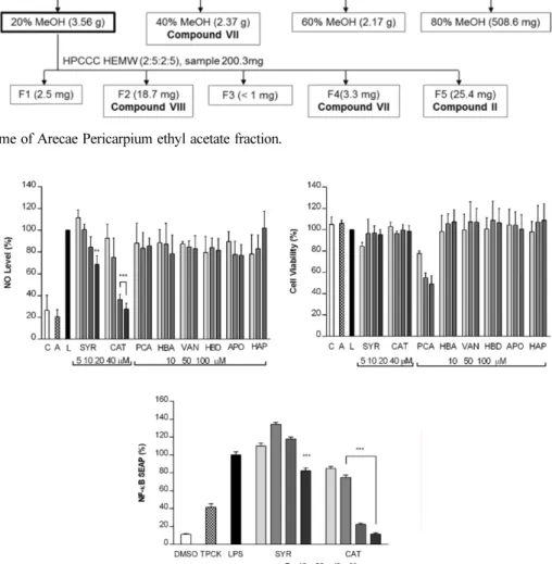
mL). Methylene chloride and ethyl acetate fractions similarly inhibited mushroom tyrosinase activity. Methanol extract exhibited strongest reduction of melanin content in B16F10 melanoma cells. Based on the bioactivity assay results, methylene chloride and ethyl acetate fractions were further separated. Eight phenolic compounds were isolated, which are dimeric syringol (1), catechol (2), 4-hydroxybenzaldehyde (3), vanillin (4), 4-hydroxya- cetophenone (5), apocynin (6), protocatechuic acid (7) and 4-hydroxybenzoic acid (8). Among the isolated compounds tested, catechol showed the strongest inhibition of LPS-induced NO production in RAW 264.7 cells.
Catechol also showed the concentration-dependent NF- κB inhibition activity. Arecae Pericarpium might have potentials to be developed as anti-inflammatory agent or dermatological product for skin-whitening agent.
Keywords − Arecae Pericarpium, Anti-inflammatory, Antioxidative, Melanogensis Inhibition, Bioassay-guided isolation
Introduction
Arecae Pericarpium is the fruit husk of Areca catechu L. (Arecaceae). According to the Compendium of Materia Medica, this herb has been used in Traditional Chinese Medicine (TCM) for abdominal distension, constipation, and edema treatment. Its combination with other herbs in Huo Xiang Zheng Qi ( 藿香正 片 ) formula is used to treat summer heat-dampness diseases and gastrointestinal cold, to cure abdominal distension, vomiting, and diarrhea.
1Modern investigations showed that Arecae Pericarpium has a fungicidal activity against Colletotrichum gloeos- porioides Penz. in vitro and in mango fruit medium.
2Arecae Pericarpium also showed the dose-dependent antioxidative activity in human hepatocarcinoma HepG
2cell line. The methanol extract of Arecae Pericarpium showed the stronger antioxidant activity compared to
other parts of the Areca catechu (L.) plant.
3The limited number of studies on Arecae Pericarpium suggests that Arecae Pericarpium has high potential to be further studied for its bioactivity. Thus, in this present report, we investigated the anti-inflammatory, free radical scavenging, and mela- nogenesis inihibition activity of Arecae Pericarpium.
The potential anti-inflammatory activity of Arecae Pericarpium was screened by examining the ability of its fractions in suppressing LPS-induced nitric oxide (NO) production in RAW 264.7 murine macrophage cells.
Macrophage plays a central role in a host’s defense system against bacterial infection through phagocytosis, cytotoxicity, and intracellular killing.
4,5Stimulation of murine macrophages by LPS results in the expression of iNOS and increased NO production which plays a critical role in macrophage activation and is associated with acute and chronic inflammations.
5The elevated NO production is examined with Griess reaction by quantifying the nitrite level in the conditioned medium of RAW 264.7 cells treated with LPS. This cell-based assay has been used for drug screening and the evaluation of potential inhibitors
*Author for correspondence
Yeong Shik Kim, College of Pharmacy and Natural Products Research Institute, Seoul National University, Gwanak-ro 1, Gwanak- gu, Seoul 08826, Republic of Korea.
Tel: +82-2-880-2479; E-mail: [email protected]
of the pathways leading to the induction of iNOS and NO production.
6Melanin is secreted by melanocytes and determines the color of skin and hair in mammalians. It protects the skin by absorbing UV sunlight and removing reactive oxygen species (ROS).
7The excessive level of melanin pigmentation causes various dermatological disorders including hyper- pigmentations such as senile lentigo, melasma, postin- flammatory melanoderma, freckles, ephelide, age spots and sites of actinic damage which can give rise to esthetic problems.
8Many studies have been conducted to find depigmenting agents from natural resources that can be used for the long-term without side effects. Arecae Semen has been reported to have anti-inflammatory and melano- genesis inhibitory activity.
9Thus, in this study Arecae Pericarpium was also tested to investigate whether it also has potential to inhibit melanogenesis.
Experimental
General experimental procedures – All organic solvents used for extraction, column chromatography and high- performance countercurrent chromatography (HPCCC) were of analytical grades. HPLC-grade acetonitrile and methanol were used for HPLC analysis and preparative HPLC. Distilled water was used for all solutions and dilutions. Silica gel 60 (0.063 - 0.200 mm) was used for open column silica chromatography separation. Silica gel 60 F254 20 × 20 cm was used for thin-layer chromato- graphy. Diaion
®-HP20 (polystyrene adsorption resin) was used for open column chromatography separation.
HPLC analyses were carried out on Hitachi L-6200 and Agilent 1100 instruments. Low resolution electrospray ionization source (ESI) LC/MS data were recorded on Agilent Technologies 1200 HPLC coupled with Agilent Technologies 6130 Quadrapole mass spectrometer. Columns used for analysis are iNNO C18 column, LUNA C18 column and Zorbax SB-C18 column. Spectrum high- performance countercurrent chromatography (HPCCC) from Dynamic Extractions Ltd with tubing id 1.6 mm, total volume 135.5 mL, and sample loop 6 mL was used for CCC separation. Preparative HPLC separation was performed using a Hitachi JP/L-7100 equipped with Hitachi L-4000 UV detector. The column used for semi- preparative separation was RSil C18 column (250 mm × 10 mm id, 10 μm particle size). The NMR analyses were recorded on Bruker Avance 500 and 600 spectrometers.
1
H and
13C NMR spectra were measured in a DMSO-d
6solution at 500 and 125 MHz or 600 and 150 MHz, respectively.
Plant materials – Arecae Pericarpium cultivated in China was purchased at a local herb market in Seoul (October, 2013). Arecae Pericarpium was identified by Professor Young Bae Suh, College of Pharmacy, Seoul National University. A voucher specimen (AP201510a) was deposited in Natural Products Research Institute, College of Pharmacy, Seoul National University.
Extraction – The dry Arecae Pericarpium (5 kg) was macerated in 28 L of methanol for 3 days. The process was repeated for 3 times. The macerate was dried with a rotary evaporator under 50 ºC. The extract was then suspended with 1 L of 10% methanol and partitioned with hexane, methylene chloride (MC), ethyl acetate (EA), and n-butanol (BuOH) in 1:1 ratio for 3 times and each fraction was dried with a rotary evaporator. The final state of all dried fractions was viscous state.
NO inhibition and cell viability assay − The ability of tested samples in inhibiting NO production was deter- mined by Griess reagent assay. RAW 264.7 cells were plated at density of 1 × 10
5cells/well in a 24-well plate with 500 µL of culture media and incubated for 24 h.
Then, cells were treated with samples in various con- centration for 2 h and stimulated with LPS (1 µg/ mL) for another 18 h. An aliquot of cell-free medium (100 µl/
well) was then removed to a 96-well plate and Griess reagent (100 µl/well) was added. To quantify nitrite con- centration, standard nitrite solutions were prepared and the absorbance of the mixture was determined at 540 nm with a microplate reader (Molecular Devices, Emax, Sunnyvale, CA, USA). Cell viability was measured by 3- (4,5-dimethylthiazol-2-yl)-2,5-diphenyl tetrazolium bromide (MTT)-based colorimetric assay. AMT, an iNOS inhibitor, was used as a positive control (10 µM). Cells treated with vehicle alone were used as a control.
NF- κB secretory alkaline phosphatase (SEAP)
reporter gene assay − Reporter enzyme activity was
measured by cell-based assay system for NF- κB activity
monitoring.
10The pNF- κB-SEAP-NPT plasmid that permits
expression of the SEAP reporter gene in response to the
NF- κB activity and contains the neomycin phospho-
transferase (NPT) gene for geneticin resistance in host
cells was constructed and transfected into murine
macrophages. Transfected RAW 264.7 cells were plated
at density of 1 × 10
5cells/well in a 24-well plate with 500
µL of geneticin-added culture medium and incubated at
37
oC for 24 h. Cells were treated with samples in various
concentrations for 2 h and stimulated with LPS (1 µg/mL)
for 18 h. Aliquots of cell-free medium (120 µL) of each
treatment was transferred to 1.5 mL vial and heated at
65
oC for 6 min and given an assay buffer (2 M dietha-
nolamine, 1 mM MgCl
2, 500 µM 4-methylumbelliferyl phosphate (MUP)) in the dark at 37
oC for 1 h. The fluorescence from the product of the SEAP/MUP was measured using a 96-well microplate fluorometer at an excitation of 360 nm and emission of 449 nm. TPCK (20 µM) was used as a positive control.
DPPH radical scavenging assay − DPPH (1,1-diphenyl- 2-picrylhydrazine) radical scavenging activity was deter- mined with methods as described by Prieto et al.
11Ascorbic acid was used as a positive control. Samples dissolved in DMSO were diluted with MeOH in 96-well plate with total volume 100 µl/well. DPPH 0.2 mM solution (100 µl) was added to each well and incubated in dark room for 30 minutes. Diluted compounds without DPPH were used as blank samples. The absorbance was measured at 540 nm with a microplatereader.
Mushroom tyrosinase assay − Various concentrations of samples dissolved in DMSO were diluted 500-fold in 0.1 M sodium phosphate buffer (pH 6.8). One hundred microliters of diluted samples and 50 µl of 1.5 mM L- tyrosin were transferred to a 96-well plate and pre-incubated for 10 min. Then, 50 µl of mushroom tyrosinase (125 unit/mL in 0.1 M sodium phosphate buffer) was added to each well and incubated at 37
oC for 30 min. The absorbance was determined at 490 nm with a microplate reader. Kojic acid was used as positive control.
Cell viability assay − The effect of prepared samples on B16F10 melanoma cell viability was measured with MTT-based colorimetric assay. In brief, cells were seeded at density of 3 × 10
3cells/well in 96-well plate and incubated for 24 h. Cells were treated with samples in various concentrations and further incubated for 3 days.
On the last day, the medium was removed, replaced with MTT solution and incubated for 2 h. Mitochondrial succinate dehydrogenase in living cells are known to convert MTT into visible formazan crystals during incu- bation. The formazan crystals were then solubilized in DMSO and the absorbance was measured at wavelength 595 nm using a microplate fluorometer.
Melanin content assay − B16F10 melanoma cells were seeded at density of 2 × 10
4cells/well in a 24-well plate and incubated for 24 h. The cells were treated with samples in various concentrations and 1 mM theophylline for 3 days. To determine the extracellular melanin content, 200 µl of cell-free medium was transferred to 96-well plate and the absorbance was determined at 490 nm with a microplate reader. To determine the intracellular melanin content, the remaining medium was removed and the cells were harvested by trypsinization (0.25% trypsin/0.02%
EDTA in PBS). The harvested cells were centrifuged and
solubilized in 100 µl of 1 M NaOH and heated at 60 ºC for 1 h. The absorbance was determined at 405 nm with a microplate reader.
Bioassay-guided isolation of MC fraction − The MC fraction (18 g) was subjected to silica gel column chro- matography with a gradient elution of chloroform – ethyl acetate (9:1, 7:3, 5:5, 3:7, 1:9 v/v) continued with ethyl acetate – methanol (9:1, 7:3, 5:5 v/v). Six fractions were collected and the MC1 fraction was separated with silica gel column chromatography. The sample (3.3 g) was eluted with hexane-ethyl acetate-n-butanol with ratio (3:2:0.05) and (2.75:2.25:0.05). Seven fractions were collected from this separation (MC1A – MC1D were eluted with 3:2:0.05, and MC1E - MC1G were eluted with 2.75:2.25:0.05). MC1G was identified as dimeric syringol (1) (yield: 9.2 mg). MC1A fraction was subjected to Diaion HP-20 column chromatography and eluted with aqueous methanol from 20% - 100% MeOH. Catechol (2) (yield: 25.4 mg) was obtained from 20 and 30% MeOH elution. MC1B fraction was separated with combination of high performance counter-current chromatography (HPCCC) and preparative HPLC. HPCCC was run using HEMW solvent system (2:5:1:4 v/v/v/v). The stationary phase retention was 70.11%. Sample injection was 130.3 mg in 6 mL of solvent system mixture and detected with UV detector at 280 nm for 200 minutes. Preparative HPLC was performed with isocratic elution 18% MeOH with 0.05% formic acid under 246 nm UV detection. 4- Hydroxybenzaldehyde (3) (yield: 1.0 mg), vanillin (4), 4- hydroxyacetophenone (5) (yield of 4 and 5 mixture: 5.0 mg), and apocynin (6) (yield: 0.6 mg) were obtained from MC1B separation process.
Bioassay-guided isolation of EA fraction − The ethyl acetate fraction was subjected to Diaion
®HP-20 resin column chromatography and eluted with aqueous methanol in increasing concentration from 20%, 40%, 60%, and 80% methanol. The 20% MeOH fraction showed NO inhibition activity and it was further separated by HPCCC.
Several solvent systems were tested to estimate the optimum K values and HEMW system (2:5:2:5 v/v/v/v) was selected for HPCCC operation. The operation of HPCCC separation was conducted with UV detection set at 280 nm. 200.3 mg of sample was weighed and dissolved in 6 mL of upper phase and lower phase mixture. The operation was performed for 200 minutes. Protocatechuic acid (7) (yield: 18.7 mg) and 4-hydroxybenzoic acid (8) (yield: 3.3 mg) were obtained from the HPCCC separation.
Identification of isolated compounds − The isolated
compounds were identified by NMR 500 or 600 MHz in
DMSO-d
6. 1D (1H and 13C) and 2D NMR (HSQC and
HMBC) was performed at SNU National Center for Inter- University Research Facilities.
Statistical analysis − All data were derived from three independent experiments and are expressed as mean ± standard deviation (SD). Statistically significant differences between the control and experimental groups were calculated by Student’s t test. Notes are given as (*) if P < 0.05, (**) if P < 0.01, and (***) if P < 0.001.
Dimeric syringol (1) − slightly yellow amorphous powder. ESI-MS m/z 307.1 [M+H]
+, m/z 305.1 [M −H]
−.
1