저작자표시-동일조건변경허락 2.0 대한민국 이용자는 아래의 조건을 따르는 경우에 한하여 자유롭게
l 이 저작물을 복제, 배포, 전송, 전시, 공연 및 방송할 수 있습니다. l 이차적 저작물을 작성할 수 있습니다.
l 이 저작물을 영리 목적으로 이용할 수 있습니다. 다음과 같은 조건을 따라야 합니다:
l 귀하는, 이 저작물의 재이용이나 배포의 경우, 이 저작물에 적용된 이용허락조건 을 명확하게 나타내어야 합니다.
l 저작권자로부터 별도의 허가를 받으면 이러한 조건들은 적용되지 않습니다.
저작권법에 따른 이용자의 권리는 위의 내용에 의하여 영향을 받지 않습니다. 이것은 이용허락규약(Legal Code)을 이해하기 쉽게 요약한 것입니다.
Disclaimer
저작자표시. 귀하는 원저작자를 표시하여야 합니다.
동일조건변경허락. 귀하가 이 저작물을 개작, 변형 또는 가공했을 경우 에는, 이 저작물과 동일한 이용허락조건하에서만 배포할 수 있습니다.
20 1 3 년 8 월 석사학위논문
고위험군 인유두종 바이러스 감염 환자에서의 자궁경부
편평상피 병변의 진단
보조인자로서의 p16과 Ki -67의 효율성
조 선 대 학 교 대 학 원
의 학 과
임 샤 론
[UCI]I804:24011-200000263943
고위험군 인유두종 바이러스 감염 환자에서의 자궁경부
편평상피 병변의 진단
보조인자로서의 p16과 Ki -67의 효율성
Ef f i cacy ofp16andKi -67i mmunost ai ni ng i n t he det ect i on ofsquamousi nt r aepi t hel i all esi on i n hi gh r i sk
HPV gr oup
2 0 1 3 년 8 월 2 3 일
조 선 대 학 교 대 학 원
의 학 과
임 샤 론
고위험군 인유두종 바이러스 감염 환자에서의 자궁경부
편평상피 병변의 진단
보조인자로서의 p16과 Ki -67의 효율성
지도교수 이 미 자
이 논문을 의학석사학위신청 논문으로 제출함.
2 01 3 년 4 월
조 선 대 학 교 대 학 원
의 학 과
임샤론의 석사학위논문을 인준함
위원장 조선대학교 교수 기 근 홍 印 위 원 조선대학교 교수 임 성 철 印 위 원 조선대학교 교수 이 미 자 印
2 0 1 3 년 5 월
조 선 대 학 교 대 학 원
목 차
표목차 ‥‥‥‥‥‥‥‥‥‥‥‥‥‥ i i
도목차 ‥‥‥‥‥‥‥‥‥‥‥‥‥‥ i i i
초 록 ‥‥‥‥‥‥‥‥‥‥‥‥‥‥ 1
I .서론 ‥‥‥‥‥‥‥‥‥‥‥‥‥‥ 2
I I .연구재료 및 방법 ‥‥‥‥‥‥‥‥‥‥ 3
I I I .결과 ‥‥‥‥‥‥‥‥‥‥‥‥‥‥ 6
I V.고찰 ‥‥‥‥‥‥‥‥‥‥‥‥‥‥ 8
V.결론 ‥‥‥‥‥‥‥‥‥‥‥‥‥‥‥ 1 1
참고문헌 ‥‥‥‥‥‥‥‥‥‥‥‥‥‥‥ 1 2
표 ‥‥‥‥‥‥‥‥‥‥‥‥‥‥‥‥‥ 1 6
그림 ‥‥‥‥‥‥‥‥‥‥‥‥‥‥‥‥ 2 2
표 목 차
Table1.Thep16expressioninHR-HPV accordingtothefinaldiagnosis
Table2.TheKi-67expressioninHR-HPV accordingtothefinaldiagnosis
Table3.Thep16expressioninnonHR-HPV accordingtothefinaldiagnosis
Table4.TheKi-67expressioninnonHR-HPV accordingtothefinaldiagnosis
Table5.DistributionoffirstandfinaldiagnosisinHR-HPV group
Table6.DistributionoffirstandfinaldiagnosisinnonHR-HPV group
-i i-
도 목 차
Fig.1.A,p16immunostaininginCIN 1withaparabasaldiffusepattern.C,p16 immunostaining in CIN 2 with a intermediate diffuse pattern.E,p16 immunostaining in CIN 3 with a superficialdiffuse pattern.B,Ki-67 immunostaining in CIN 1 with a basal one third layer. D,Ki-67 immunostaining in CIN 2 with a upper two third layer.F,Ki-67 immunostaininginCIN 3withafullthicknessdiffusepattern.
Fig.2.Comparison ofsquamous cellcarcinoma cases representing HR-HPV (rightcolumn)and nonHR-HPV (leftcolumn)by H&E (A,B),p16(C, D)and Ki-67(E,F).p16immunostaining showsstrong,diffusepattern in only HR-HPV squamouscellcarcinoma.Ki-67showspositivity with diffusepatterninbothcases.
- 1 -
초 록
고위험군 인유두종 바이러스 감염 환자에서의 자궁경부 편평상피 병변의 진단 보조인자로서의 p16과 Ki -67의 효율성
임샤론
지도교수 :이미자
조선대학교 대학원 의학과
배경:자궁경부 편평상피 병변을 위축성 변화 또는 비정형 편평상피의 화생과 감별진 단을 하는데 있어서 p16과 Ki-67면역화학염색이 보조적인 방법으로 사용되어져 왔다.
본 연구의 목적은 자궁경부 생검의 정확한 진단에 위의 두가지 면역화학염색 인자의 효율성을 평가하고 또한 인유두종 바이러스 감염 상태와의 상관성을 알아보고자 하였 다.방법:각각 인유두종 바이러스 검사를 시행한 123명 환자의 자궁경부 생검 조직을 이용하여 p16과 Ki-67 면역화학염색을 시행하였다.헤마토실린 및 에오신(H&E)염색 을 한 슬라이드,p16그리고 Ki-67염색 슬라이드를 판독하였다.결과:고위험군 인유 두종 바이러스에 감염된 상태에서 편평상피 병변의 등급이 높을수록 p16과 Ki-67의 양 성도가 증가하였다.H&E 슬라이드 판독 결과 각각 CIN 2와 CIN 3로 진단되었던 3개 의 경우에서 p16면역염색에서 음성 소견,Ki-67에서 매우 드물게 염색되는 결과를 보 였으며 이는 다시 비정형 편평상피 화생으로 진단되었다.인유두종 바이러스에 음성인 28명의 환자 중 2명의 환자에서 p16음성인 소견이 나타났으나 조직학적 소견으로 고 등급 자궁경부 편평상피병변과 편평상피암종의 기준을 만족하였다.이들은 각각 CIN 2 과 편평상피종양으로 진단되었다.결론:고위험 인유두종 바이러스에 감염된 그룹에서 p16과 Ki-67 면역화학염색에 미만성으로 강하게 동시에 염색이 되어 실제로 CIN 2, CIN 3그리고 편평상피암종을 나타냈다.그러므로 p16과 Ki-67의 면역염색이 자궁경부 편평상피병변을 정확하게 진단하는데 많은 도움을 주는 표지자로서 효율성이 증명되었 으나 고위험군 인유두종 바이러스 감염 상태가 아닌 그룹에서는 이러한 면역염색 결과 를 진단에 적용하는데 있어서 엄격한 고려가 필요하다고 판단된다.
I .I nt r oduct i on
Cervicalcancerrepresents the second mostcommon cancerboth incidence andmortality inwomenworldwide,withanincreasedincidenceinlow resource countries.1Virtually allcervicalcancersarecausedby persistentinfectionswith high risk-human papilloma virus (HR-HPV)types which may cause cervical intraepithelialneoplasia (CIN) and squamous cellcarcinoma (SCC).2 Cervical biopsy,used in conjunction with Pap cytology testing,HPV DNA testing,and colposcopy,hasan importantrolein theevaluation andmanagementofwomen with cervicaldysplastic lesions,which is crucialforthe prevention and early detection ofcervicalcancer.3Buttherecan bea misinterpretation ofhistologic changes in association with various situations such as atrophy, immature metaplasia,transitional metaplasia,repair or inflammatory atypia,inadequate sample size and tissue artifact,which may eventually cause an interobserver variationandapoorintraobserverreproducibility.4-6Recently,severalbiomarkers have been evaluated fortheirpotentialto improve the diagnostic consistency and accuracy ofcervicalbiopsy interpretation.6,7 Many ofsuch studies8,9 and other studies10,11 endorse the use ofp16 and Ki-67 immunostains and more recently,ProExC immunostain11-13asvery usefuladjuncttechniquestoconfirm a diagnosisofCIN 2/3 and to distinguish itfrom its mimics.With regard to p16,ithasbeen shown thatalmost100% ofCIN 2/3andSCC associatedwith HR-HPV expresshigh levelsofp16,whereasnondysplasticcervicalepithelium oflow-grade CIN associated with low-risk HPV (LR-HPV) types does not expressp16.Toimprovethediagnosticaccuracy,othermarkersincludingKi-67 havebeenusedalongwithp16inahistologicalassessmentofCIN andSCC of theuterinecervix.5,15Similartop16,Ki-67isoverexpressedinCIN 2/3,SCC.16
Given the above knowledge, we investigated the efficacy of immuno- histochemicalstain forp16andKi-67in thepathologicalassessmentofuterine cervicalbiopsysampleswithviralandhistopathologicalcorrelation.
- 3 -
I I .Mat er i al sandMet hods
A.Mat er i al s
Thepathology dataprogram ofChosun university hospitalwassearched for final report including the terms low grade squamous intraepithelial lesion (LSIL),high grade squamous intraepitheliallesion (HSIL)and squamous cell carcinomaofallgynecologicspecimen during January 2012toMarch 2013.We selectedatotalof123specimenswiththefollowingpathologicdiagnoses:LSIL, HPV,32cases;LSIL,CIN 1,27cases;HSIL,CIN 2,19cases;HSIL,CIN 3,30 cases;SCC,15cases.Thepatientshadundergoneeithercervico-vaginalsmear and HPV DNA test.By reviewing themedicalrecordsofthemedicalrecords and pathologicreports oftheselected patients,weconfirmed thepatientsage, underlying disease,othertumorhistory,cytologic diagnosis and HPV infected status.
B.Met hods
1.HPV DNA testing andgenotyping
HPV DNA testforHR-HPV was achieved by polymerase chain reaction– based HPV genotyping assay (HPV 9G DNA chip,Fammed Co.,Ltd.).DNA chipcandetect28HPV genotypes(14HR-HPVs:16,18,31,33,39,45,51,52, 54,56,58,59,66and 68;and 7LR-HPVs:6,11,34,40,42,43 and 44).The HPV genotyping were classified as HR-HPV or LR-HPV according to the schemeproposedbyDunneetal.17
2.Immunohistochemicalstudies
For the investigation, 3 μm-thick serial sections were prepared from representative formalin-fixed,paraffin-embedded tissue blocks and mounted on positively charged glass slides for the immunohistochemical studies.
Immunostaining were performed by autostainer using an Ventana BenchMark XT instrument (Ventana MedicalSystem,Tucson,AZ,USA).The primary antibodies were p16 (1:100,mouse monoclonalantibody,clone E6H4,Ventana Medical System, Tucson, AZ, USA) and Ki-67 (1:50, mouse monoclonal antibody,clone MM1, Novocastra, Newcastle, UK). Primary antibodies and enzyme complexes were visualized by a brown 3,3’-diaminobenzidine and abbreviation (DAB)reaction and the slides were counterstained with Mayer's hematoxylin.
3.Immunohistochemicalinterpretation
Theinterpretation ofp16ispositivewithobservedinnuclearornuclearand cytoplasmic,strong and continuous staining beginning from basalcelllayerof epithelium in contrast of either focal and sporadic pattern or completely negative. The scoring of p16 classified in four groups as 0 (basal), 1 (parabasal)(Fig.1A),2 (intermediate)(Fig.1C)and 3 (superficial)(Fig.1E). The expression ofKi-67 was also categorized into fourgroups based on the distribution and proportionsofnuclearstaining positivecells.score0,<10% of the cells,restricted to the parabasalcelllayers;score 1,10 to <30% ofthe cells,restricted to the lowerthird ofthe epithelium (Fig.1B);score 2,30 to
<70% ofthecells,reachingtheupperthirdoftheepithelium (Fig.1D);score3, 70% ormoreoftheepithelialcellsincluding fullthicknessexpression ofKi-67 (Fig.1F).18
- 5 - 4.Evaluation oftheslides
Two observers evaluated the hematoxylin and eosin (H&E)-stained slides, andthenmadeainitialdiagnosisbasedontheonly histologicalfeaturesonhis orher own criteria.These diagnoses include normal,benign lesions such as squamous metaplasia orLSIL with viralcytopathic change (HPV effect)and precancerouslesion such as CIN 1,CIN 2 and CIN 3 and malignantlesion - SCC.Then,each observers had second look ofH&E slide included p16 and Ki-67.Then,thediagnosiswasrevised andthen,theconsensusdiagnosiswas reached.Thewholeslideswerereviewed independently by twoobservers,who wereblindedtoalltheclinicopathologicinformation.
5.Statisticalanalysis
SPSS ver.12.0 (SPSS,Inc.,Chicago,IL,USA) was used for statistical analysis.Multivariate analysis was performed using hit ratio between initial diagnosis and revised diagnosis then these value compared with the HPV infected status: HR-HPV and nonHR-HPV. P-value less than 0.05 was considered statistically significant.Thecorrelation between initialdiagnosisand reviseddiagnosiswereevaluatedbyPearson'scorrelationcoefficient.
I I I .Resul t s
A.Expr essi onofp16andKi -67i n HR-HPV
Using PCR based HPV genotyping assay,high risk HPV genotypes were detected in 62% (76/123).In the 12 cases of benign lesion - normal or squamous metaplasia,allofthem are p16 negative.Ofthe 4 cases outof9 casesLSIL with viralcytopathicchangeshowed1scorein p16staining.Allof the7casesofCIN 1lesionsshowedap16expressionscoreof1,but13cases ofCIN 2lesionsshowedap16expressionscoreof2.Inadditiontherewere25 casesoutof25casesCIN 3and10casesofallSCC casesshowedexpression ofscore3(Table1).
Twelve cases were diagnosed as benign lesion - normal or squamous metaplasia,only onecaseshowed Ki-67expression scoreof1.Ofthe3cases outof9casesLSIL with viralcytopathicchangeshowed scoreof1in Ki-67 staining.6 casesofthe7 casesofCIN 1 lesionsshowed a Ki-67 expression score of 1,but 10 cases out of 13 cases CIN 2 lesions showed a Ki-67 expression scoreof2.In addition therewere24casesoutof25casesCIN 3 and10casesofallSCC casesshowedexpressionofscore3(Table2).
B.Expr essi on ofp16andKi -67i n nonHR-HPV
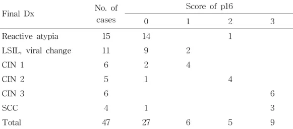
HPV genotyping assay detected in 38%(47/123)casesofnon-high risk HPV genotypes.Inthe14casesoutof15casesbenignlesion-normalorsquamous metaplasia,except1caseofscore2showedexpressionscore0.Ofthe9cases outof11 cases LSIL with viralcytopathic change showed 0 score in p16 staining andtherest2casesarescore1.Inthe4casesoutof6casesCIN 1,
- 7 -
except2caseofscore0showedexpression score1andCIN 2lesionsshowed onecasep16score0and fourcasesp16score2.But,therewere6casesout of6casesCIN 3showedexpression ofscore3.Among 4casesofSCC lesions, onepatientshowedcompletelylossinp16stainingexceptionally(Table3).
Ofthe14casesoutof15casesbenign lesionsshowed scoreof0in Ki-67 staining.6casesofthe11casesofLSIL with viralcytopathicchangeshowed aKi-67expression scoreof1,butonly 3casesoutof6casesCIN 1lesions showedaKi-67expression scoreof1.4casesofCIN 3lesionsexcept1case showedofscore2.Similarly,5casesofCIN 3lesionsexcept1caseshowedof score3.And4casesofallSCC casesshowedexpressionofscore3(Table4).
C.Ef f i cacy ofp16and Ki -67i n accor di ng t oHPV genot ypes
There were 3 cases ofatypicalsquamous metaplasia (n=3)with previously CIN 2 (n=1)/CIN 3 (n=2)with H&E showed loss ofp16 and rare Ki-67 index.And wefound adiscordancein nonHR-HPV group.Completely negative in p16 stain cases (n=2) in nonHR-HPV group,they were fulfilled ofhigh grade CIN pathologic criteria even carcinoma features in H&E slides.So they were each diagnosed finally in CIN 2 and squamous cellcarcinoma based on histopathologicfindings.
Thepositivityofp16andKi-67increasedwiththeseverityofcervicallesion in both HR-HPV and nonHR-HPV groups. Following a review of the immunostained slides forp16 and Ki-67,we reviewed the firstdiagnosis.In nonHR-HPV group, frequency of diagnostic revision was higher than in HR-HPV group(Table5,6).Thereweresignificantdifferencesinthehitratios betweentheHR-HPV andnonHR-HPV groups(p<0.05).
I V.Di scussi on
Diagnostic interpretation of dysplasia in the cervix is usually approached followed,hypercellularity,significant atypia,mitotic figures and disorientation involvingmorethanfrom parabasaltoupperlayers.However,unusualhistologic features- mildly increased cellularity,absencemitoticfiguresand questionable atypia can be observed in the lesion.In daily practicalfield,the accurate diagnosis ofcervicaldysplastic lesions results is variable by these several factorsbetweeninterobserverandintraobserver.
Immunostain can improvethediagnosticreproducibility and accuracy ofthe CIN lesion.Previousstudy haveshown thatp16and Ki-67arecoexpressed in virtually 100% ofcases ofhigh grade squamous and glandular lesions,and these markers are rarely coexpressed in normalorbenign lesions ofcervical epitheliallesion.19,20,21-23 Immunohistocemicalstaining forp16 is investigated in cervical pathology as a marker for HPV-transformed lesion and it has demonstratedinmany studiesthatp16showing diffuseandcontinuousstaining beginning in thebasalandparabasalcelllayers.7,24,25Basedon theconceptthat HPV-mediated transformation in triggered by dysregulated expression ofthe viral oncogene E6 and E7 in basal and parabasal cells, p16 immuno- histochemistry was hypothesized to distinguish between transforming and nontransforming HPV infections,and only the diffuse p16 expression pattern wasdefined as hallmark ofHPV-dependenttransfomation and thusconsidered asp16positive.24,26,27
Ki-67isdetectedbytheMIB-1monoclonalantibodyandisanuclearprotein thatisassociatedwithRNA transcriptionandcellcycleprogression.28Similarto p16INK4a,Ki-67 isoverexpressed in CIN 2/3,SCC,adenocarcinoma in situ,and adenocarcinoma.16 However, in contrast with p16INK4a, Ki-67 is also overexpressed in the basalcells ofnormalsquamous mucosa and in benign
- 9 -
proliferativelesions,including basalcellhyperplasiaofthesquamousmucosa.20 So combination of p16 and Ki-67 is recommended for specificity in distinguishing CIN from its mimickers ratherthan using each immunostaining markersalone.11
While reviewing of previous articles, we had found that these immunostaining markers applied in selective HR-HPV group and high grade dysplasticlesion.23,24Thus,inthisstudyweinvestigatedtoevaluatetheefficacy oftheseimmunostaining markersin accurateinterpretationofcervicalbiopsyin comparison between HR-HPV or nonHR-HPV groups.The p16 and Ki-67 showedthatdiffuseand strong staining CIN 3and SCC in both HR-HPV and nonHR-HPV groups.AllCIN 3/SCC casesinHR-HPV groupwerep16score3.
The obvious expression ofp16 has been linked to high-risk HPV infection, whichwealsofoundinourstudy.Wealsofound3casesofatypicalsquamous metaplasia(n=3)withpreviously CIN 2(n=1)/CIN 3(n=2)withH&E showed loss of p16 and low Ki-67 index in both HR-HPV and nonHR-HPV.But, severalcasesin nonHR-HPV groupshaveshown thatdiscordancyin H&E and immunohistochemical findings. We found unusual three cases that showed unexpected immunostaining result of p16 out of 47 nonHR-HPV infected patients.Thefirstcasesshowed p16-negative(Fig.2D)and Ki-67scoreof3 (Fig.2F)with consistentwith invasive SCC histopathologic feature (Fig.2B). So final diagnosis was SCC. The patient had radiation therapy and chemotheraputic regimen 2 times but she expired due to pancytopenia and multiple metastatic nodular progression in 9 months later.Nextcases initial diagnosis was HSIL,CIN 2 with p16-negative and Ki-67 score of1 in this case the firstdiagnosis was notrevised.This case was finally confirmed in hysterectomy specimen in 3 weeks later.We found thatthe clinicopathologic identification limited on H&E stain. The last case, showed abnormal overexpression in both markersbuthad nodysplasticlesion butonly observed with atypicalsquamous metaplasia.In 12 month later,she had rebiopsy in
uterinecervix anddiagnosedinchroniccervicitis.Itiscurrentlyunknownwhat causes true-negative p16 immunostaining in CIN 3 or SCC in nonHR-HPV group.In nonHR-HPV group,frequency ofdiagnosticrevision washigherthan in HR-HPV group (Table 5,6).There were significantdifferences in the hit ratiosbetweentheHR-HPV andnonHR-HPV groups(p<0.05).
- 11 -
V.Concl usi on
In conclusion,simultaneously diffuse and strong overexpression ofp16 and Ki-67virtually implicationofCIN 3andSCC inHR-HPV group.Bothmarkers are efficient in advancing the diagnostic accuracy of cervical biopsy, but application in daily sign-outprocessing ofa immunostain findings should be discreetlyconsideredinnonHR-HPV group.
This is a pilot study with a smallnumber ofcases,butwe hope that pathologistshouldbeawareofthatunusualimmunostain resultofnonHR-HPV groups imply factors other than high risk HPV infection to negative p16 staining CIN 3 orSCC.In the future,new immunostaining markers orother methodswhich arecould beapplicablein nonHR-HPV group areneeded tobe assessedforreproduciblediagnosisofcervix.
Ref er ences
1. Ferlay J, Shin HR, Bray F, Forman D, Mathers C and Parkin DM.
GLOBOCAN 2008,CancerIncidenceand Mortality Worldwide:IARC Cancer BaseNo.10[Internet].Lyon,France:InternationalAgency forResearch on Cancer;2010.Availablefrom:http://globocan.iarc.fr.
2.Schiffman M,Castle PE,Jeronimo J,Rodriguez AC,WacholderS.Human papillomavirusandcervicalcancer.Lancet2007;370:890-907.
3.WrightTC Jr,Massad LS,Dunton CJ,etal.2006consensusguidelinesfor the management of women with cervical intraepithelial neoplasia or adenocarcinomainsitu.JLow GenitTractDis.2007;11:223-39.
4. Wright TC Jr, Cox JT, Massad LS, Twiggs LB, Wilkinson EJ; ASCCP-Sponsored ConsensusConference.2001 Consensus guidelines forthe managementofwomen with cervicalcytologicalabnormalities.JAMA 2002;
287:2120-9.
5.Kalof AN,Cooper K.p16INK4a immunoexpression: surrogate marker of high-risk HPV and high-grade cervicalintraepithelialneoplasia.Adv Anat Pathol2006;13:190-4.
6.Klaes R,Benner A,Friedrich T,et al.p16INK4a immunohistochemistry improvesinterobserveragreementin thediagnosisofcervicalintraepithelial neoplasia.Am JSurgPathol2002;26:1389-99.
7.HornLC,ReichertA,OsterA,etal.Immunostainingforp16INK4ausedasa conjunctivetoolimprovesinterobserveragreementofthehistologicdiagnosis ofcervicalintraepithelialneoplasia.Am JSurgPathol.2008;32:502-12.
8.Ma L,Fisk JM,Zhang RR,etal.Eosinophilic dysplasia ofthe cervix:a newly recognized variantofcervicalsquamousintraepithelialneoplasia.Am JSurgPathol2004;28:1474-84.
9.Aoyama C,Liu P,Ostrzega N,etal.Histologic and immunohistochemical
- 13 -
characteristic of neoplastic and nonneoplastic subgroups of atypical squamous lesions of the uterine cervix. Am J Clin Pathol 2005; 123:
699-706.
10. Benevolo M, Mottolese M, Marandino F, et al. Immunohistochemical expression of p16INK4A is predictive of HR-HPV infection in cervical low-gradelesions.ModPathol2006;19:384-91.
11.Pinto AP,SchlechtNF,Woo TYC,etal.Biomarker(ProExC,p16INK4A, andMiB-1)distinctionofhigh-gradesquamousintraepitheliallesionfrom its mimics.ModPathol2008;21:1067-74.
12. Sanati S, Huettner P, Ylagan LR. Role of ProExC: a novel immunoperoxidase marker in the evaluation of dysplastic squamous and glandularlesionsincervicalspecimens.IntJGynecolPathol2010;29:79-87.
13.WaltsAE,BoseS.p16,Ki-67,andBD ProExC immunostaining:apractical approachfordiagnosisofcervicalintraepithelialneoplasia.Hum Pathol2009;
40:957-64.
14.Keating JT,Ince T,Crum CP.Surrogate biomarkers ofHPV infection in cervicalneoplasiascreeninganddiagnosis.AdvAnatPathol2001;8:83-92.
15.McCluggage WG.Immunohistochemistry as a diagnostic aid in cervical pathology.Pathology2007;39:97-111.
16.Cina SJ,Richardson MS,Austin RM,etal.Immunohistochemicalstaining for Ki-67 antigen,carcinoembryonic antigen,and p53 in the differential diagnosisofglandularlesionsofthecervix.ModPathol.1997;10:176–80.
17.DunneEF,UngerER,SternbergM,McQuillanG,SwanDC,PatelSS,etal. Prevalence ofHPV infection among females in the United States.JAMA 2007;297:813–9.
18.ReuschenbachM,etal.Evaluationofcervicalconebiopsiesforcoexpression ofp16INK4aandKi-67inepithelialcellsInt.J.Cancer2012;130:388–94.
19.Cameron RI,MaxwellP,Jenkins D,etal.Immunohistochemicalstaining with MIB1,bcl2 and p16 assists in the distinction of cervicalglandular
intraepithelialneoplasiafrom tubo-endometrialmetaplasia,endometriosisand microglandularhyperplasia.Histopathology2002;41:313–21.
20.AgoffSN,Lin P,Morihara J,etal.p16INK4a expression correlates with degree of cervical neoplasia: a comparison with Ki-67 expression and detectionofhigh-riskHPV types.ModPathol.2003;16:665–73.
21.Liang J,MittalKR,WeiJJ,etal.Utility ofp16INK4a,CEA,Ki67,P53and ER/PR in the differentialdiagnosis ofbenign,premalignant,and malignant glandularlesionsoftheuterinecervix andtheirrelationshipwith Silverberg scoring system for endocervicalglandular lesions.Int J GynecolPathol. 2007;26:71–5.
22. Pavlakis K,Messini I, Athanassiadou S,et al. Endocervical glandular lesions:a diagnosticapproach combining a semiquantitativescoring method totheexpressionofCEA,MIB-1andp16.GynecolOncol.2006;103:971–6.
23. Qiao X, Bhuiya TA, Spitzer M. Differentiating high-grade cervical intraepitheliallesion from atrophy in postmenopausalwomen using Ki-67, cyclinE,andp16immunohistochemicalanalysis.Low GenitTractDis.2005;
9:100–7.
24.Bergeron C,OrdiJ,SchmidtD,TrunkMJ,KellerT,RidderR.Conjunctive p16INK4a testing significantly increases accuracy in diagnosing high-grade cervicalintraepithelialneoplasia.Am JClinPathol2010;133:395–406.
25.Sayed K,Korourian S,Ellison DA,KozlowskiK,Talley L,Horn HV, SimpsonP,Parham DM.Diagnosingcervicalbiopsiesinadolescents:theuse of p16 immunohistochemistry to improve reliability and reproducibility.J Low GenitTractDis2007;11:141–6.
26. von Knebel Doeberitz M, Vinokurova S. Host factors in HPV-related carcinogenesis:cellular mechanisms controlling HPV infections.Arch Med Res2009;40:435–42.
27.von KnebelDoeberitz M.New markers forcervicaldysplasia to visualise thegenomiccreated by aberrantoncogenicpapillomavirusinfections.EurJ
- 15 - Cancer2002;38:2229–42.
28.HitchcockCL.Ki-67staining asameanstosimplify analysisoftumorcell proliferation.Am JClinPathol.1991;96:444–6.
FinalDx No.of cases
Scoreofp16
0 1 2 3
Reactiveatypia 12 12
LSIL,viralchange 9 5 4
CIN 1 7 7
CIN 2 13 13
CIN 3 25 25
SCC 10 10
Total 76 17 11 13 35
Table1.Thep16expressioninHR-HPV accordingtothefinaldiagnosis
HR-HPV, high risk human papilloma virus; LSIL, low-grade squamous intraepitheliallesion;CIN,cervicalintraepithelialneoplasia;SCC,squamouscell carcinoma
- 17 - FinalDx No.of
cases
ScoreofKi-67
0 1 2 3
Reactiveatypia 12 11 1 LSIL,viralchange 9 6 3
CIN 1 7 6 1
CIN 2 13 3 10
CIN 3 25 1 24
SCC 10 10
Total 76 17 13 12 34
Table2.TheKi-67expressioninHR-HPV accordingtothefinaldiagnosis
HR-HPV, high risk human papilloma virus; LSIL, low-grade squamous intraepitheliallesion;CIN,cervicalintraepithelialneoplasia;SCC,squamouscell carcinoma
FinalDx No.of cases
Scoreofp16
0 1 2 3
Reactiveatypia 15 14 1 LSIL,viralchange 11 9 2
CIN 1 6 2 4
CIN 2 5 1 4
CIN 3 6 6
SCC 4 1 3
Total 47 27 6 5 9
Table3.Thep16expressioninnonHR-HPV accordingtothefinaldiagnosis
nonHR-HPV,non-high risk human papillomavirus;LSIL,low-gradesquamous intraepitheliallesion;CIN,cervicalintraepithelialneoplasia;SCC,squamouscell carcinoma
- 19 - FinalDx No.of
cases
ScoreofKi-67
0 1 2 3
Reactiveatypia 15 14 1 LSIL,viralchange 11 5 6
CIN 1 6 1 3 1 1
CIN 2 5 1 4
CIN 3 6 1 5
SCC 4 4
Total 47 20 10 7 10
Table4.TheKi-67expressioninnonHR-HPV accordingtothefinaldiagnosis
nonHR-HPV,non-high risk human papillomavirus;LSIL,low-gradesquamous intraepitheliallesion;CIN,cervicalintraepithelialneoplasia;SCC,squamouscell carcinoma
FinalDx First(%) Final(%) Difference(%) Reactiveatypia 0(0.00) 12(15.79) 15.79 LSIL,viralchange 17(22.37) 9(11.84) -10.53 CIN 1 15(19.74) 7(9.21) -10.53 CIN 2 13(17.11) 13(17.11) 0.00 CIN 3 19(25.00) 25(32.89) 7.89 SCC 12(15.79) 10(13.16) -2.63 Total 76(1) 76(1) 0.00 Table5.DistributionoffirstandfinaldiagnosisinHR-HPV group
HR-HPV, high risk human papilloma virus; LSIL, low-grade squamous intraepitheliallesion;CIN,cervicalintraepithelialneoplasia;SCC,squamouscell carcinoma
- 21 -
FinalDx First(%) Final(%) Difference(%) Reactiveatypia 0(0.00) 15(31.92) 31.92 LSIL,viralchange 16(34.04) 11(23.40) -10.64 CIN 1 12(25.53) 6(12.77) -12.77 CIN 2 5(10.64) 5(10.64) 0.00 CIN 3 10(21.28) 6(12.77) -8.51 SCC 4(8.51) 4(8.51) 0.00 Total 47(1) 47(1) 0.00 Table6.DistributionoffirstandfinaldiagnosisinnonHR-HPV group
HR-HPV, high risk human papilloma virus; LSIL, low-grade squamous intraepitheliallesion;CIN,cervicalintraepithelialneoplasia;SCC,squamouscell carcinoma
Fig.1.
A
C
B
D
E F
- 23 - Fig.2.
E C
F D
A B