THE PROGNOSTIC SIGNIFICANCE OF P16, KI-67, P63, AND CK17 EXPRESSION DETERMINED BY IMMUNOHISTOCHEMICAL STAINING IN CERVICAL INTRAEPITHELIAL NEOPLASIA 1
Su Mi Kim, MD
1, Jeong Uee Lee, MD
2, Dae Woo Lee, MD
1, Min Jung Kim, MD, PhD
1, Hae Nam Lee, MD, PhD
11Department of Obstetrics and Gynecology, Bucheon St. Mary’s Hospital, The Catholic University of Korea School of Medicine, Bucheon; 2Department of Pathology, Daejeon St. Mary’s Hospital, The Catholic University of Korea School of Medicine, Daejeon, Korea
Objective
To evaluate the prognostic signifi cance of p16, Ki-67, p63, and cytokeratin (CK) 17 expression determined by immunohistochemical staining in cervical intraepithelial neoplasia (CIN) 1.
Methods
Biopsy tissue samples from 33 patients diagnosed with CIN 1 were stained immunohistochemically for p16, Ki-67, p63, and CK17.
The staining results were correlated with the clinical course of the disease.
Results
Seventeen of 18 (94.4%) p16-negative patients experienced regression, and only 1 patient (5.6%) developed persistent disease.
Fifteen of the 16 (93.8%) Ki-67-negative patients experienced regression, and 1 patient (6.3%) developed persistent disease.
Negative p16 and Ki-67 expression correlated signifi cantly with disease regression ( P=0.004 and P=0.017, respectively). Fourteen of 15 (93.3%) patients negative for both p16 and Ki-67 experienced regression, and 1 patient negative for both p16 and Ki-67 (6.7%) developed persistent disease. The expression levels of p63 and CK17 were not signifi cantly associated with disease regression or persistence ( P=0.149 and P=0.642, respectively). Ten of the 13 (76.9%) p16-positive patients had a high-risk HPV infection. High- risk HPV infection was signifi cantly associated with p16 expression ( P=0.049).
Conclusion
CIN 1 with p16- or Ki-67-negative immunohistochemical staining was associated with spontaneous disease regression. The p63 and CK17 expression patterns were not related to the behavior of CIN 1.
Keywords:
Cervical intraepithelial neoplasia 1, p16, Ki-67, p63, Cytokeratin 17
Received: 2010.10.12. Revised: 2011. 3. 8. Accepted: 2011. 3.16.
Corresponding author: Hae Nam Lee, MD, PhD
Department of Obstetrics and Gynecology, Bucheon St. Mary’s Hospital, Sosa-dong, Wonmi-gu, Bucheon 420-717, Korea Tel: +82-10-8875-8551 Fax: +82-32-340-7083
E-mail: [email protected]
Th is is an Open Access article distributed under the terms of the Creative Commons Attribution Non-Commercial License (http://creativecommons.org/licenses/
by-nc/3.0/) which permits unrestricted non-commercial use, distribution, and reproduction in any medium, provided the original work is properly cited.
Cervical intraepithelial neoplasia (CIN) is a precursor to invasive squamous cell carcinoma. CIN is classifi ed as CIN 1, CIN 2, and CIN 3; CIN 2 and CIN 3 have a high progression rate to a higher grade of CIN or invasive squamous cell carcinoma [1]. CIN 2 frequently progresses to CIN 3 or invasive cancer, and CIN 3 to invasive cancer, but the progression rate from CIN 1 to a higher- grade lesion or to cancer is low (e.g., 9% from CIN 1 to CIN 3 and 1% to invasive cancer) [1-3]. The 2006 American Society for Col- poscopy and Cervical Pathology guidelines recommend observa- tion for the management of CIN 1 [4]. However, as a few reported
pISSN 2233-5188 · eISSN 2233-5196
cytological follow-up and biopsies are often performed in women with CIN 1. This can lead to overtreatment and unnecessary con- trols during the follow-up of patients whose CIN does not prog- ress (most cases), creating an inefficient burden for the health- care system. The ability to predict the behavior of CIN 1 would be valuable in clinical practice because it would allow individualized management of cervical lesions according to the risk of progres- sion.
Recently developed biomarkers may be useful for evaluating the biological potential of early CIN lesions. p16 is a tumor suppressor protein whose main biological function involves regulation of cell cycle progression at the G1/S boundary [5]. The value of p16 as a diagnostic marker for cervical dysplasia and cervical carcinoma has been demonstrated. p63 is a homologue of the tumor sup- pressor gene p53 and is expressed in the embryonic, adult murine, and human basal squamous epithelium. Previous studies have shown that p63 has potential as a marker for grading CIN. Ki-67 is a nuclear proliferation-associated antigen and a well-known cell proliferation marker. Cytokeratin (CK) is a cytoskeletal intermedi- ate fi lament protein. The CK isotype depends on the cell type and the localization of CK in the cytoplasm. In this study, we used im- munohistochemical staining to study the expression of p16, Ki-67, p63, and CK17, and we investigated the prognostic potential of the staining pattern to predict the progression of CIN 1.
Materials and Methods
1. Patients
CIN 1 patients who underwent a colposcopy-directed punch bi- opsy at Daejeon St. Mary’s Hospital between 2000 and 2009 were recruited retrospectively and their medical records were reviewed.
Eighty-seven patients were enrolled, of whom 54 were excluded for the following reasons: 26 patients were lost to follow-up, 15 patients underwent conization and hysterectomy instead of obser- vation after their punch biopsy, and 13 patients had an insuffi cient biopsy sample to perform immunohistochemical staining or no CIN 1 tissue when reviewed by the pathologists. We performed immunostaining on the tissue samples from the remaining 33 CIN 1 patients. This study was approved by the hospital’s institutional review board.
We retrieved the samples from the formalin-fi xed, paraffi n-embed- ded archives of the Department of Pathology at Daejeon St. Mary’s Hospital. Tests for high-risk human papillomavirus (HPV) infection were performed through vaginal swab at the time of the biopsy.
An oligonucleotide microarray DNA chip (MyGene Inc., Seoul, Ko- rea) or an HPV hybrid capture II kit (Digene/Abbott, Gaithersburg, MD, USA) was used to detect high-risk HPV. Follow-up tests were performed between 6 months and 18 months after the initial punch biopsy. Patients who had an abnormal colposcopic fi nding received a punch biopsy or conization of the cervix in addition to a Pap smear. We defi ned disease as persistent if a sample showed CIN 1 or a higher-grade lesion at the follow-up punch biopsy or conization of the cervix.
Spontaneous regression of disease was defi ned as a normal col- poscopic fi nding and within the normal limits or reactive changes Pap smear. All patients who had CIN 1 or a higher-grade lesion at the follow-up test received conization of the cervix.
2. Immunohistochemical staining
All tissue samples were examined pathologically by 2 experts.
Four-micrometer-thick sections were cut from the paraffi n blocks, and these sections were mounted on positively charged glass slides for immunohistochemistry. The paraffin sections were de- paraffi nized, rehydrated, and subjected to antigen retrieval using a vapor lock. The primary antibodies were as follows: p16 (1:100;
Dako, Glostrup, Denmark), Ki-67 (1:100; Dako), p63 (1:100;
Dako), and CK17 (1:50; Epitomics, Burlingame, CA, USA). Immu- nostaining for p16, Ki-67, p63, and CK17 was performed.
3. Evaluation of p16, Ki-67, p63, and CK17 expression
The immunoreactivity of p16 and p63 was judged as positive when more than 50% and 10% of the tumor cell nuclei showed a strong intensity, respectively. We considered that the expression of CK17 had been lost when the cells that were reactive for CK17 represented less than 10% of the cells evaluated in a tumor. The Ki-67 proliferative index was defi ned as the percentage of Ki-67- positive cells in a total of 1,000 dysplastic cells counted. Ki-67 was considered positive when the Ki-67 proliferative index was more than 10%.
4. Statistical analysis
The associations between the CIN 1 behavior and the p16, Ki-67,
p63, and CK17 expression levels were evaluated using Fisher’s
exact test. Pearson’s chi-square test and Fisher’s exact test were
used to analyze the associations between the p16, Ki-67, p63, and
CK17 immunohistochemical expression levels and the presence of
a high-risk HPV infection. SPSS ver. 12.0 (SPSS Inc., Chicago, IL, USA)
was used to analyze the data. A P-value<0.05 was regarded as
signifi cant.
Results
The mean age of the 33 CIN 1 patients was 40.0 ± 11.6 years.
From the results of the initial Pap smear, 7 patients were within the normal limits or showed reactive changes, 14 patients had atypical squamous cells of undetermined signifi cance, 2 patients had atypical squamous cells that cannot exclude a high-grade le-
sion, and 10 patients had a low-grade squamous intraepithelial lesion. All patients whose Pap smear was within the normal limits or showed reactive changes had abnormal colposcopic findings and received a punch biopsy. Spontaneous regression of CIN 1 oc- curred in 24 patients (72.7%), and persistence of CIN was noted in 9 patients (27.3%) during the follow-up. Progression to CIN 2 was seen in 3 patients, and maintenance of CIN 1 was noted in 6
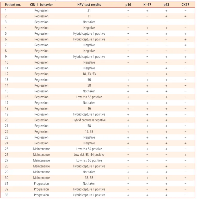
Table 1. The behavior of CIN 1 and the expressions of p16, Ki-67, p63, and CK17
Patient no. CIN 1 behavior HPV test results p16 Ki-67 p63 CK17
1 Regression 31 - + + -
2 Regression 31 - - + +
3 Regression Not taken - - - -
4 Regression Negative - + + -
5 Regression Hybrid capture II positive - - + +
6 Regression Hybrid capture II positive - - - +
7 Regression Negative - - - +
8 Regression Negative - - - -
9 Regression Hybrid capture II positive - - + +
10 Regression Negative - - - -
11 Regression Negative - - + -
12 Regression 18, 33, 53 - - + -
13 Regression 56 + + + -
14 Regression 58 + + + -
15 Regression Not taken + + + -
16 Regression Low risk 55 positive + - + -
17 Regression Not taken + + + -
18 Regression 16 + + + -
19 Regression Hybrid capture II positive + + + -
20 Regression Hybrid capture II negative + + + -
21 Regression 58 + + + -
22 Regression 16, 33 + + + -
23 Regression Negative + + + -
24 Regression Negative + + + +
25 Maintenance Low risk 54 positive - + + -
26 Maintenance Low risk 53, 44 positive - - - +
27 Maintenance Low risk 66 positive - - - -
28 Maintenance Hybrid capture II positive - - + -
29 Maintenance Not taken + + + -
30 Maintenance 33, 58 + + + -
31 Progression Not taken - - + -
32 Progression Hybrid capture II positive - - + -
33 Progression Hybrid capture II positive + + + -
CIN, cervical intraepithelial neoplasia 1; HPV, human papillomavirus; CK17, cytokeratin 17.
of the 9 patients with persistent CIN. There was no progression to CIN 3.
The CIN 1 behavior, HPV test results, and p16, Ki-67, p63, and CK17 expression levels are summarized in Table 1 and Fig. 1.
Among the 18 p16-negative patients, 17 patients (94.4%) expe- rienced regression, and 1 patient (5.6%) had persistent disease.
p16-negative expression correlated significantly with disease
regression (P=0.004). Fifteen of the 16 Ki-67-negative patients (93.8%) experienced regression, and 1 patient (6.2%) had persis- tent disease. Ki-67-negative expression also correlated signifi cant- ly with disease regression (P=0.017). Fourteen of the 15 patients (93.3%) negative for both p16 and Ki-67 experienced regression, and 1 patient (6.7%) had persistent disease. p63 was expressed in 26 patients (78.8%). All of the 7 p63-negative patients expe- rienced disease regression. However, the expression of p63 did not correlate significantly with disease regression and persistence (P=0.149). Six of the 7 CK17-positive patients (85.7%) experienced regression, and 1 patient (14.3%) had persistent disease. However, CK17 expression was not signifi cantly associated with disease re- gression or persistence (P=0.642) (Table 2).
The HPV test was performed in 28 of the 33 patients. Five pa- tients refused the HPV test or had received a punch biopsy before the HPV test was introduced at our hospital. The samples for 8 patients were analyzed using the HPV hybrid capture II test and those for 20 patients were analyzed using HPV DNA genotyping.
Sixteen of the 28 patients who underwent the HPV test (57.1%) had a high-risk HPV infection. Twenty-one patients in the regres- sion group and 7 patients in the persistent disease group received the HPV test; the rates of a high-risk HPV infection were 57.1% (12 of 21 patients) and 57.1% (4 of 7 patients), respectively (P=1.000).
We evaluated the associations between the p16, Ki-67, p63, and CK17 expression levels measured immunohistochemically and the presence of a high-risk HPV infection in the 28 patients who had undergone the HPV test (Fig. 2). Ten of the 13 p16-positive patients who had had the HPV test (76.9%) had a high-risk HPV infection. Having a high-risk HPV infection was significantly as- sociated with p16 expression (P=0.049). In the 13 p16-positive
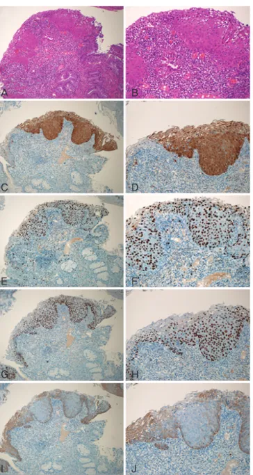
A B
C D
E F
G H
I J
Fig. 1. (A) H&E, ×100, (B) H&E, ×200. The expression of p16 (C, D), Ki- 67 (E, F), and p63 (G, H) in the lower one third of the cervical epithelium (×100, ×200). Note the loss of the CK17 expression in the lower one third of the cervical epithelium (I, J) ×100, ×200. (C-J) immunohisto- chemical stain.
(P=0.049)a
(P=0.067)
(P=0.624)
(P=0.133)b n=10
n=3
n=4
n=9
n=1 n=4 n=14
n=11
Fig. 2. The association of positive P16, Ki-67, p63, and CK17 immunohis- tochemical expressions with high risk HPV infection. aPearson’s chi-square test, bFisher’s exact test.
patients, 4 of the 10 high-risk HPV-positive patients (40.0%) and 2 of the 3 high-risk HPV-negative patients (66.7%) experienced persistent disease. In the p16-positive patients, having a high-risk HPV infection was not associated with the behavior of CIN 1. Only 1 of 18 p16-negative patients experienced persistent disease, and this patient did not have an HPV infection.
The Ki-67, p63, and CK17 expression levels were not signifi cantly associated with having a high-risk HPV infection ( P =0.067, P=0.624, and P=0.133, respectively). In the 15 Ki-67-positive pa-
tients, 4 of the 11 high-risk HPV-positive patients (36.4%) and 2 of the 4 high-risk HPV-negative patients (50.0%) experienced per- sistent disease. In the 23 p63-positive patients, 4 of the 14 high- risk HPV-positive patients (28.6%) and 3 of the 9 high-risk HPV- negative patients (33.3%) experienced persistent disease.
Discussion
Many studies have focused on the prognostic factors for CIN, including p16, Ki-67, p63, HPV L1, CK17, and CK8, but the value of these factors is controversial. A diffuse p16-staining pattern is detected immunohistochemically in almost all high-grade precan- cerous cervical lesions and cervical carcinoma, whereas a reactive condition-p16-negative staining or in the lesion cells-shows only sporadic staining [6-9]. Negri et al performed p16 immunohisto- chemical staining on CIN1 lesions and found that the CIN1 cases with diffuse p16 staining had a significantly higher tendency to progress to a higher-grade lesion than did the p16-negative cases [10,11]. Murphy et al. [12] studied p16
INK4Aas a marker for cervi- cal dyskaryosis and found that all formalin-fi xed, paraffi n wax-em- bedded sections with either strong nuclear or cytoplasmic staining were considered positive; a certifi ed pathologist then graded all sections qualitatively according to the following arbitrary scale: 0 (no positive staining), 1 (<10% positive staining), 2 (10-50% pos-
itive staining), and 3 (>50% positive staining). Most studies have used this scale, and we initially tried to use it to score the p16- stained slides. However, p16 exhibited a strong diffusely stained pattern in our study, and instead of the 0-3 scale, we considered grade 3 as positive in the statistical analysis of our small number of specimens. Ki-67 expression is greater in CIN samples than in those from normal and metaplastic epithelium, and Ki-67 staining is stronger in high-grade CIN than in low-grade CIN. Therefore, Ki- 67 is usually used to differentiate and grade CIN [13,14]. Kruse et al. [15,16] applied Ki-67 immunoquantifi cation using a comput- erized image analysis system to predict the progression of early CIN. They classifi ed the early CIN biopsies as low risk or high risk according to a Ki-67 model, which comprised the stratifi cation in- dex and the percentage of positive nuclei in the middle-third layer of the epithelium. In our current study, the number of p16- or Ki- 67-positive patients was not signifi cantly greater in the persistent group than in the spontaneous regression group, indicating that the p16- and Ki-67-expression patterns did not predict the high- risk group. However, the number of patients with p16- or Ki-67- negative expression was significantly greater in the regression group than in the persistent group. Thus, p16- or Ki-67-negative expression was helpful in identifying the low-risk group CIN1 pa- tients whose lesions did not progress to a higher grade.
A few previous studies have analyzed p63 expression in CIN and reported that p63 expression increases in high-grade CIN [17,18].
Quade et al. [17] performed p63 immunohistochemical staining on samples from CIN patients and reported that p63 typically localized to the basal and parabasal cells in CIN1 and that the p63-staining pattern extended to the middle and upper layers in samples from patients with CIN 2 or CIN 3. Martens et al. [18] re- ported similar results for the p63 immunohistochemical staining of samples from CIN patients. In our current study of CIN 1 patients, the p63 expression pattern did not differ signifi cantly between the regression group and the persistent group, suggesting that p63
Table 2. The behavior of CIN 1 as related to the expression statuses of p16, Ki-67, p63 and CK17Behavior Total (n)
p16 Ki‐67 p63 CK17 p16/Ki‐67
- + - + - + - + -/- -/+, +/- +/+
Regression 24 17 (94.4) 7 (46.7) 15 (93.8) 9 (52.9) 7 (100) 17 (65.4) 18 (69.2) 6 (85.7) 14 (93.3) 4 (100) 6 (42.9) Persistence 9 1 (5.6) 8 (53.3) 1 (6.3) 8 (47.1) 0 9 (34.6) 8 (30.8) 1 (14.3) 1 (6.7) 0 8 (57.1) Total 33 18 (54.6) 15 (45.4) 16 (48.5) 17 (51.5) 7 (21.2) 26 (78.8) 26 (78.8) 7 (21.2) 15 (45.4) 4 (12.1) 14 (42.4)
P‐valuea 0.004 0.017 0.149 0.642
Values are presented as number (%).
CIN, cervical intraepithelial neoplasia 1; CK17, cytokeratin 17.
aFisher’s exact test.
was of no value as a prognostic factor for early CIN; although all 7 patients who were negative for p63 expression were in the spon- taneous regression group, this was not statistically signifi cant.
Several studies have used immunohistochemistry to evaluate the expression of CK17 in the uterine cervix and its prognostic value, but these studies have produced diverse results. In the study of Re- gauer and Reich [19], all the CIN patients showed negative CK17 immunohistochemical staining Martens et al. [18] also reported that the CK17 expression in CIN was confi ned to the basal com- partment. However, Smedts et al. [20,21] found a clear increase in the expression of CK17 during the progression of CIN. We found no signifi cant difference in the CK17 expression pattern between the regression group and the persistent group. As observed for p63 expression, CK17 expression was not a prognostic factor for CIN 1 progression.
HPV induces cervical cancer through uncontrolled G1/S transition.
The inactivation of pRb by E7 causes p16 overexpression because p16 is regulated by the negative feedback of pRb [22]. Previous studies reported that p16 is associated with high-risk HPV infec- tion and that p16 expression increases in patients infected with high-risk HPV [6,23]. We also found a signifi cant association be- tween high-risk HPV infection and p16 expression. However, high- risk HPV infection in the p16-, Ki-67-, p63-, or CK17-positive pa- tients was not associated with the behavior of CIN 1, and the rate of high-risk HPV infection did not differ between the regression group and persistent disease group. In patients who showed a positive biomarker, the persistent disease rate was higher in high- risk HPV-negative patients than in high-risk HPV-positive patients.
These paradoxical results might be explained by the small number of patients who had an HPV test and the poor quality of the HPV DNA chip.
The current study did not show many signifi cant results for several reasons. This study was a small, retrospective study, and the differ- ences in the immunohistochemical staining patterns of expression between the CIN 1 and normal epithelium was less clear than those observed between high-grade CIN or cervical cancer and the normal epithelium. This may have limited our ability to detect signifi cant differences. Follow-up using only colposcopy or a Pap smear is less accurate than follow-up including pathology testing of samples obtained by conization or hysterectomy. Our results do not provide suffi cient evidence to conclude whether the expression patterns of p16, Ki-67, p63, or CK17 are of value as prognostic factors for early CIN.
In conclusion, CIN 1 with negative p16 and Ki-67 immunohistochemical staining expression was associated with spontaneous regression,
and p63 and CK17 expression patterns were not related to the behavior of CIN 1. The presence of a high-risk HPV infection was associated with p16 expression but not with the behavior of CIN1 in p16-, Ki-67-, p63-, or CK17-positive patients.
Acknowledgments
Funding support for this study was provided by Daejeon St. Mary Hospital, College of Medicine, Catholic University of Korea.
References
1. Ostör AG. Natural history of cervical intraepithelial neoplasia:
a critical review. Int J Gynecol Pathol 1993;12:186-92.
2. Schlecht NF, Platt RW, Duarte-Franco E, Costa MC, Sobrinho JP, Prado JC, et al. Human papillomavirus infection and time to progression and regression of cervical intraepithelial neo- plasia. J Natl Cancer Inst 2003;95:1336-43.
3. Nobbenhuis MA, Helmerhorst TJ, van den Brule AJ, Rozendaal L, Voorhorst FJ, Bezemer PD, et al. Cytological regression and clearance of high-risk human papillomavirus in women with an abnormal cervical smear. Lancet 2001;358:1782-3.
4. Wright TC Jr, Massad LS, Dunton CJ, Spitzer M, Wilkinson EJ, Solomon D, et al. 2006 consensus guidelines for the manage- ment of women with cervical intraepithelial neoplasia or ad- enocarcinoma in situ. Am J Obstet Gynecol 2007;197:340-5.
5. Ruas M, Peters G. The p16INK4a/CDKN2A tumor suppressor and its relatives. Biochim Biophys Acta 1998;1378:F115-77.
6. Klaes R, Friedrich T, Spitkovsky D, Ridder R, Rudy W, Petry U, et al. Overexpression of p16 (INK4A) as a specifi c marker for dysplastic and neoplastic epithelial cells of the cervix uteri. Int J Cancer 2001;92:276-84.
7. Keating JT, Cviko A, Riethdorf S, Riethdorf L, Quade BJ, Sun D, et al. Ki-67, cyclin E, and p16INK4 are complimentary sur- rogate biomarkers for human papilloma virus-related cervical neoplasia. Am J Surg Pathol 2001;25:884-91.
8. Negri G, Egarter-Vigl E, Kasal A, Romano F, Haitel A, Mian C.
p16INK4a is a useful marker for the diagnosis of adenocarci- noma of the cervix uteri and its precursors: an immunohisto- chemical study with immunocytochemical correlations. Am J Surg Pathol 2003;27:187-93.
9. O’Neill CJ, McCluggage WG. p16 expression in the female
genital tract and its value in diagnosis. Adv Anat Pathol
2006;13:8-15.
10. Negri G, Vittadello F, Romano F, Kasal A, Rivasi F, Girlando S, et al. p16INK4a expression and progression risk of low-grade intraepithelial neoplasia of the cervix uteri. Virchows Arch 2004;445:616-20.
11. Negri G, Bellisano G, Zannoni GF, Rivasi F, Kasal A, Vittadello F, et al. p16 ink4a and HPV L1 immunohistochemistry is helpful for estimating the behavior of low-grade dysplastic lesions of the cervix uteri. Am J Surg Pathol 2008;32:1715-20.
12. Murphy N, Ring M, Killalea AG, Uhlmann V, O’Donovan M, Mulcahy F, et al. p16INK4A as a marker for cervical dyskaryo- sis: CIN and cGIN in cervical biopsies and ThinPrep smears. J Clin Pathol 2003;56:56-63.
13. al-Saleh W, Delvenne P, Greimers R, Fridman V, Doyen J, Boniver J. Assessment of Ki-67 antigen immunostaining in squamous intraepithelial lesions of the uterine cervix. Correla- tion with the histologic grade and human papillomavirus type.
Am J Clin Pathol 1995;104:154-60.
14. Isacson C, Kessis TD, Hedrick L, Cho KR. Both cell proliferation and apoptosis increase with lesion grade in cervical neoplasia but do not correlate with human papillomavirus type. Cancer Res 1996;56:669-74.
15. Kruse AJ, Baak JP, de Bruin PC, Jiwa M, Snijders WP, Boodt PJ, et al. Ki-67 immunoquantitation in cervical intraepithe- lial neoplasia (CIN): a sensitive marker for grading. J Pathol 2001;193:48-54.
16. Kruse AJ, Baak JP, Janssen EA, Kjellevold KH, Fiane B, Lovslett
K, et al. Ki67 predicts progression in early CIN: validation of a multivariate progression-risk model. Cell Oncol 2004;26:13- 20.
17. Quade BJ, Yang A, Wang Y, Sun D, Park J, Sheets EE, et al. Ex- pression of the p53 homologue p63 in early cervical neopla- sia. Gynecol Oncol 2001;80:24-9.
18. Martens JE, Arends J, Van der Linden PJ, De Boer BA, Helmer- horst TJ. Cytokeratin 17 and p63 are markers of the HPV tar- get cell, the cervical stem cell. Anticancer Res 2004;24:771-5.
19. Regauer S, Reich O. CK17 and p16 expression patterns distin- guish (atypical) immature squamous metaplasia from high- grade cervical intraepithelial neoplasia (CIN III). Histopathol- ogy 2007;50:629-35.
20. Smedts F, Ramaekers FC, Vooijs PG. The dynamics of keratin expression in malignant transformation of cervical epithelium:
a review. Obstet Gynecol 1993;82:465.
21. Smedts F, Ramaekers F, Troyanovsky S, Pruszczynski M, Link M, Lane B, et al. Keratin expression in cervical cancer. Am J Pathol 1992;141:497-511.
22. Dyson N, Howley PM, Münger K, Harlow E. The human papil- loma virus-16 E7 oncoprotein is able to bind to the retino- blastoma gene product. Science 1989;243:934-7.
23. Song SH, Park HM, Eom DW, Lee JK, Lee NW, Kim AR, et
al. The expression of p16 (INK4a) and Ki-67 in relation to
high-risk human papilloma viral load and residual disease
after conization with positive margins. Int J Gynecol Cancer
2007;17:858-67.
경증 자궁경부상피내종양의 p16, Ki-67, p63, CK17 면역조직화학염색양상과 예후와의 관계
가톨릭대학교 의과대학 1부천성모병원 산부인과, 2대전성모병원 병리과 김수미1, 이정의2, 이대우1, 김민정1, 이해남1
목적
경증 자궁경부상피내종양(cervical intraepithelial neoplasia [CIN] 1)의 p16, Ki-67, p63, cytokeratin (CK) 17 면역조직화학염색양상과 임상 예후와의 관계를 평가하고자 하였다.
연구방법
33명의 CIN 1 환자들로부터 얻은 자궁경부조직을 p16, Ki-67, p63, CK17 면역조직화학염색을 하였다, 면역염색양상과 CIN 1의 임상예후 를 비교하였다.
결과
p16 면역염색에 음성인 환자 18명 중 17명(94.4%)에서 이후 CIN 1 병변이 소실되었으며 나머지 한 명(5.6%)에서만 병변이 지속되었 다. Ki-67 면역염색에 음성인 환자 16명 중 15명(93.8%)에서 이후 병변이 소실되었고 한 명(6.3%)에서만 병변이 지속되었다. P16과 Ki- 67 면역염색 음성은 병변의 소실과 유의한 관계를 보였다(P=0.004와 P=0.017). p16과 Ki-67 면역염색에 모두 음성인 환자 15명 중 14명 (93.3%)에서 병변이 소실되었고 나머지 한 명(6.7%)에서만 병변이 지속되었다. P63과 CK17 면역염색양상은 질환의 임상양상과 는 유의한 관계가 없었다(P=0.149와 P=0.642). p16 면역염색 양성인 13명의 환자 중 10명(76.9%)이 고위험 인유두종바이러스 (human papillomavirus, HPV)에 감염된 상태였다. 고위험 HPV 감염은 p16 면역염색 양성과 유의한 관계가 있었다(P=0.049).
결론
CIN 1이 p16이나 Ki-67 면역염색 음성을 보일 경우 병변이 저절로 소실되는 경향을 보였다. 반면 P63과 CK17은 CIN 1의 임상양상과 관 계가 없었다.
중심단어: 경증 자궁경부상피내종양, p16, Ki-67, p63, CK17