저작자표시-비영리-동일조건변경허락 2.0 대한민국 이용자는 아래의 조건을 따르는 경우에 한하여 자유롭게
l 이 저작물을 복제, 배포, 전송, 전시, 공연 및 방송할 수 있습니다. l 이차적 저작물을 작성할 수 있습니다.
다음과 같은 조건을 따라야 합니다:
l 귀하는, 이 저작물의 재이용이나 배포의 경우, 이 저작물에 적용된 이용허락조건 을 명확하게 나타내어야 합니다.
l 저작권자로부터 별도의 허가를 받으면 이러한 조건들은 적용되지 않습니다.
저작권법에 따른 이용자의 권리는 위의 내용에 의하여 영향을 받지 않습니다. 이것은 이용허락규약(Legal Code)을 이해하기 쉽게 요약한 것입니다.
Disclaimer
저작자표시. 귀하는 원저작자를 표시하여야 합니다.
비영리. 귀하는 이 저작물을 영리 목적으로 이용할 수 없습니다.
동일조건변경허락. 귀하가 이 저작물을 개작, 변형 또는 가공했을 경우 에는, 이 저작물과 동일한 이용허락조건하에서만 배포할 수 있습니다.
A DISSERTATION
FOR THE DEGREE OF MASTER
Retinal Function and Histopathological Assessment after Intravitreal Cidofovir Injection in Normal Dogs
정상 개에서 유리체내
Cidofovir 주사후의 망막기능 및 조직병리학적평가
by
Sung Won Park
MAJOR IN VETERINARY CLINICAL SCIENCES DEPARTMENT OF VETERINARY MEDICINE
GRADUATE SCHOOL
SEOUL NATIONAL UNIVERSITY
February, 2015
Retinal Function and Histopathological Assessment after Intravitreal Cidofovir Injection in Normal Dogs
by
Sung Won Park
Supervised by Professor Kangmoon Seo
Thesis
Submitted to the Faculty of the Graduate School of Seoul National University
in partial fulfillment of the requirements for the degree of Master
in Veterinary Medicine October, 2014
Major in Veterinary Clinical Science Department of Veterinary Medicine
Graduate School Seoul National University
December, 2014
ii
Retinal Function and Histopathological Assessment after Intravitreal Cidofovir Injection in Normal Dogs
Supervised by Professor Kangmoon Seo
Sung Won Park
Major in Veterinary Clinical Sciences, Department of Veterinary Medicine Graduate School, Seoul National University
ABSTRACT
This study was performed to evaluate effect of an intravitreal injection of cidofovir in beagle dogs.
Nine beagle dogs (18 eyes) were used and randomly assigned to three groups of various dosage: 100, 500, and 1,000 µg. Aqueoucentesis was followed by an intravitreal injection of cidofovir. Intraocular pressure (IOP) was measured twice a week and electroretinography (ERG) and ophthalmoscopic examination were performed every week during the study. At the end of the study, all eyes were enucleated for histopathologic evaluation after euthanasia.
The lowest IOP was observed 10 days after the injection in all groups. Irreversible
reductions in IOP and reduced amplitudes of ERG recordings were identified in the eyes injected with higher dose groups than the 100 µg group. Histopathologic examination revealed that there were dose-related toxicities to the ciliary body and the retina.
These results suggest that intravitreal cidofovir had dose-dependent IOP lowering effects associated with ciliary body destruction, but had the potential to cause retinal toxicity in beagle dogs.
____
Keywords: cidofovir, intravitreal injection, retina, IOP, dog Student Number: 2012 – 23568
iv
CONTENTS
INTRODUCTION···1
MATERIALS AND METHODS···3
1. Experimental animals···3
2. Preparation of the drug···4
3. Electroretinography···5
4. Intravitreal injection ···6
5. Histopathologic evaluation ···8
6. Statistical analyses···9
RESULTS···10
1. Intraocular pressure changes···10
2. Ophthalmic examination findings···12
3. Electroretinographic changes···15
4. Histopathologic changes···17
DISCUSSION··· ···20
REFERENCES···23
ABSTRACT IN KOREAN···26
Introduction
Glaucoma is a group of diseases affecting the optic nerve and accompanied by the loss of retinal ganglion cells in a characteristic pattern. Elevated intraocular pressure is an important factor in the development of glaucoma. Chronically glaucomatous and irreversibly avisual eyes are common and are a source of patient discomfort related to elevated intraocular pressure (IOP) and complications, including buphthalmos and exposure keratopathy (Gelatt et al., 2013; Maggs et al., 2013). Long-term therapy with topical hypotensives has rarely been successful because the response to the drugs varies from patient to patient and the efficacy of the medicines can diminish over time (Gelatt et al., 2013; Maggs et al., 2013).
Effective management methods currently used to alleviate discomfort and to provide a cosmetically acceptable appearance are enucleation, evisceration with an intraocular prosthesis, or chemical cycloablation by an intravitreal gentamicin injection (Gelatt et al., 2013). Recently, one study reported the hypotensive effects of intravitreal cidofovir injections in dogs with chronic glaucoma (Low et al., 2014).
Cidofovir [S-1-(3-hydroxy-2-phosphonylmethoxypropyl)cytosine; S-HPMPC]
has been an anti-viral medication administered to human patients as a form of therapy for acquired immune deficiency syndrome (AIDS)-associated cytomegalovirus retinitis (Kirsch et al., 1995; Rahhal et al., 1995). Intravitreal cidofovir injections have been utilized in humans to decrease systemic exposure and renal toxicity rather than intravenous injections (Masur et al., 1996). Twenty micrograms of intravitreal cidofovir have been reported to be well tolerated by the
human eye (Taskintuna et al., 1997b). Intravitreal injections of 20 µg of cidofovir have been found to have a statistically significant hypotensive effect compared with baseline (Banker et al., 1997; Taskintuna et al., 1997c).
Intravitreal cidofovir was determined to be safe in studies of its histopathologic effects in various animals (Banker et al., 1998; Taskintuna et al., 1997a). Intravitreal injections of cidofovir have been found to be toxic to the ciliary body and retina in guinea pigs and rabbits (Taskintuna et al., 1997a). Although several studies showed that intravitreal cidofovir decreased IOP, presumably because of its destruction of the ciliary body, its effect on the retina has not been investigated in dogs yet.
Therefore, the aim of this study was to investigate dose-related changes in retinal function and histopathology in normal beagle eyes after intravitreal injection of cidofovir.
Materials and Methods
1. Experimental animals
Both eyes of 9 healthy beagle dogs were included in this study. The mean age was 3.4 ± 0.2 years. Seven of the dogs were male and 2 were female. The eyes were randomly divided into 3 groups that were given different doses of cidofovir; 100 (A), 500 (B), and 1,000 µg (C). Prior to beginning the experiment, the dogs were underwent complete ophthalmic examinations including slit-lamp biomicroscopy (SL-D7®, Topcon, Japan), indirect ophthalmoscopy (Vantage Plus®, Keeler Ltd., UK), rebound tonometry (Tonovet®, Icare Finland, Finland), and electroretinogram (ERG; Reti-com®, Roland Instrument, Germany) to ensure clinically normal eyes. All care and experimental procedures were confirmed to the Institutional Animal Care and Use Committee (IACUC) of Seoul National University (SNU-131213-1).
2. Preparation of the drug
The cidofovir (C5874, Sigma-Aldrich, USA) was dissolved in sterile, nonbacteriostatic, and nonpyrogenic 0.9% sodium chloride solution (NaCl, Daihan Pharm. Co. Ltd., South Korea). Sodium hydroxide (NaOH) was added to solubilize the drug leading to the final pH of 7.0. Then, 0.9% NaCl was added to the solution to reach the calculated volume (final concentrations of 1, 5, and 10 mg/ml) and filtered through a sterile syringe with a 0.22 µm nylon, sterile, and nonpyrogenic syringe filter (Minisart®, Sartorius Stedim Biotech, Germany). The prepared solution was aliquot into 0.5 ml insulin syringes and randomly injected into the beagles’ eyes. In all groups, the volume of solution injected was 0.1 ml.
3. Electroretinography
Electroretinography (ERG) was performed under a dim red light in a dark room. After producing mydriasis with 1% tropicamide (Mydriasil®, Alcon, South Korea), the animals were adapted to the dark for 20 min. All of the dogs were anesthetized with 5 µg/kg of medetomidine (Domitor®, Orion Pharm, Norway) and 1.25 mg/kg of a combination of zolazepam/tiletamine (Zoletil®, Virbac, Australia) by intravenous injections prior to ERG. The ground electrode was positioned over the external occipital protuberance, and the reference electrode was positioned at approximately 2 cm caudal to the lateral canthus. The contact lens electrode (ERG-jet®, Fabrinal SA, Switzerland) was located on the cornea after topical anesthesia with 0.5% proparacaine (Alcaine®, Alcon, Belgium) and lubrication with 2% hypromellose (Hycell®, Samil, South Korea). ERG was performed on bilateral eyes at equal intensity (2.5 cd·s/m2). Responses were analyzed for a- and b-wave amplitudes and implicit times. The amplitude of the a-wave was calculated from the baseline to the first negative deflection, and that of the b-wave was measured from the trough of the a-wave to the positive peak of the b-wave. Implicit times of a- and b-waves were calculated from the onset of the light stimulus to the peak of the a- and b-waves.
4. Intravitreal injection
The trial was designed as a prospective, randomized, observer-masked study. The IOP and ERG were recorded at baseline for the beagle dogs. A complete ophthalmic examination was performed on all eyes at the time of injection using dilated indirect ophthalmoscopy and fundus photography.
All dogs were intravenously injected with tramadol 2 mg/kg (Tradol®, BTO pharm, Korea) for analgesia. Topical proparacaine was instilled before the intravitreal injection. Each eye was aseptically prepared by rinsing the ocular surface with 0.5% povidone-iodine solution. Aqueoucentesis was performed to decrease IOP for minimizing ocular pain during the injection and postinjection leakage through the vitreous injection site. A syringe with a 30- gauge needle was inserted into the cornea adjacent the superior limbus with the needle parallel and anterior to the plane of the iris. Cidofovir was injected 5 mm posterior to the superior limbus through the bulbar conjunctiva, sclera, and pars plana into the vitreal chamber using a 30-gauge needle directed toward the posterior pole. After the intravitreal injection, 4 mg of triamcinolone (Rheudenolone®, Kukje Pharma, South Korea) and gentamicin (Gentapro®, Huons, Korea) were administered subconjunctivally. All dogs were treated with neomycin-polymyxin B-dexamethasone solution (Maxitrol®, Alcon, Belgium) every 12 h for 2 weeks. Twice a week, IOPs were measured between 6 and 7 PM. Visual assessment was determined by the abnormality of the menace response, visual placing, dazzle reflex, and pupillary light reflex.
ERG and slit-lamp examination were performed once a week with dilated pupils. At the end of 4 weeks, the beagles were euthanized and their eyes were enucleated for histopathologic evaluation.
5. Histopathologic evaluation
Enucleated eyes were placed in a mixture of freshly prepared Davidson’s solution (2% paraformaldehyde, 35% ethanol, 10% glacial acetic acid, and 53% distilled water). After 48 h, the solution was changed to 70% ethanol. The eyes were processed through graded alcohols and xylene and embedded in paraffin. For histopathologic analysis, 5 µm vertical sections of the pupil and optic nerve were obtained every 200 µm and stained with hematoxylin and eosin.
6. Statistical analyses
The results are expressed as mean ± standard deviation (SD). All analyses were performed using statistical software (SPSS® ver.21 for Windows, IBM, USA). In the event that significance was achieved by repeated analysis of variance (ANOVA) measurements, the Bonferroni post-hoc test was applied, considering values of P < 0.05 statistically significant. P values lesser than 0.001 was considered extremely statistically significant.
Results
1. Intraocular pressure changes
No significant IOP changes were observed compared with baseline in Group A. The IOPs in Groups B and C were significantly lower than baseline values three days during experimental periods (Table 1). In Groups B and C, the lowest IOP was measured on day 10 (25.2% and 49.8% of baseline, P <
0.001, respectively). IOP was estimated as a lowest value on day 10 and increased slightly thereafter in all groups (Table 1). There were no statistically significant differences between Group B and C.
Table 1. Intraocular pressure (IOP) of baseline and post cidofovir injections in beagle eyes
* mean ± S.D.
** Percentage drop in IOP from baseline.
† Significantly lower than baseline (P < 0.05).
‡ Significantly lower than baseline (P < 0.001).
Groups
Days
A (100 µg/eye)
B (500 µg/eye)
C (1000 µg/eye)
IOP (mmHg)
% drop**
IOP
(mmHg) % drop IOP
(mmHg) % drop
Baseline 16.8 ± 2.1* 16.1 ± 2.0 15.8 ± 0.9
3 days 14.4 ± 1.9 14.2 14.2 ± 2.2† 11.7 12.4 ± 1.1‡ 21.7 7 days 14.4 ± 2.5 13.9 12.8 ± 1.2‡ 20.3 9.6 ± 2.3‡ 39.7 10 days 14.2 ± 2.7 15.6 12.1 ± 1.9‡ 25.2 7.9 ± 2.9‡ 49.8 14 days 14.5 ± 4.0 13.6 12.6 ± 0.9‡ 21.7 8.8 ± 2.7‡ 44.2 17 days 15.2 ± 4.1 9.6 12.7 ± 0.9‡ 21.4 9.1 ± 2.0‡ 42.5 21 days 15.4 ± 1.9 8.3 13.6 ± 1.1‡ 15.8 10.0 ± 2.5‡ 36.8 24 days 15.8 ± 3.1 5.6 14.1 ± 0.7† 12.8 10.3 ± 2.7‡ 35.1 28 days 15.9 ± 2.6 5.3 14.2 ± 1.3† 12.1 10.3 ± 2.4‡ 34.7
2. Ophthalmic examination findings
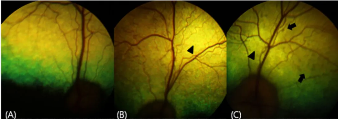
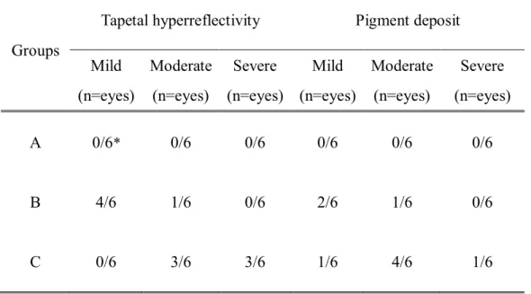
Intravitreal injections in Group A produced no remarkable findings during the study period (Fig. 1A). In Group B, intravitreal cidofovir caused moderate conjunctival hyperemia with mild aqueous flare until 7 days. Fundus examination showed mild tapetal hyperreflectivity and pigment deposit at 28 days in Group B (Fig. 1B and Table 2). Furthermore, at 28 days, some of the eyes showed a delayed response to visual assessments such as the menace and visual placing, but vision was present until the end of the study. In Group C, intravitreal cidofovir caused more toxic effects than were observed in the other groups. The eyes developed moderate to severe conjunctival hyperemia, aqueous flare, and hyalitis at 7 days, followed by the moderate to severe peripheral deposit of pigment and tapetal hyperreflectivity at 28 days (Fig. 1C and Table 2). Among them, three eyes had no menace responses and visual placing reactions. There were no cataractogenic findings in any of the eyes treated with cidofovir. A conjunctival hemorrhage caused by injection trauma was present in 3 eyes, but it was not dose-related, and the blood was naturally absorbed within a week.
Fig. 1. Fundus examination on 28 days. (A) Fundus image of Group A showed no significant abnormalities. (B) There is mild tapetal hyperreflectivity (arrowhead) in Group B. (C) There is dispersed light-brownish pigment (arrow) on the retina and moderate hyperreflectivity (arrowhead) of the tapetum in Group C.
Table 2. Fundus image changes on day 28 after intravitreal cidofovir injection in beagle eyes
Groups
Tapetal hyperreflectivity Pigment deposit Mild
(n=eyes)
Moderate (n=eyes)
Severe (n=eyes)
Mild (n=eyes)
Moderate (n=eyes)
Severe (n=eyes)
A 0/6
*0/6 0/6 0/6 0/6 0/6
B 4/6 1/6 0/6 2/6 1/6 0/6
C 0/6 3/6 3/6 1/6 4/6 1/6
* Number of eyes showing fundic changes / Total number of eyes (n=6)
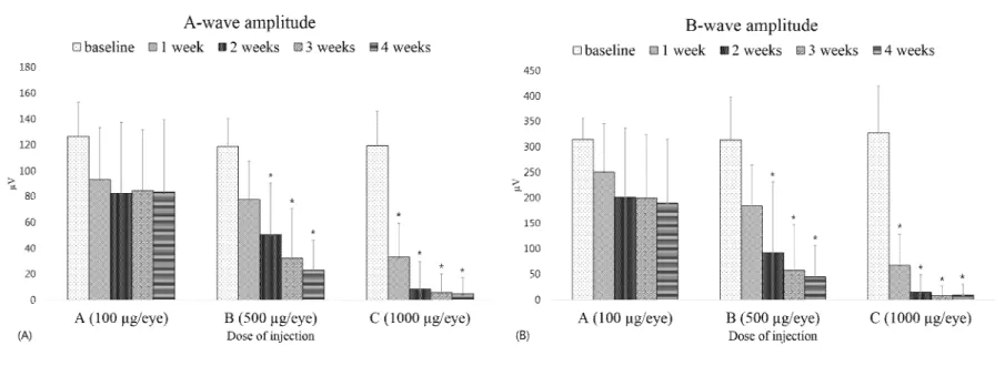
3. Electroretinographic changes
There was no statistically significant change in Group A. Intravitreal injections of cidofovir in group B statistically reduced both a- and b-wave amplitudes at 14 days (P < 0.001, Fig. 2). Group C showed that amplitude had been significantly decreased after 7 days compared to baseline (P < 0.001, Fig.
2). Most of the eyes in Group C had remarkably flat amplitudes of both a- and b-waves at the end of study despite no statistically significant differences in Groups B and C. The amplitude of waves in Group A were statistically different from other groups. There were no significant changes in the implicit time of waves during the study period in all groups.
* P < 0.001
Fig. 2. Changes in the amplitude of electroretinogram (ERG) in each group after intravitreal cidofovir injections in beagle eyes.
(A) = Amplitude of a-wave; (B) = Amplitude of b-wave.
4. Histopathologic changes
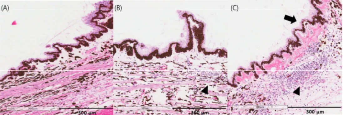
The ciliary body and retina of the eyes in Group A were within normal limits until 28 days (Fig. 3A and 4A). Injections of cidofovir in Group B caused mild attenuation of the pars plicata, disruption of the pigment epithelium (PE) in the ciliary process, infiltration of monocytes in the stroma of the ciliary body (Fig. 3B) and mild migration of pigment cells into the photoreceptor layer (Fig. 4B). In Group C, intravitreal injections of cidofovir caused moderate disruption of the PE and the infiltration of monocytes and pigment-laden cells in the ciliary body and fibrous materials in the vitreous body (Fig. 3C and 4C). Furthermore, there was moderate-to-severe disruption of the retinal pigment epithelium (RPE), pigmented cell migration, and degeneration of outer retinal architecture with retinal fold (Fig. 4C).
Fig. 3. Histopathologic changes of the ciliary body after intravitreal cidofovir injections in beagle eyes. H&E stain. (A) There were no remarkable findings in Group A. (B) Histopathology in Group B showed a mild disruption of PE and infiltration of monocytes in the stroma of the ciliary body (arrow head). (C) There is a moderate disruption of pigment epithelium in the ciliary processes (arrow head), moderate-to-severe infiltration of monocytes (arrow), and dispersed pigment-laden cells in the stroma of the ciliary body in Group C.
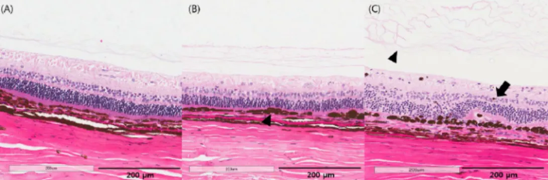
Fig. 4. Histopathologic changes of the retina after cidofovir injection. H&E satin.
(A) There were no remarkable findings in Group A. (B) Group B showed mild migration of pigment into the photoreceptor layer (arrow head). (C) There was moderate-to-severe migration of pigment cells into the outer retina segment, photoreceptor layer degeneration with retinal fold (arrow), and fibronous materials in the vitreous body (arrow head) in the Group C.
Discussion
Even though cidofovir has been used in antiviral medication for viral retinitis in humans, cidofovir has an intraocular hypotensive effect (Banker et al., 1997;
Taskintuna et al., 1997c). Furthermore, one study showed that intravitreal injections of 561.5 µg of cidofovir had an IOP-lowering effect (78% lower than baseline) in dogs with chronic glaucoma (Low et al., 2014).
A study of vitreous volume in canine and equine eyes suggested that the mean vitreous volume of dog eyes was 1.7 ± 0.86 ml (Gilger et al., 2005). In this study, injections of 100, 500, and 1000 µg of cidofovir (final vitreous concentrations of 58.8, 294, and 588 µg/ml, respectively) were performed intravitreally. In all groups, the lowest IOP at day 10 might be associated with postoperative inflammation because IOP rises with reduced intraocular inflammation. Doses of 500 and 1000 µg of cidofovir (final vitreous concentrations of 294 and 588 µg/ml, respectively) resulted in the permanent reduction of IOP. In guinea pigs, intravitreal injections of 50 and 156.25 µg of cidofovir (final vitreous concentrations of 200 and 625 µg/ml, respectively) statistically decreased IOP (53 and 70% lower than baseline, respectively) (Taskintuna et al., 1997a). In rabbit eyes, injections of 875 and 2800 µg of cidofovir intravitreally (final vitreous concentrations of 625 and 2000 µg/ml, respectively) lowered IOP (26 and 11% of baseline, respectively), which did not reach statistical significance (Taskintuna et al., 1997a). These results suggest that cidofovir’s IOP-lowering effect was stronger in guinea pigs than beagles or rabbits.
Furthermore, glaucomatous eyes were more susceptible to the hypotensive effect of cidofovir than normal eyes (Low et al., 2014).
The loss of both a- and b-wave amplitudes in all eyes suggests that cidofovir is toxic to the retinal structure. The amplitude of a- and b-waves relates to the function of photoreceptors and müller cells, respectively (Ekesten, 2013). Thus, the loss of amplitude in this study indicates that intravitreal cidofovir decreased the retinal function associated with retinal degeneration. However, one study showed that subcutaneous injections of cidofovir prevented the loss of a- and b-wave amplitudes of ERG, limited viral replication, and reduced disease pathology in a murine model of ocular cytomegalovirus disease (Garneau et al., 2003). These paradoxical results might be related to differences in subjects and routes of drug administration.
Histopathologic evaluation in this study showed that intraocular pathologic changes occurred in a dose-dependent manner. Disruption of PE in ciliary processes and the infiltration of monocytes suggested that intravitreal cidofovir had destructive effects on the ciliary body. Retinal degeneration in Groups B and C was related to the loss of both a- and b-wave amplitudes in the electroretinography (Ekesten, 2013). Intravitreal injections of cidofovir in Group C (final vitreous concentration of 588 µg/ml) markedly exacerbated the degeneration of retinal architecture and the accumulation of pigment in the photoreceptor layer. Taskintuna et al. showed that 156.25 µg of cidofovir (final vitreous concentration of 625
µg/ml) caused no remarkable changes in the ciliary body and retina in rabbits (Taskintuna et al., 1997a). Thus, rabbit retinas appear more resistant to the toxicity of cidofovir than those of beagles or guinea pigs.
The accumulation of irregular light-brownish pigment spots in the tapetal fundus and hyperreflectivity have been well-documented typical fundus
appearances of central progressive retinal atrophy (CPRA) in dogs (Aguirre et al., 1976). Retinal degeneration in the CPRA has been related to retinal pigment epithelium (RPE) disruption (Bedford, 2009). Vitamin E deficiency might play a role in the development of CPRA (Davidson et al., 1998), and the pathogenesis is quite different from pigment accumulation in this study. The RPE photoreceptor complex could be exposed to oxidative stress-associated free radical formation and lipid peroxidation (Zabrin, 2012). Photoreceptor outer segments contain a high concentration of fatty acids and are exposed to a high partial pressure of oxygen, a condition that predisposes the macular region to lipid peroxidation in humans (Young, 1987). Pathological studies showed that the main source of vision loss was at the level of the photoreceptor; however, photoreceptor loss appears to be secondary to damage to the RPE cells, suggesting that the RPE cells would be the site of primary injury (Young, 1987). Thus, dose-related degeneration of the retinal outer segment in Groups B and C might be related to the RPE disruption and lipid peroxidation caused by oxidative stress related to intravitreal cidofovir.
This study showed that intravitreal injections of cidofovir (more than 500 µg) had not only a hypotensive effect resulting from the destruction of the ciliary body, but also caused vision loss due to retinal toxicity in beagle eyes. Thus, intravitreal injections of cidofovir for hypotensive effects in clinical cases might require doses greater than 500 µg and should be applied irreversibly blind dogs because of the drug’s retinal toxicity.
References
Aguirre GD, Laties A, 1976. Pigment epithelial dystrophy in the dog. Experimental Eye Research 23, 247-256.
Banker AS, Arevalo JF, Munguia D, Rahhal FM, Ishimoto B, Berry C, De Clercq E, Ochabski R, Taskintuna I, Freeman WR, 1997. Intraocular pressure and aqueous humor dynamics in patients with AIDS treated with intravitreal cidofovir (HPMPC) for cytomegalovirus retinitis. American Journal of Ophthalmology 124, 168-180.
Banker AS, De Clercq E, Taskintuna I, Keefe KS, Bergeron-Lynn G, Freeman WR, 1998. Influence of intravitreal injections of HPMPC and related nucleoside analogues on intraocular pressure in guinea pig eyes.
Investigative Ophthalmology & Visual Science 39, 1233-1242.
Bedford PG, 2009. Retinal pigment epithelial dystrophy in the Briard. Veterinary Record 12, 377.
Davidson MG, Geoly FJ, Gilger BC, McLellan GJ, Whitley W, 1998. Retinal degeneration associated with vitamin E deficiency in hunting dogs.
Journal of the American Veterinary Medical Association 5, 645-651.
Ekesten B, 2013. Ophthalmic Examination and Diagnostics. In: Veterinary ophthalmology. Ames, John Willey & Sons, 684-685.
Garneau M, Bolger GT, Bousquet C, Kibler P, Tremblay F, Cordingley MG, 2003.
HPMPC therapy of MCMV-induced retinal disease in the SCID mouse measured by electroretinography, a non-invasive technique. Antiviral Research 59, 193-200.
Gelatt KN, Plummer CE, Regnier A, 2013. The Canine Glaucoma. In: Veterinary ophthalmology. Ames, John Willey & Sons, 1050-1129.
Gilger BC, Reeves KA, Salmon JH, 2005. Ocular parameters related to drug delivery in the canine and equine eye: aqueous and vitreous humor volume and scleral surface area and thickness. Veterinary Ophthalmology 8, 265-269.
Kirsch LS, Arevalo JF, Chavez de la Paz E, Munguia D, de Clercq E, Freeman WR, 1995. Intravitreal cidofovir (HPMPC) treatment of cytomegalovirus retinitis in patients with acquired immune deficiency syndrome.
Ophthalmology 102, 533-543.
Low MC, Landis ML, Peiffer RL, 2014. Intravitreal cidofovir injection for the management of chronic glaucoma in dogs. Veterinary Ophthalmology 17, 201-206.
Maggs DJ, Miller PE, Ofri R, 2013. Slatter’s fundamentals of veterinary ophthalmology. St. Louis, Elsevier, 251-271.
Masur H, Whitcup SM, Cartwright C, Polis M, Nussenblatt R, 1996. Advances in the management of AIDS-related cytomegalovirus retinitis. Annals of Internal Medicine: Journal 125, 126-136.
Rahhal FM, Arevalo JF, Munguia D, Taskintuna I, Chavez de la Paz E, Azen SP, Freeman WR, 1996. Intravitreal cidofovir for the maintenance treatment of cytomegalovirus retinitis. Ophthalmology 103, 1078-1083.
Taskintuna I, Banker AS, Rao NA, Wiley CA, Flores-Aguilar M, Munguia D, Bergeron-Lynn G, De Clercq E, Keefe K, Freeman WR, 1997a. An animal model for cidofovir (HPMPC) toxicity: Intraocular pressure and
histopathologic effects. Experimental Eye Research 64, 795-806.
Taskintuna I, Rahhal FM, Arevalo JF, Munguia D, Banker AS, De Clercq E, Freeman WR, 1997b. Low-dose intravitreal cidofovir (HPMPC) therapy of cytomegalovirus retinitis in patients with acquired immune deficiency syndrome. Ophthalmology 104, 1049-1057.
Taskintuna I, Rahhal FM, Rao NA, Willey CA, Mueller AJ, Banker AS, De Clercq E, Arevalo JF, Freeman WR, 1997c. Adverse events and autopsy findings after intravitreous cidofovir (HPMPC) therapy in patients with acquired immune deficiency syndrome (AIDS). Ophthalmology 104, 1827-1837.
Young RW, 1987. Pathophysiology of age-related macular degeneration. Survey of Ophthalmology 31, 291-306.
Zarbin MA, 2012.Pathogenesis of Age-Related Macular Degeneration. In: Medical Retina vol 1. Basel, Karger, 125-133.
국 문 초 록
정상 개에서 유리체내
Cidofovir 주사후의 망막기능 및 조직병리학적 평가
지도교수 서 강 문
박 성 원
서울대학교 대학원 수의학과 임상수의학 전공
본 연구에서는 건강한 개를 대상으로 유리체내 Cidofovir 주사의 효과를 평가하였다.
각 9마리(18안구)는 100, 500 그리고 1000 µg 용량으로 나뉜 군에 무작위로 설정되었다. 유리체내 Cidofovir 주사에 앞서 앞방방수천자를 실시하였다. 연구기간 동안, 안압은 1주일에 두 번씩 일정한 시간에 측정하였고, 망막전위도(electroretinography)와 검안경 검사는 매주
실시되었다. 4주후에는 주사를 실시한 안구를 적출한 뒤 조직검사를 실시하였다.
모든 그룹에서 주사한 후 10일뒤의 안압이 가장 낮게 측정되었다.
투여한 용량이 500 ug 이상인 그룹에서 비가역적인 안압감소와 망막전위도의 진폭(amplitude) 감소가 확인되었다. 조직검사에서는 모양체와 망막에 용량의존성 독성이 나타나는것을 알 수 있었다.
이러한 결과는 유리체네 Cidofovir 가 비글에서 용량에 의존적으로 모양체 파괴와 관련된 안압하강 효과가 있지만, 안압하강을 유의적으로 일으키는 Cidofovir 의 용량은 망막에 독성을 일으킬 가능성이 있다는 것을 제시하였다.
주요어 : Cidofovir, 유리체내주사, 망막, 안압, 개 학 번: 2012 - 23568