www.ogscience.org 190
Received: 2012.9.29. Revised: 2012.12.17. Accepted: 2012.12.17.
Corresponding author: Hye-Sung Won
Department of Obstetrics and Gynecology, University of Ulsan College of Medicine, Asan Medical Center, 88 Olympic-ro 43-gil, Songpa-gu, Seoul 138-736, Korea
Tel:+82-2-3010-3644 Fax: +82-2-3010-6944 E-mail: [email protected]
Articles published in Obstet Gynecol Sci are open-access, distributed under the terms of the Creative Commons Attribution Non-Commercial License (http://creativecommons.
org/licenses/by-nc/3.0/) which permits unrestricted non-commercial use, distribution, and reproduction in any medium, provided the original work is properly cited.
Copyright © 2013 Korean Society of Obstetrics and Gynecology
Introduction
Isochromosomes are supernumerary chromosomes that are made up of two copies of the same arm on a chromosome [1-3]. Tet- rasomy 18p was first reported by Froland et al. in 1963 [4].
It is a very rare chromosomal anomaly with a prevalence of 1/140,000-180,000 but is also one of the most commonly observed isochromosomes, and affects both genders equally [1-4]. Tetrasomy 18p syndrome is characterized by nonspecific morphologic features; low birth weight, microcephaly, low- set ears, strabismus, abnormalities in muscle tone and deep tendon reflex [1,3,5,6]. Feeding difficulties and developmental retardation are also followed [2,6]. Cardiac and renal malfor- mations are rare, therefore, mortality rate is low [1,5,7]. Be- cause of its very low prevalence rate, tetrasomy 18p has not yet been reported in Korea. Herein, we report the first case of prenatally diagnosed tetrasomy 18p.
Case report
A 28-year-old primi gravid woman was referred to our fetal treatment center because of suspected fetal congenital heart disease at 32+4 weeks of gestation. The ultrasonography showed asymmetric intrauterine growth retardation (IUGR)
with 5-week smaller abdominal circumference. The fetal echocardiography demonstrated dextrocardia with cardio- megaly (cardio-thoracic ratio, 0.64), mild pericardial effusion, and decreased left ventricular function (modified myocardial performance index, 0.68). There was no intracardiac abnor- mality. Doppler findings in the middle cerebral artery revealed increased peak systolic velocity (71 cm/sec, 1.3-1.5 MoM), which suggested fetal anemia. Imperforate anus was also sus- pected. The cordocentesis was performed at 33+2 weeks of gestation for identifying the karyotype, hemoglobin, and the presence of viral infection. The karyotyping confirmed tetra- somy 18p by G-banding and fluorescence in situ hybridization
A case report of prenatally diagnosed tetrasomy 18p
Phill-Seung Jung
1, Hye-Sung Won
1, In-Ji Cho
1, Min-Kyung Hyun
2, Jae-Yoon Shim
1, Pil-Ryang Lee
1, Ahm Kim
1Department of Obstetrics and Gynecology, 1University of Ulsan College of Medicine, Asan Medical Center, Seoul; 2Inha University Hospital, Inha University College of Medicine, Incheon, Korea
Tetrasomy 18p, one of the most commonly observed isochromosomes, consists of two copies of the p arms on chromosome 18[i(18p)]. It is known as a de novo occurrence of non-disjunction or centromeric mis-division during meiosis II in the vast majority of cases. It has a prevalence of 1/140,000-180,000 live births and affects both genders equally.
A 28-year-old woman was referred at 33+2 weeks gestation to rule out fetal congenital heart disease. Her prenatal ultrasonography showed intrauterine growth retardation, cardiomegaly, and imperforate anus. Doppler ultrasonographic finding showed fetal anemia. Tetrasomy 18p was confirmed by conventional karyotyping and fluorescence in situ hybridization. Because of its very low prevalence rate, only several cases of tetrasomy 18p has been reported worldwide and it has not yet been reported in Korea before. Therefore, we report a case of prenatally diagnosed tetrasomy 18p.
Keywords: Isochromosome 18p; Prenatal diagnosis; Tetrasomy 18p
Case Report
Obstet Gynecol Sci 2013;56(3):190-193 http://dx.doi.org/10.5468/ogs.2013.56.3.190 pISSN 2287-8572 · eISSN 2287-8580
www.ogscience.org 191 Phill-Seung Jung, et al. A prenatally diagnosed tetrasomy 18p
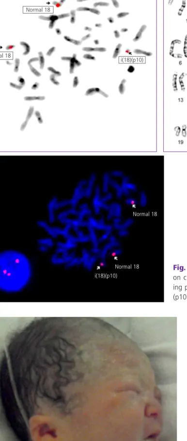
(Fig. 1). Fetal hemoglobin level was 9.7 g/dL and there was no evidence of viral infection such as toxoplasma, rubella, cy- tomegalovirus, and herpes simplex virus.
The male infant was delivered at 36+6 weeks of gestation;
weighing 2,256 g, which was below 10 percentile. Apgar score was 6, 8 at 1, 5 minutes, respectively. Because of low oxygen saturation (76%), the baby was admitted to the neo- natal intensive care unit. Initial hemoglobin level was 11.4 g/
dL, and after transfusion of packed red blood cells, his oxygen saturation got over 97% without applying oxygen. Low-set ears with small auricles were shown (Fig. 2) and muscle tone was increased. The postnatal echocardiography and cardiac computerized tomography revealed dextrocardia and mild cardiomegaly without pericardial effusion, and the cardiac function was within normal range. As prenatally suspected, Fig. 2. Low-set ears with small auricles in the neonatal period.
Fig. 1. (A, B) Conventional karyogram showing two copies of the p arm on chromosome 18. (C) Fluorescence in situ hybridization results us- ing probes for the centromeric region of chromosome 18 (47,XY,+i(18) (p10)).
Normal 18
Normal 18
Normal 18
Normal 18 i(18)(p10)
i(18)(p10)
A B
C
www.ogscience.org 192
Vol. 56, No. 3, 2013
the baby had a low type of imperforate anus, and underwent anoplasty. Swallowing difficulty was also found and after feeding rehabilitation therapy, the baby could be fed. There were no other abnormalities on further evaluations.
At the age of 3 months, the brainstem auditory evoked potentials and auditory brainstem response revealed right sensorineural hearing loss due to the peripheral conduction defect. The follow-up echocardiography still showed dextro- cardia without cardiomegaly. The baby was scheduled for the developmental test.
Discussion
Tetrasomy 18p is a duplication of the short arm and dele- tion of the long arm on chromosome 18 [3,5]. It is a very rare chromosomal abnormality with a prevalence of one in 140,000 to 180,000 live births, affecting males and females equally [2,3,7]. Most cases are reported to be de novo for- mation from parents with normal karyotypes, however, there are also some case of mosaic type [1,3,4,6-8]. In the present report, the karyotypes of the parents were normal, indicat- ing that the affected baby also had a de novo formation. In- creased paternal or maternal age is considered to be relevant to tetrasomy 18p because of higher rates of centromeric nondisjunction or misdivision of chromosome 18 during the second phase of meiosis [3,4,9-11].
Tetrasomy 18p demonstrates various characteristic features;
low birth weight, microcephaly, low-set ears, short palpebral fissures, high nasal bridge, abnormal muscle tone, feeding dif- ficulties, developmental delay, and mental retardation [2,8].
There could be occurrence of strabismus, recurrent otitis me- dia, cryptorchidism, scoliosis/kyphosis [2-5,8,9]. In the present report, the fetus showed IUGR, dextrocardia with cardiomeg- aly, anemia and imperforate anus. The baby also had low birth weight, low-set ears, feeding difficulties and increased muscle tone. Because of its variety of clinical features and its rarity, it is difficult to diagnose tetrasomy 18p prenatally.
Combined cardiac anomaly has been reported to be rare in tetrasomy 18p. According to the review of tetrasomy 18p by Sebold et al. [2], which reported most cases with cardiac anomaly, 12.3% of tetrasomy 18p patients had cardiac anomalies including patent foramen ovale, ventricular or atrial septal defects, and some of them also showed cardiac dys- function, such as valvular regurgitation or stenosis. However,
the present fetus did not have such cardiac anomaly or dys- function, except for the dextrocardia. Although cardiomegaly with mild pericardial effusion was found prenatally, it was resolved spontaneously after transfusion. Fetal anemia can cause volume overload and redistribution of body fluid, lead- ing to fetal cardiomegaly with pericardial effusion, ascites or hydrops [12]. Postnatal laboratory test identified that fetal anemia was due to iron deficiency from postnatal laboratory tests. Even there is no report about fetal anemia in tetrasomy 18p, based on the present case, fetal anemia could be associ- ated with tetrasomy 18p.
As shown in the present report, when the fetus showed IUGR, anemia and combined facial dysmorphism, we recom- mend performing karyotyping and study for viral infection.
After making a diagnosis chromosomal anomaly, it is crucial to refer the patient to the tertiary center and provide genetic counseling followed by subsequent medical and behavioral management in order to enhance the quality of life of the af- fected individuals and their families.
Acknowledgments
The authors have no conflicts of interest or financial ties to disclose.
References
1. Brambila Tapia AJ, Figuera L, Vazquez Cardenas NA, Ramirez Torres V, Vazquez Velazquez AI, Garcia Contre- ras C, et al. The variable phenotype in tetrasomy 18p syndrome: a propos of a subtle dysmorphic case. Genet Couns 2010;21:277-83.
2. Sebold C, Roeder E, Zimmerman M, Soileau B, Heard P, Carter E, et al. Tetrasomy 18p: report of the molecular and clinical findings of 43 individuals. Am J Med Genet A 2010;152:2164-72.
3. Dundar M, Caglayan AO, Saatci C, Cetin Z, Arslan K, Uzak AS. A case with a rare chromosomal abnormality:
isochromosome 18p. Genet Couns 2010;21:69-74.
4. Plaiasu V, Ochiana D, Motei G, Georgescu A. A rare chromosomal disorder-isochromosome 18p syndrome.
Maedica (Buchar) 2011;6:132-6.
5. Rivera H, Moller M, Hernandez A, Enriquez-Guerra MA,
www.ogscience.org 193 Phill-Seung Jung, et al. A prenatally diagnosed tetrasomy 18p
Arreola R, Cantu JM. Tetrasomy 18p: a distinctive syn- drome. Ann Genet 1984;27:187-9.
6. Ramegowda S, Gawde HM, Hyderi A, Savitha MR, Patel ZM, Krishnamurthy B, et al. De novo isochromo- some 18p in a female dysmorphic child. J Appl Genet 2006;47:397-401.
7. Bakshi SR, Brahmbhatt MM, Trivedi PJ, Chudoba I. Con- stitutional tetrasomy 18p. Indian Pediatr 2006;43:357- 60.
8. Habecker-Green JG, Naeem R, Gold H, O’Grady JP, Kanaan C, Bayer-Zwirello L, et al. Prenatal diagnosis and clinical features of an individual with tetrasomy 18p and trisomy 18q mosaicism. J Perinatol 1998;18:395-8.
9. Kotzot D, Bundscherer G, Bernasconi F, Brecevic L, Lurie
IW, Basaran S, et al. Isochromosome 18p results from maternal meiosis II nondisjunction. Eur J Hum Genet 1996;4:168-74.
10. Swingle HM, Ringdahl J, Mraz R, Patil S, Keppler-Noreuil K. Behavioral management of a long-term survivor with tetrasomy 18p. Am J Med Genet A 2006;140:276-80.
11. Boyle J, Sangha K, Dill F, Robinson WP, Yong SL. Grand- maternal origin of an isochromosome 18p present in two maternal half-sisters. Am J Med Genet 2001;101:65-9.
12. Wuttikonsammakit P, Uerpairojkit B, Tanawattanacha- roen S. Causes and consequences of 93 fetuses with cardiomegaly in a tertiary center in Thailand. Arch Gyne- col Obstet 2011;283:701-6.