Articles published in Obstet Gynecol Sci are open-access, distributed under the terms of the Creative Commons Attribution Non-Commercial License (http://creativecommons.
org/licenses/by-nc/3.0/) which permits unrestricted non-commercial use, distribution, and reproduction in any medium, provided the original work is properly cited.
Copyright © 2014 Korean Society of Obstetrics and Gynecology http://dx.doi.org/10.5468/ogs.2014.57.6.427
pISSN 2287-8572 · eISSN 2287-8580
Introduction
Although most obstetric ultrasound examinations are per- formed to make antenatal diagnoses of fetal abnormalities, the use of ultrasound for women in labor is increasing as a re- sult of accumulating evidence of its usefulness for objectively evaluating the progress of labor.
When progress failure or fetal distress occurs, obstetricians should choose between an operative vaginal delivery or a ce- sarean section. For the fetus successful instrumental delivery is safer than emergency cesarean delivery. Because the fetal head is impacted deeply in the maternal pelvis, second-stage cesarean section is in association with increased maternal risks such as major hemorrhage, bladder injury, and extension tears of the uterine angle resulting in broad ligament hematoma.
Furthermore, cesarean section after failed vacuum extraction is also associated with an increased risk of fetal trauma. The incidence of intracranial hemorrhage with cesarean section following failed operative vaginal delivery is 1/334 compared to 1/860 with successful vacuum delivery [1]. Therefore, it remains to be determined if intrapartum ultrasound can be used to diagnose the presence of abnormal labor and predict the possibility of successful instrumental delivery, consequently improving maternal and fetal outcomes.
As determinants of normal vs. abnormal labor, the specific targets of intrapartum ultrasound are the engagement of the fetal head into the maternal pelvis, fetal head station, the detection of occiput posterior position during labor, and pla-
cental separation. In this review, we discuss the usefulness of intrapartum ultrasound for the evaluation of labor progress and predicting successful operative vaginal delivery.
Fetal head position
During labor, the fetus exhibits the cardinal movements of labor, which include engagement, descent, flexion, internal rotation, extension, external rotation, and expulsion. Regard- ing cardinal movement, ultrasound examination may provide more accurate information about fetal position than digital examination.
In a study of 496 single pregnancies in labor at term, digital examination failed to detect the fetal head position in 166 cases (33.5%); moreover, the digital and sonographic findings were
Received: 2014.6.25. Revised: 2014.8.18. Accepted: 2014.9.3.
Corresponding author: Min-Jeong Oh
Department of Obstetrics and Gynecology, Korea University Guro Hospital, Korea University College of Medicine, 148 Gurodong-ro, Guro-gu, Seoul 152-703, Korea
Tel: +82-2-2626-3141 Fax: +82-2-838-1560 E-mail: [email protected]
Intrapartum ultrasound: A useful method for evaluating labor progress and predicting operative vaginal delivery
Ki Hoon Ahn, Min-Jeong Oh
Department of Obstetrics and Gynecology, Korea University College of Medicine, Seoul, Korea
The last step of a successful pregnancy is the safe delivery of the fetus. An important question is if the delivery should vaginal or operative. In addition to the use of conventional antenatal ultrasound, the use of intrapartum ultrasound to evaluate fetal head station, position, cervical ripening, and placental separation is promising. This review evaluates and summarizes the usefulness of intrapartum ultrasound for the evaluation of labor progress and predicting successful operative vaginal delivery.
Keywords: Ultrasound; Obstetric labor; Obstetrical extraction
concordant in only 163 cases [2]. Similarly, another study of 112 patients in the second-stage of labor at term with normal single cephalic-presenting fetuses and membrane rupture demon- strated a high rate of error (65%) of the vaginal digital assess- ment of fetal head position compared to ultrasound assessment as the gold standard [3]. Interestingly, in that study, there was no difference in the technique of senior residents and attending physicians. However, during active labor, attending physicians were almost twice as successful at measuring the correct fetal head position by physical examination. Nevertheless, the discor- dance rate between vaginal examination and ultrasound assess- ment was also high (76%) during active labor [4].
The main reason for using sonography to define fetal posi- tion is to diagnose persistent occiput posterior position (POPP).
POPP is a well-known cause of abnormal labor and occurs in approximately 5% of deliveries and 20% at labor onset. POPP is associated with approximately 4- and 13-fold higher rates of operative vaginal and cesarean deliveries, respectively [5,6].
Occiput posterior position during labor mostly changes to the anterior position even at full cervical dilatation. Nevertheless, most occiput posterior positions at delivery are the initial oc- ciput posterior position rather than misrotation from an origi- nal occiput anterior or transverse position [5,6]. Several recent studies support this notion of POPP as the main cause of oc- ciput posterior delivery, although the concept of malrotation of the initial occiput anterior or transverse position for POPP prevailed even until approximately a decade ago [5,7,8].
Blasi et al. [7] suggest that the positions of the head and spine during the second stage of labor could be practical indi- cators for predicting the occiput posterior position at delivery.
In their prospective cohort study, 100 singleton pregnant women underwent intrapartum ultrasound during the first and second stages of labor, and the positions of the fetal head and spine were defined. The rate of occiput posterior posi- tion during the first stage of labor was 51%, but the majority of these cases rotated to an anterior position before delivery.
There were 6 cases of occiput posterior position at delivery; all of these were among the 23 fetuses in an occiput posterior position during the second stage of labor. With occiput poste- rior and spine anterior position on ultrasound, none of the ba- bies was born in the occiput posterior position. On the other hand, the fetuses presenting occiput posterior position at delivery also had a posterior spine position during the second stage of labor. If the results of this study are confirmed with larger sample sizes, they could be very helpful for managing POPP pregnancies.
Furthermore, digital pelvic examination is inferior to ultra- sound for assessing the fetal head transverse position during labor. In particular, the caput succedaneum related to the deep transverse position decreases the diagnostic accuracy of vaginal digital examination. The transverse position of the fetal head can interrupt fetal descent through asynclitism. A recent study by Malvasi et al. [9] shows that “squint sign” and “sunset of thalamus and cerebellum signs” are 2 simple ultrasono- graphic signs for anterior and posterior asynclitism, respec- tively. The most frequent transverse position was the left one, while the most frequent asynclitism was the anterior one. The transverse head positioning with anterior or posterior asyn- clitism is unlikely to be promoted by drug- or performance- related mechanisms but should rather be a consequence of cephalopelvic disproportion. Furthermore, epidural analgesia does not raise the rate of dystocic labor [10].
Fetal head station
According to the classification of the American College of Ob- stetricians and Gynecologists, station divides the pelvis above and below the ischial spines into fifths at 1-cm intervals. Zero station means that the lowermost fetal presenting part is at the level of the spines. Meanwhile, station +5 is the status of the fetal head being visible at the introitus. However, the digital examination of fetal head station is unreliable. Dupuis et al. [11]
investigated the reliability of digital examination of fetal head station assessed by 32 residents and 25 attending physicians using a newly designed birth simulator mannequin. The error rates of residents and attending physicians were 50% to 88%
and 36% to 80%, respectively. Furthermore, caput succedane- um, which forms during labor, can also hinder accurate digital examination of fetal station. Therefore, objective measurements for the engagement and station in labor are required.
Currently used ultrasonographic markers to measure the fe- tal station during labor include head-perineum distance, angle of progression, fetal head-symphysis distance, intrapartum translabial ultrasound station, and fetal direction.
1. Head-perineum distance
In 2006, Eggebo et al. [12] proposed the head-perineum dis-
tance for evaluating fetal head engagement, the time from
premature membrane rupture to delivery, and the need for
operative delivery. The head-perineum distance is measured by
calculating the shortest distance from the perineal skin surface
to the outmost bony limit of the fetal skull in a transverse view (Fig. 1). A shorter head-perineum distance was significantly as- sociated with shorter time to delivery, fewer cesarean deliver- ies, and decreased use of epidural analgesia. The authors state that this parameter is easy to measure even by non-experts and is relatively safe for women with membrane rupture.
2. Angle of progression
The angle of progression is defined as the angle between a line drawed the midline of the pubic symphysis and a line running from the inferior apex tangentially to the fetal skull (Fig. 2) [13].
The level of the ischial spine is a clinically important indicator of zero station. Barbera et al. [14] developed a geometric model from computed tomographic images of 70 non-pregnant women and measured the angle between the mid-point of the line connecting the 2 ischial spines and the long axis of the symphysis pubis. They found that a transperineal ultrasono- graphic angle of 99° is correlated with zero station.
Furthermore, Barbera et al. [13] assessed the reproduc- ibility of the angle of progression (they use the term “angle of head descent” instead) in transperineal ultrasound. The analysis of 75 subjects with repeated measurements by the same observer concurrently showed the average standard deviation of intraobserver variability was 2.9°; meanwhile, the interobserver error estimate calculated from 15 assessments with repeated measurements by a second observer was 1.24°.
Thus, they conclude that transperineal ultrasound provides an objective, accurate, and reproducible methods for assessing of
fetal head station by angle of progression during labor.
The measurement of the angle of progression may be a novel predictor of spontaneous onset of labor at term. Our data of 77 nulliparous women show that women who experi- enced spontaneous onset of labor within 7 days had a signifi- cantly larger angle of progression than those who underwent labor after 7 days [15]. Logistic regression analysis shows that a larger angle of progression is an independent indicator of spontaneous labor in next 7 days. Furthermore, the angle of progression is negatively correlated with cervical length and positively correlated with gestational age (Fig. 3) [15].
The concept of angle of progression to predict the mode of delivery has been actively researched over the last decade and will be discussed separately in a later section.
3. Intrapartum translabial ultrasound station
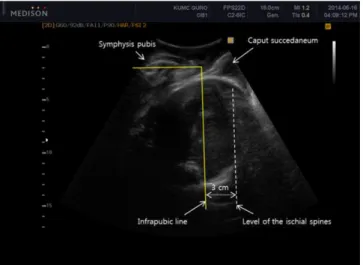
Tutschek et al. [16] suggest using intrapartum translabial ul- trasound station to measure fetal station. They measured the longest visible axis of the fetal head between the intersections with the deepest bony part of the fetal head and the infrapu- bic line; they subtracted 3 cm for the level of the ischial spines, because the infrapubic line indicates the plane 3 cm cranial to a parallel plane crossing through the ischial spines (Fig. 4).
The intrapartum translabial ultrasound station is strongly and linearly correlated with the angle of progression according to the following function: head station=angle of progres- sion×0.0937−10.911 [17].
Fig. 1. Head-perineum distance on transperineal ultrasound. Transverse perineal ultrasound image showing measurement of the shortest fetal head-perineal distance of 4.77 cm.
Fig. 2. Angle of progression on transperineal ultrasound. Transperineal ultrasound image (sagittal view) depicting the long axis of the pubic symphysis (a), angle of progression (b), and line extending from the low- ermost point of the symphysis tangentially to the fetal skull contour (c)
4. Head-symphysis distance
Youssef et al. [18] introduced a new parameter measured by three-dimensional ultrasound: fetal head-symphysis distance.
The head-symphysis distance is the distance between the low- est margin of the symphysis pubis and the nearest part of the fetal skull along a line crossing perpendicular to the long axis of the symphysis pubis (Fig. 5). The fetal head-symphysis dis- tance is significantly negatively correlated with both fetal head station assessed by digital examination and angle of progres- sion. Thus, fetal head-symphysis distance is another simple and reliable indicator of fetal head descent in labor. The mea- surement of head-symphysis distance has high intraobserver reliability (r=0.995, P<0.001) and interobserver reliability
(r=0.991, P<0.001).
In a recent study, several ultrasound parameters including intrapartum transperineal ultrasound head station, angle of progression, head-symphysis distance, and head-perineum distance showed good correlations with each other as well as moderate correlations with digital examination for assessing fetal head station [17].
In summary, updated data on intrapartum ultrasound for fe- tal head engagement and station support its use as a supple- mentary or alternative tool to digital examination in labor.
Fig. 4. Correlation between the infrapubic line and ischial spine: the par- allel line running through the projected level of the ischial spines (dotted line) lies 3 cm caudal to the infrapubic line.
Fig. 5. Head-symphysis distance on transperineal ultrasound. Ultrasound images demonstrating fetal head-symphysis distance measurements (head-symphysis distance, 32 mm). The head-symphysis distance is the distance between the inferior edge of the symphysis pubis to the nearest point of the fetal skull along a line passing perpendicular to the long axis of the symphysis pubis.
Fig. 3. Relationships among the angle of progression, cervical length, and gestational age. (A) Angle of progression and gestational age. (B) Cervical length and gestational age. (C) Angle of progression and cervical length (From Cho GJ, et al. J Perinat Med 2014 Jun 17 [Epub], with permission from Professor Oh MJ) [15].
A B C
5. Head direction
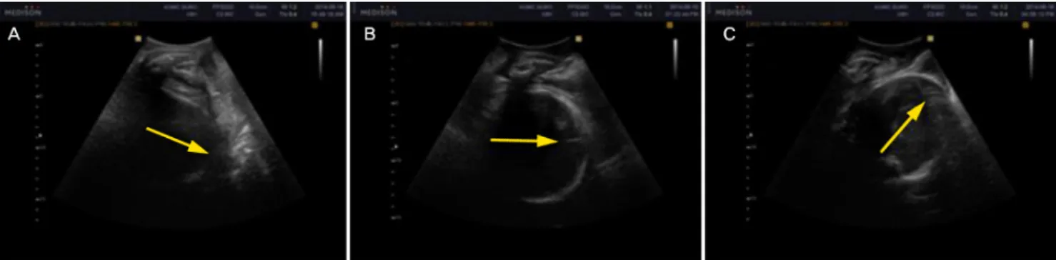
Many authors have aimed to describe fetal head station by using ultrasound landmarks including the head direction with respect to the long axis of the symphysis. Considering the cardinal movement of the fetal head and curved pathway of the pelvis, the concept of head direction is reasonable for assessing station (Fig. 6). As mentioned above, horizontal or downward head direction is associated with poor success for operative vaginal delivery [19]. Ghi et al. [20] report results consistent with previous the previous data. In their study of 60 patients at term gestation with vertex presentation fetuses in the second stage of labor, serial transperineal ultrasound in a sagittal section was performed using digital examination for assessing station. The downward, horizontal, and upward directions of the fetal head were ≤+1, ≤+2, and ≥+3 cm from the ischial spine, respectively. The probability of a station ≥+3 cm was especially high with an upward direction of the head, combined with a rotation <45°.
6. Prediction of normal vaginal delivery vs. operative delivery
One major question in obstetric history is who should un- dergo unplanned operative interventions. Many researchers have tried to elucidate the predictors of operative delivery.
Intrapartum ultrasound has recently been the focus of many investigations. Earlier, we mentioned the angle of progression is a useful tool for assessing fetal station. An interesting study determined if the narrow angle of progression in nulliparous non-laboring women is associated with a higher rate of cesar- ean delivery at term. Levy et al. [21] performed transperineal ultrasound in pregnant women with no labor at 39 or more weeks of gestation. They compared the angle of progression between women who underwent cesarean section and vaginal delivery as well as between nulliparous and parous women. A
narrow angle of progression <95° in nulliparous non-laboring women at term was associated with an increased rate of ce- sarean delivery. Parous women had a narrower angle of pro- gression than nulliparous women; however, in parous women, this was not associated with cesarean delivery. Our prospective observational study also produced similar results. We measured the angle of progression in nulliparous pregnant women with no labor at ≥37 weeks of gestation who delivered within 1 week of sonography. The angle of progression was compared between women who underwent cesarean and vaginal deliv- ery. The median angle of progression before labor onset was narrower in women who underwent cesarean section than those who delivered vaginally (86.81±5.49° vs. 95.21±10.86°,
P<0.001). An angle of progression ≥99° (derived from clinicalstation “0 or more”) was associated with vaginal delivery in 100% of women. This result suggests the angle of progression is an objective and noninvasive method for predicting the de- livery mode before labor [22]. Kalache et al. [23] prospectively studied 41 women at term ≥37 weeks with progress failure to the second stage of labor in comparison to the angle of progression on transperineal ultrasound imaging. An angle of progression of 120° lead to the probability of an easy and suc- cessful vacuum or spontaneous vaginal delivery in 90%.
Some studies have evaluated the predictors for successful operative vaginal delivery. Henrich et al. [19] used head station and head direction assessed by transperineal sonography dur- ing maternal pushing in the second stage of labor as predic- tors of successful operative vaginal delivery. They propose 3 landmarks with the transducer placed infrapubically: 1) the in- frapubic line which extends dorsally from its inferior margin in a mid-sagittal plane, 2) the widest fetal head diameter and its movement regarding the infrapubic line during pushing, and 3) the head direction in relation to the long axis of the symphy-
Fig. 6. Head direction on intrapartum translabial ultrasound. Categorization of fetal head direction (indicated by arrows) in longitudinal translabial so- nograms: (A) downward, (B) horizontal, and (C) upward direction.
sis. They show that head-up sign and objective descent of the fetal head under the infrapubic line during maternal pushing result in successful operative delivery. Ghi et al. [20] used the direction and rotation of the fetal head to define fetal station in the second stage of labor in uncomplicated singleton preg- nancies at term gestation. Head rotation was categorized as
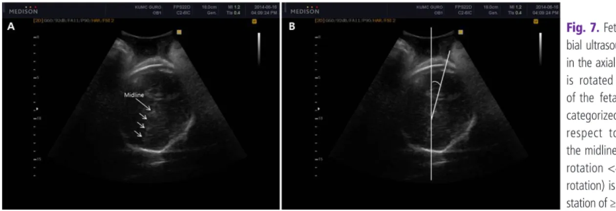
≥45° or <45° with respect to the angle formed by the echo- genic midline of the fetal head and anteroposterior diameter of the pelvis (Fig. 7) [20]. On the sonogram, when the fetal head direction was downward, horizontally, and upward, the most frequent stations were ≤+1, ≤+2, and ≥+3 cm from the ischial spines, respectively (44/57, 77.2%; 53/59, 89.8%; and 46/52, 88.5% of cases; respectively). Failure to detect the ce- rebral midline or a rotation ≥45° was associated with a station of ≤+2 cm in 98/103 (95.1%) examinations. In comparison, a rotation <45° was associated with a station of ≥+3 cm in 45/65 (69.2%) examinations. Most vacuum extractions were only performed when the clinical station was ≥+2 cm; mostly,
the fetal head was directed upward (9/11) in these cases. In the 6 cases with cesarean section, the head direction was horizontal and downward in 4 and 2 cases, respectively; rota- tion was undetectable or ≥45° in all 6 cesarean cases. Given the simplicity and good predictability for successful vacuum delivery, the head direction and rotation can be easily applied to diagnose obstructed labor.
It was recently revealed that pubic arch angle in prolonged second stage of labor is another significant predictor of delivery mode. For the measurement of pubic arch angle, the transducer is tilted 45° on the perineum to obtain an image showing the pubic symphysis and the 2 symmetrical inferior pubic rami. The angle between the lowermost borders of the pubic rami that converge at the middle of the pubic symphysis is measured. A study of 62 women at ≥37 weeks of gestation who fail to prog- ress in the second stage of labor showed that the probability for operative delivery increased with decreasing pubic arch angle [24].
Obstetricians can choose various ultrasonographic tech-
Table 1. Intrapartum ultrasonographic techniques according to various situations of women in labor
Clinical situations Ultrasonographic methods
1st stage of labor
To detect occiput posterior position Check position of fetal head and spine 2nd stage of labor
When an occiput posterior position is suspected To detect asynclitism in head transverse position To measure fetal station or in case of abnormal labor
(prolongation disorders, protraction disorders, and arrest disorders) For a successful vacuum delivery
Check position of fetal head and spine
Check “squint sign” and “sunset of thalamus and cerebellum signs”
Check angle of progression, head-perineum distance, fetal head- symphysis distance, intrapartum translabial ultrasound station, and fetal direction and rotation
Check the followings: an angle of progression >120°; upward fetal head direction; and a rotation <45°
3rd stage of labor
In case of abnormal placental separation Check placental remnants and continuous flow between myometrium and placenta using color Doppler
Fig. 7. Fetal head rotation. Transla- bial ultrasound of the maternal pelvis in the axial plane. (A) The transducer is rotated to visualize the midline of the fetal head. Head rotation is categorized as ≥45° or <45° with respect to the angle formed by the midline of the fetal head. (B) A rotation <45° (successful internal rotation) is associated with a lower station of ≥+3 cm [20].