접 수 일:2010. 5. 5.
수 정 일:2010. 7. 8.
채 택 일:2010. 7. 14.
교신저자:장병우
E-mail:[email protected]
*이 논문은 2007년도 건양대학교 명곡학술연구비의 (부분적인) 지원에 의해 이루어진 것임.
황체화 과립막세포에서 연령에 따른 mitochondrial DNA deletion에 관한 연구
건양대학교병원 산부인과1, 건양대학교 명곡의과학 연구소2, 대전마리아의원3 김남희1․ 이성기1,2․ 장병우1,2․ 이동훈3
Interrelationship of aging and mitochondrial DNA deletion in luteinized granulosa cells
Nam Hee Kim, M.D.1, Sung Ki Lee, M.D., Ph.D.1,2, Byung Woo Jang, M.D., Ph.D.1,2, Dong Hun Lee, M.D.3
1Department of Obstetrics and Gynecology, Konyang University Hospital;
2Konyang University Myunggok Medical Research Institute; 3Daejeon Maria Infertility Clinic, Daejeon, Korea
Objective: Many clinical trials have proven the close relationship between the loss of human mitochondrial DNA and aging process. The purpose of this study was to evaluate the different types of mitochondrial DNA deletion and its frequency in luteinized granulosa cells in different aged groups of women undergoing in vitro fertilization (IVF).
Methods: The ovum pick up was done in 51 women who participated in Konyang University IVF program, and mitochondrial DNAs extracted from luteinized granulosa cells, were screened to search for different types of deletion and its frequency. The deleted mitochondrial DNA were analyzed by polymerase chain reaction method. DNA sequencing was performed to reveal exact deletion point.
Results: Three different types of deletions (4,977 bp, 7,150 bp, and 5,777 bp) were confirmed. To find the difference between the aged groups, we have divided women into groups younger than 32 years, between 32 to 37 years, and older than 37 years. The deletion of 4,977 bp was 60.9% (14/23) in younger than 32 years, 46.2% (6/13) in 32 to 37 years, 46.7% (7/15) in older than 37 years. There was no statistical significance between aged groups and the incidence of the deletion. The deletion of 7150 bp was 34.8% (8/23), in younger than 32 years, 30.8% (4/13) in 32 to 37 years, 40% (6/15) in older than 37 years. We investigated relationship between the frequency of deletion and the aging, but there was no statistical significance. In case of 5,777 bp, the deletion was 43.5% (10/23) in younger than 32 years, 30.8% (4/13) in 32 to 37 years, 53.3% (8/15) in older than 37 years. It showed no statistical significance as well as other types.
Conclusion: In this study we have found three different types of deletion of mitochondrial DNA obtained from luteinized granulosa cells in women with infertility. There were no significant differnces of each type of deletion in 3 different aged groups of infantile women. The limitation of this study is that the analyze were done qualitatively. If we could provide the quantitative analyze it could be applied clinically.
Key Words: Ovary, Aging, Luteinized granulosa cells, Mitochondrial DNA deletion
인간 미토콘드리아의 인산화 능력 감소와 다양한 조직에 서의 노화 과정이 밀접한 관련이 있는 것은 이미 알려져 있 으나 기전에 대하여는 명확하지 않다. 지난 수세기 동안 연 령이 증가하는 것에 비례하여 뇌조직, 근육, 심장근 등에서 미토콘드리아 DNA 결손이 축적, 증가한다는 연구결과가 있는 반면 다른 연구에서는 그렇지 않았다는 상반된 결과를 보고하기도 하였다. 최근에는 미토콘드리아 DNA 결손이 노화뿐 아니라 세포자멸사 (apoptosis)의 주요한 기전으로
주목받고 있다.
중합효소반응을 이용하여 다양한 조직에서 5 kb,1,2 6 kb,3 7.4 kb4 크기의 미토콘드리아 DNA의 결손이 확인되었 고 이의 축적으로 염기의 부적절한 조합에 기인하는 임상적 병변이 보고되고 있다.4
미토콘드리아는 인산화 과정을 통해 ATP 생산이 이루어 지는 세포 내 소기관이며 일반적으로 90% 이상의 산소가 대 사되고 1~4%는 산소 과산화물로 전환된다.5,6
각각의 체세포는 700~1,000여 개의 미토콘드리아를 가 지고 있으며 각각의 미토콘드리아는 자체의 핵을 가지고 있 다. 뿐만 아니라 유전자의 multiple copy를 가지고 있다. 그 러므로 미토콘드리아 DNA 결손이 있는 경우라도 모두 임상 적 표현이 있는 것이 아니라 결손의 정도와 수준이 어떤 역 치에 있는가에 따라 표현이 다양하다.
전자현미경 구조를 보면 두 겹의 인지질 막과 단백질로 구성된 내 외막상 구조물을 사이에 두고 기능적으로 5개의 분획으로 나누어져 있다. 가장 바깥쪽의 외막, 외막과 내막 사이의 공간 (intermembrane space), 내막, 내막이 안쪽으 로 주름 잡힌 crista 공간, 내막 안쪽의 기질부분이 그것이 다. 외막 (outer membrane)은 전체 미토콘드리아를 싸고 있는 구조물로 porins라는 단백질과 인지질이 1:1의 비율로 분포하며 5,000 dalton 이하의 물질들을 통과시키는 기능 이 있다.
내막 (inner membrane)에 존재하는 단백질은 5가지 기 능을 담당하는데 인산화 기능, adenosine triphosphate (ATP) 생산, 기질 안과 밖으로의 대사산물 이동, 단백질 이 동수단, 미토콘드리아 자체의 융합과 분열 과정에 필요한 단백질로서의 역할을 한다. 기질은 미토콘드리아 전체 2/3 을 차지하는 내막 안쪽의 부분으로 미토콘드리아 ribo- some, tRNA, 2~20 copies의 미토콘드리아, 유전정보와 ATP 생산에 중요한 효소를 다량 포함하는 부위이다.
미토콘드리아가 세포 내 산소대사 산물을 생성하는 주요 기관이지만 산화 대사물에 의해 손상받는 1차 목표 세포기 관이 되기도 한다. 따라서 미토콘드리아 손상의 축적을 노 화에 따른 기능 저하의 주요 원인으로 보고 있는 이유이다.7 난소 기질 세포의 기능 저하와 스테로이드 생산의 감소 등 일련의 변화가 노화 과정의 기전으로 생각되는 세포의 apoptosis와 연관성은 아직 확실하지 않다. 특히 난소 과립 막 세포에서 진행되는 apoptosis는 세포내 미토콘드리아
유전자 결손의 빈도가 증가하고 특히 나이가 증가할수록 그 빈도가 높은 것으로 추정하고 있다.1
본 연구는 황체화 과립막 세포에서 미토콘드리아 유전자 결손의 종류와 빈도가 연령의 증가에 따라 영향을 받는지 알아보고자 하였다.
연구 대상 및 방법
1. 연구 대상
2000년 3월부터 2007년 8월까지 건양대병원 시험관아 기 프로그램에 참여하였던 여성 51명을 대상으로 난자 채취 후 황체화 과립막 세포를 얻었다. 연구 참여자 나이에 따라 3군 (<32세, 32~37세, ≥37세)으로 나누었다. 연구에 사용 된 세포는 난소 과자극 프로토콜이나 불임의 원인에 무관하 게 난자 채취 후 얻어진 배양액을 원심 분리하여 세포군을 얻었으며 혈액 세포의 오염을 배제하기 위하여 멸균된 PBS 배양액과 혼합하여 ‐70℃ 냉동 보관하였다. 이 연구는 건양 대학교 병원 IRB로부터 승인을 받았다.
2. 방법
1) 유전자분리
세포의 DNA 분리에 QIAamp DNA Mini Kit (QIAGEN, Venlo, Netherland)를 사용하였다. 보관된 과립막 세포 덩 어리를 해동시켜 세포 희석액 200 μL와 20 μL protease (QIAGEN)를 섞고 RNase A (QIAGEN)가 포함된 AL buf- fer (QIAGEN) 400 μL를 넣었다. 56℃에서 10분간 반응 후 상층액을 제거하고 100% ethanol 200 μL를 첨가하여 QIAamp spin culumn에 넣어 원심 분리하였다. AW buffer (QIAGEN)로 세척 후 50 μL의 elution buffer (QIAGEN)를 넣어 column에서 DNA를 분리하였다. 260 nm에서 흡광도 를 측정하여 DNA농도를 계산하였다.
2) 중합효소연쇄반응 (Polymerase chain reaction, PCR)
DNA 100 ng에 HotStart DNA polymerase (Bioneer, Deajeon, Korea) 1 unit, PCR buffer (Bioneer) (contains 1.5 mM MgCl2), dNip mixture (Bioneer) (250 μM each),
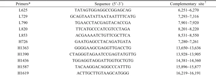
Primers* Sequence (5’-3’) Complementary site†
L625 TATAGTGGAGGCCGGAGCAG 6,251~6,270
L729 GCAGTAATATTAATAATTTTCATG 7,293~7,316
L790 TGAACCTACGAGTACACCGA 7,901~7,920
L820 TTCATGCCCATCGTCCTAGA 8,201~8,220
L853 ACGAAAATCTGTTCGCTTCA 8,531~8,550
H726 GAATGAGCCTACAGATGATA 7,280~7,261
H1363 GGGGAAGCGAGGTTGACCTG 13,650~13,636
H1390 CTAGGGTAGAATCCGAGTATGTTG 13,928~13,905
H1436 TGGAGGTAGGATTGGTGCTGTG 14,381~14,360
H1587 TACAAGGACAGGCCCATTTG 15,896~15,877
H1619 ACTTGCTTGTAAGCATGGG 16,219~16,191
*Primers L625, L729, L790, L820 and L853 were used for amplification of the light strand of mitochondrial DNA. And primers H726, H1363, H1390, H1436, H1587, and H1619 were used for amplification of the heavy strand of mitochondrial
†DNA.Numbering of mitochondrial DNA is according to that of Anderson et al27.
Table 1. Synthesized primers used for polymerase chain reaction, cloning and sequencing
Primer (Bioneer) (10 pmole/μL each) DW들을 각각 넣어 반응시켜 mitochondrial DNA fragment를 증폭하였다.
PCR primer는 합성주문 (Bioneer)하여 사용하였다. 염기 서열은 Table 1과 같다.
PCR은 94℃에서 5분간 전처리 후 94oC에서 5분간 DNA 를 변성 (denaturation)한 후, 58oC에서 1분간 primer를 붙 이고 (annealing), 72oC에서 1분간 합성 신장 (extension) 하는 과정을 35회 반복한 후 72oC에서 7분간 신장 (extension)하고 4oC에서 반응을 끝냈다.
PCR의 산물은 ethidium bromide (Sigma‐Aldrich, St.
Louis, MO, USA)가 포함된 1% agarose gel (Sigma‐Ald- rich)에 DNA size marker (Bioneer)와 함께 전기영동하여 확인하였고 해당 band를 잘라 Gel Purification Kit (Bioneer)로 정제하였다. 정확한 결손위치를 규명하기 위하 여 결손이 확인된 PCR 산물 중 크기가 800 bp 이상인 경우 는 원래의 PCR 산물 내에 존재하는 염기서열을 가진 PCR primer쌍을 이용하여 nested PCR 을 시행하여 800 bp 이 하 크기의 PCR 산물을 얻었고, 정제된 PCR 산물의 염기서 열분석을 위하여 TA cloning에 insert로 사용하였다.
3) TA cloning과 유전자 염기서열 확인
정제된 DNA 50 ng에 1 μL salt solution, 1 μL Topo vector (Invitrogen, Carlsbad, CA,, USA)를 넣고, 증류수 를 첨가하여 최종부피를 6 μL로 맞춘 후 실온에서 5분 li-
gation 시킨 후 one shot
E. coli
cell (Invitrogen)에 li- gation mixture (Invitrogen) 2 μL를 넣어 잘 섞고 얼음 위 에서 30분간 유지한 후, 42oC에서 30초간 heat shock을 주 었다. 250 μL SOC 배지 (Invitrogen)를 넣고 37oC에서 1시 간 동안 배양한 세포 50 μL를 X‐gal (Sigma‐Aldrich), IPTG (Sigma‐Aldrich), kanamycin (Sigma‐Aldrich) 50 μg/mL 이 포함된 LB plate (Sigma‐Aldrich)에 도포하여 37oC에서 하룻밤 배양하였다. Colony를 키워 10개 가량의 white colony와 blue colony를 따서 kanamycin (Sigma‐Ald- rich, 50 μg/mL)이 포함된 LB 배지 (Sigma‐Aldrich)에서 각각 12시간 배양하였다. AccuPrep Nano‐Plus Plasmid mini Extraction kit (Bioneer)를 사용하여 DNA를 추출하 였으며 EcoRI으로 잘라 insert가 들어간 것을 확인하였다.유전자 염기서열 분석은 Bioneer에 의뢰하여 M13 For- ward primer를 사용하여 single extension으로 분석하였다.
4) 통계분석
통계적 유의성 검증을 위하여 SPSS ver. 13.0 (SPSS Inc., Chicago, IL, USA) 통계프로그램을 이용하여 피셔의 정확검정을 시행하였고, 유의성은
P
<0.05 미만인 경우를 통계적 유의성 있음으로 판정하였다. 1030 bp 4,000-
2,000- 1,000- 500-
Fig. 1. The existence of a sufficient amount of mitochondrial DNA for the PCR reaction. Mitochondrial DNA of cells in follicular fluid was subjected to PCR using primer L625 and H726. Amplified 1,030 bp bands that indicated similar density were detected in all samples. Marker, DNA molecular weight marker.
*Group A: women under 32 years of age.
†Group B: women between 32 and 37 years of age.
‡Group C: women over 37 years of age.
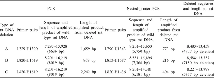
PCR Nested-primer PCR
Deleted sequence and length of mt
DNA
Type of mt DNA deletion
Primer pairs
Sequence and length of amplified
product of wild type mt DNA
Length of amplified product
from deleted mt DNA
Primer pairs
Sequence and length of amplified product of wild
type mt DNA
Length of amplified product from
deleted mt DNA A L729-H1390 7,293~13,928
(6636 bp) 1,659 bp L790-H1363 8,201~13,650
(5,750 bp) 773 bp 8,483~13,459 (4977 bp deletion) B L820-H1619 8,201~16,219
(8019 bp) 869 bp L853-H1587 8,531~15,896
(7,366 bp) 216 bp 8,588~15,737 (7150 bp deletion) C L820-H1619 8,201~16,219
(8019 bp) 2,242 bp L820-H1436 8,201~14,381
(6,181 bp) 404 bp 8,221~13,997 (5777 bp deletion) PCR: polymerase chain reaction.
Table 2. Primers, sequences and length of amplified product of wild type and deleted mitochondrial DNA
A770 bp 2,000-
1,000- 500-
Fig. 2. Detection of deleted mitochondrial DNA in human follicular fluid cells by PCR using primer L729 and H1390.
Mitochondrial DNA of three groups was sujected to PCR, separated on a 1% agarose gel, and stained with ethidium bromide. Amplification of the 6.6-kb region resulted in a 0.77- kb band (arrow A) with 5-kb deletion of mitochondrial DNA.
Marker, DNA molecular weight marker.
*Group A: women under 32 years of age.
†Group B: women between 32 and 37 years of age.
‡Group C: women over 37 years of age.
결 과
1. 미토콘드리아 유전자의 확인
과립막 세포에서 추출한 총 DNA 중에서 미토콘드리아 DNA에 국한된 증폭이 이루어졌는지 알아보기 위하여 유전 자 결손에 대한 보고가 없었던 부위에 중합효소 연쇄반응을 실시하였다.
PCR primer L625와 H726으로 얻어진 과립막 세포에서 PCR을 시행한 결과 1,030 bp의 PCR product가 결손이 없 는 것이 확인되어 미토콘드리아 DNA가 특이적으로 증폭된
조건임을 알 수 있었다 (Fig. 1).
2. 미토콘드리아 유전자 결손의 종류
연령별로 분류한 세 군의 과립막 세포에서 미토콘드리아 DNA 결손을 알아보기 위하여 세 가지 서로 다른 PCR pri- mer쌍 (Table 1)을 이용하여 중합효소 연쇄반응을 시행하 였다. L729와 H1390 primer쌍을 이용한 경우 5.0 kb 정도의 결손 (Fig. 2)을 확인하였으며, 이를 다시 L7290과 H1363 primer쌍을 이용하여 nested PCR을 시행한 후 TA cloning 하여 염기서열을 분석한 결과 ATPase 8유전자와 ND5유전
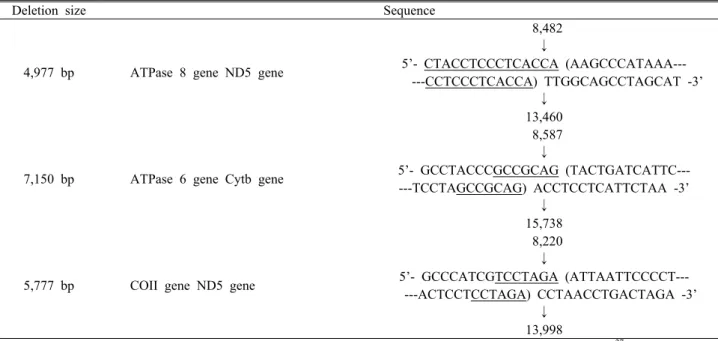
Deletion size Sequence
4,977 bp ATPase 8 gene ND5 gene
8,482
↓
5’- CTACCTCCCTCACCA (AAGCCCATAAA--- ---CCTCCCTCACCA) TTGGCAGCCTAGCAT -3’
↓ 13,460
7,150 bp ATPase 6 gene Cytb gene
8,587
↓
5’- GCCTACCCGCCGCAG (TACTGATCATTC--- ---TCCTAGCCGCAG) ACCTCCTCATTCTAA -3’
↓ 15,738
5,777 bp COII gene ND5 gene
8,220
↓
5’- GCCCATCGTCCTAGA (ATTAATTCCCCT--- ---ACTCCTCCTAGA) CCTAACCTGACTAGA -3’
↓ 13,998
Direct repeat sequences are underlined. The numbering of nucleotides is according to that of Anderson et al.27 The breakpoints are indicated by parentheses and the nucleotides shown inside the parentheses (indicated by italics) were deleted.
Table 3. Nucleotide sequences of the deletion breakpoint regions of mitochondrial DNA
B440 bp
C220 bp 2,000-
1,000- 500- 200-
Fig. 3. Detection of deleted mitochondrial DNA in human follicular fluid cells by polymerase chain reaction (PCR) using primer L820, H1619 and nested-PCR using primer L853, H1587, L820 and H1436. mitochondrial DNA of three groups was subjected to PCR, separated on a 1% agarose gel, and stained with ethidium bromide. Amplification of the 6.2 kb region resulted in a 0.4 kb band (arrow B) with 5.8 kb dele- tion of mitochondrial DNA. And amplification of the 7.4 kb reg- ion resulted in a 0.22 kb band (arrow C) with 7.2 kb deletion of mitochondrial DNA. Marker, DNA molecular weight marker.
*Group A: women under 32 years of age.
†Group B: women between 32 and 37 years of age.
‡Group C: women over 37 years of age.
자 사이인 nucleotide 8483~13459 위치의 4,977 bp결손을 확인하였다 (Table 2).
L820과 H1619 primer쌍으로 7.1 kb 정도의 결손과 5.8 kb 정도의 두 가지 결손을 확인하였고 (Fig. 3) 각각에 대하
여 L853과 H1587과, L820과 H1436 primer쌍으로 염기서 열을 분석한 결과 7,150 bp결손은 APTase 6과 cytochrome 유전자 사이의 nucleotide 8,588~15,737 위치였고, 5,677 bp 결손은 cytochrome C oxidase II 유전자와 ND5유전자 사이의 nucleotide 8,221~13,997이다 (Table 2).
4,977 bp 결손, 7,150 bp 결손 및 5,677 bp 결손 위치의 break point에서 direct repeat 염기서열이 있음을 확인하 였다 (Table 3).
3. 과립막세포에서 연령에 따른 유전자 결손의 발현빈도
시험관아기클리닉 프로그램에 참여한 여성으로부터 얻 어진 황체화 과립막세포 총 51예에 대하여 32세 미만 23예, 32세에서 37세 미만인 군 13예, 37세 이상 15예에서 추출한 미토콘드리아 유전자에서 4,777 bp 결손, 7150 bp 결손, 5,777 bp 결손을 확인하였고 빈도를 비교한 결과 4,977 bp 결손의 경우 32세 미만의 연령군에서 14예 (60.9%), 32세부 터 37세 미만 연령군에서 6예 (46.2%), 37세 이상 연령군에 서 7예 (46.7%)로 연령에 따른 통계적 유의성은 없었다.
7,150 bp결손의 경우 32세 미만의 연령군에서 8예 (34.8%), 32세부터 37세 미만 연령군에서 4예 (30.8%), 37
Age (yr)
Deletion
4,977 bp 7,150 bp 5,777 bp
<32 (23)
14 (60.9%)
8 (34.8%)
10 (43.5%) 32~37
(13)
6 (46.2%)
4 (30.8%)
4 (30.8%)
≥37 (15)
7 (46.7%)
6 (40%)
8 (53.3%) Table 4. The incidence of three types of mitochondrial DNA deletion in cells from follicular fluid of three groups of women
세 이상 연령군에서 6예 (40%)로 연령에 따른 통계적 유의 성은 없었다.
5,777 bp 결손은 32세 미만의 연령군에서 10예 (43.5%), 32세부터 37세 미만 연령군에서 4예 (30.8%), 37세 이상 연 령군에서 8예 (53.5%)로 37세 이상의 연령군에서 37세 미 만 여성군에 비하여 통계적 유의성은 없었다 (Table 4).
고 찰
미토콘드리아 유전자는 16.5 kb 크기이며 2개의 rRNA와 22개의 tRNA, 그리고 인산화 과정에 필요한 13개 소단위의 효소관련 유전정보를 포함하고 있다.8
미토콘드리아의 유전정보는 모성유전을 하며9 유전자의 구조는 매우 단순하고 histone과 repairing system을 갖추 지 않았기 때문에 유리기 산소에 대한 손상에 민감하며 세 포막 손상이나 돌연변이의 빈도가 매우 높다.10 여러 형태의 미토콘드리아 유전자 손상 중에서 특정 부위의 결손은 인체 의 질환이나 노화의 진행과 깊은 연관이 있다.11
미토콘드리아 유전자가 에너지 생산과 관련한 중요효소 의 유전정보를 공유하고 있기 때문에 세포의 죽음이나 조직 의 변성 및 유축 등을 초래한다.12,13
세포 내 대사 산물인 유리기 산소는 세포구성성분에 손상 을 끼치는 물질로서 세포 내 소기관인 미토콘드리아에서 생 산되며 미토콘드리아 자체의 손상을 유발시킬 수 있다. 특 히 에너지 생산의 중심축을 이루는 미토콘드리아의 유전자 손상이나 돌연변이 등은 정상적 노화과정에서 인산화 작용능 력의 감소를 초래하여 미토콘드리아 유전자 손상을 가속화하 며14 이로 인하여 각 조직 및 기관의 기능 상실로 이어진다.
미토콘드리아 유전자 결손이 노화된 심장근, 심장근세포,1 근육세포,15 뇌,16 간조직3 등에서 높은 빈도로 발견되었고, 노화된 난소조직에서 그 상관관계가 보고되었다.17 노화와
관련된 미토콘드리아 유전자 결손은 common deletion으로 알려진 4,977 bp 결손의 빈도가 흔하며 특히 이 부위의 결 손은 미토콘드리아 전자전달체계 complex I, IV, 그리고 V 의 기능을 담당하는 부위이다.1
미토콘드리아 유전자 4,977 bp 결손의 정도는 같은 조직 에서도 부위에 따라 1,000배 이상의 다양한 분포를 보이는 데 고령의 소뇌 조직에서는 결손이 나이와 무관한 반면, Nigra, Cauda, Putamen 등의 부위에서는 나이의 증가와 함께 의미 있는 결손의 빈도를 보인다.16
최근 연구에서 263개의 다양한 미토콘드리아 DNA 결손 이 확인되었다.18 미토콘드리아 DNA 결손은 항상 hetero- plamsic form이며 생화학적 결함과 임상적 표현은 유전자 결손의 분포가 특정 임계수준까지 도달할 때까지 나타나지 않는다.19
단일세포에서 mt DNA 결손에 대한 연구가 진행되었는 데,20 100세가 넘는 4명의 사람으로부터 얻은 유전자 결손 은 각각 사람에 대하여 한 가지의 결손이 관찰되었고 그 분 포는 11~64%까지 다양하였다고 하였다. 이는 처음 시작된 단일 유전자변이가 clonal expansion된 것을 시사한다고 할 수 있다.
난소에서 진행되는 노화는 일차적으로 난포의 질적, 수 적 변화가 있다. 난포의 수가 감소하고 난포 내의 과립막 세 포수가 감소함으로써 스테로이드와 glycoprotein 성장인자 가 떨어지고 과립막 세포의 증식 또한 감소할 뿐 아니라 apoptosis의 빈도가 증가한다.21,22
난자와 배아에서 가장 흔한 미토콘드리아 유전자 결손은 4,977 bp common deletion이며 수정되지 않는 난자에서의 빈도가 비정상 수정된 수정란에서보다 높았다.23 4,977 bp 결손이 있는 미토콘드리아는 ATPase6‐8, cytochrome ox- idase III 그리고 NADH‐CO oxidoreductase (ND3, ND4, ND4L, ND5)의 인산화 효소관련 유전자의 손상이 있게 되 며 ATPase 8의 5`portion과 ND5의 3`portion의 비정상 연 결에 의한 chimeric gene을 형성한다. 그러나 난자의 능력 과 수정란 발생을 예측하는 인자로서 과립막 세포의 미토콘 드리아 유전자 결손 빈도를 확인한 연구에서 나이에 따른 차이가 없음을 보고하였다.24
한편, 한 연구에서는 황체화 과립막 세포에서 연령증가 에 따른 미토콘드리아 유전자 4,977 bp 결손의 빈도를 알아 본 다른 연구는 38세 미만의 여성에서 얻어진 과립막 세포
에서는 12예 중에서 7예에서 정상 미토콘드리아 유전자를 확인하였고, 38세 이상의 여성에서 얻어진 과립막 세포에서 는 12예 중에서 단 한 예도 정상유전자를 보여주지 못함으 로 연령이 증가할수록 의미 있는 유전자 결손 빈도를 보고 하였다.25
본 연구에서는 황체화 과립막 세포에서 미토콘드리아 유 전자 결손의 종류와 그 빈도를 알아보고자 하였는데 이미 보고된 4,977 bp common deletion뿐 아니라 7,150 bp, 5,777 bp 크기의 결손이 확인되었고 후자의 경우 모두 4977 bp 결손을 포함하는 부위였다. 연령에 따른 결손의 종류 분 포와 빈도의 차이는 통계적 의미는 없었다.
4,977 bp 결손은 이전의 연구 등에서 보고한26 ATPase 8 유전자와 Nb5유전자 사이에 위치한 결손임을 확인하였고, 7,150 bp 결손은 ATPase 6유전자와 cytochrome b 유전자 사이의 결손이고, 5,777 bp 결손은 cytochrome c oxidase II유전자와 NO5유전자 사이의 결손이다.
본 연구에서 확인된 3가지 유형의 결손된 유전정보는 respiratory complex part에서 가장 중요한 energy trans- ducing function을 갖는 부위로 protein molecular as- sembly의 장애가 초래되어 효소계의 작용을 방해하여 ATP 생산이 이루어지지 않는다.12
미토콘드리아 유전자 결손의 발생과 축적이 연령이 증가 하면서 가속화되고 조직의 기능상실과 이상을 동반하며, 질 병의 병인론으로 추정되는 많은 연구에도 불구하고,17 본 연 구자들의 연구결과에서와 같이 빈도에 차이가 없는 경우는 미토콘드리아 유전자 결손 수준의 다양성에 따라 임상적 표 현 양상이 다른 것을 알 수 있다.
본 연구는 정량적 중합효소 연쇄반응이 시행되지 않았고 결손 부위를 증폭하여 확인하는 과정이었기 때문에 극히 적 은 부위의 결손이라도 증폭되었고 미토콘드리아는 세포 내 에 수백~수천 개씩 존재하며 2~20 copies를 갖는 유전자
의 특징 때문에 연령에 따른 임상적 변화를 보이는 역치를 알아 보는 데는 제한이 있었다.
이번 연구에서 확인된 4,977 bp, 5,777 bp, 7,150 bp 결 손 중에서 5,777 bp 결손의 경우 같은 조건의 중합효소 연 쇄반응을 시행하였던 다른 결손의 부산물과 비교하여 선명 하고 진한 band를 보여줌으로써 결손 빈도는 4,977 bp 결 손에 비하여 낮지만 황체화 과립막 세포에서 다량 발현되는 결손 부위로 추정된다.
여성의 난소 구성 세포인 황체화 과립막 세포에서 미토콘 드리아 유전자 결손이 난자의 질, 배아의 성장, 호르몬불균 형과 이에 따른 임상적 변화와의 관계에 대한 연구는 아직 보고된 바 없다.
4,977 bp 크기의 유전자 결손은 세포 내 에너지 생산의 가장 핵심적 부위이며 이 부위의 결손은 장기적으로 조직의 손상을 초래한다. 에너지 부족상태가 지속되는 환경에서 생 식력의 변화는 예정된 것으로 추정 할 수 있다.
본 연구는 단일 세포를 분리하여 미토콘드리아 유전자 결 손유무와 빈도를 확인하는 과정이었다. 미토콘드리아가 각 각의 세포에 많은 수로 존재하기 때문에 결손된 유전자의 특징적 표현형이나 결손의 구성비를 알기 어려운 문제를 갖 고 있으며, 세포를 획득하는 과정에 다른 세포군의 오염 등 과 같은 기술적 제한이 있었다.
향후 보조 생식술 과정 중에 얻어진 황체화 과립막 세포 를 이용하여 미토콘드리아 유전자 결손의 정성적, 정량적 측정으로 난소의 생리적 기능평가뿐 아니라 임신의 예후를 위한 예측인자로 임상에 적용할 수 있을 것을 기대한다. 또 한 난소조직을 구성하는 세포 분획을 나누어 각 세포군에서 특징적으로 보여지는 미토콘드리아 유전자 결손의 종류와 빈도, 정량적 측정 방법이 개발되면 임상적으로 의미 있는 증상의 발현과 관련된 역치 연구가 가능할 것으로 생각된다.
참고문헌
1. Cortopassi GA, Arnheim N. Detection of a specific mitochondrial DNA deletion in tissues of older humans. Nucleic Acids Res 1990; 18: 6927‐33.
2. Cortopassi GA, Shibata D, Soong NW, Arnheim N.
A pattern of accumulation of a somatic deletion of mi- tochondrial DNA in aging human tissues. Proc Natl Acad Sci USA 1992; 89: 7370‐4.
3. Yen TC, Pang CY, Hsieh RH, Su CH, King KL, Wei YH. Age‐dependent 6kb deletion in human liver mi- tochondrial DNA. Biochem Int 1992; 26: 457‐68.
4. Hayakawa M, Sugiyama S, Hattori K, Takasawa M, Ozawa T. Age‐associated damage in mitochondrial DNA in human hearts. Mol Cell Biochem 1993; 119:
95‐103.
5. Richter C. Reactive oxygen and DNA damage in mitochondria. Mutat Res 1992; 275: 249‐55.
6. Chance B, Sies H, Boveris A. Hydroperoxide metabolism in mammalian organs. Physiol Rev 1979; 59: 527‐605.
7. Harman D. The biologic clock: the mitochondria? J Am Geriatr Soc 1972; 20: 145‐7.
8. Clayton DA. Replication and transcription of verte- brate mitochondrial DNA. Annu Rev Cell Biol 1991;
7: 453‐78.
9. Hammans SR. Mitochondrial DNA and disease.
Essays Biochem 1994; 28: 99‐112.
10. Yakes FM, Van Houten B. Mitochondrial DNA dam- age is more extensive and persists longer than nuclear DNA damage in human cells following oxidative stress. Proc Natl Acad Sci USA 1997; 94: 514‐9.
11. Grossman LI, Shoubridge EA. Mitochondrial genetics and human disease. Bioessays 1996; 18: 983‐91.
12. Ozawa T. Genetic and functional changes in mi- tochondria associated with aging. Physiol Rev 1997;
77: 425‐64.
13. Meissner C, von Wurmb N, Oehmichen M.
Detection of the age‐dependent 4977 bp deletion of mitochondrial DNA. A pilot study. Int J Legal Med 1997; 110: 288‐91.
14. Larsson NG, Clayton DA. Molecular genetic aspects of human mitochondrial disorders. Annu Rev Genet 1995; 29: 151‐78.
15. Cortopassi GA, Arnheim N. Using the polymerase chain reaction to estimate mutation frequencies and rates in human cells. Mutat Res 1992; 277: 239‐49.
16. Corral‐Debrinski M, Horton T, Lott MT, Shoffner JM, Beal MF, Wallace DC. Mitochondrial DNA dele- tions in human brain: regional variability and increase with advanced age. Nat Genet 1992; 2: 324‐9.
17. Kitagawa T, Suganuma N, Nawa A, Kikkawa F, Tanaka M, Ozawa T, et al. Rapid accumulation of de- leted mitochondrial deoxyribonucleic acid in post- menopausal ovaries. Biol Reprod 1993; 49: 730‐6.
18. Samuels DC, Schon EA, Chinnery PF. Two direct re- peats cause most human mtDNA deletions. Trends Genet 2004; 20: 393‐8.
19. Chomyn A, Attardi G. MtDNA mutations in aging and apoptosis. Biochem Biophys Res Commun 2003;
304: 519‐29.
20. Khrapko K, Bodyak N, Thilly WG, van Orsouw NJ, Zhang X, Coller HA, et al. Cell‐by‐cell scanning of whole mitochondrial genomes in aged human heart reveals a significant fraction of myocytes with clonally expanded deletions. Nucleic Acids Res 1999; 27:
2434‐41.
21. Seifer DB, Gardiner AC, Ferreira KA, Peluso JJ.
Apoptosis as a function of ovarian reserve in women undergoing in vitro fertilization. Fertil Steril 1996;
66: 593‐8.
22. Klein NA, Battaglia DE, Woodruff TK, Padmana- bhan V, Giudice LC, Bremner WJ, et al. Ovarian fol- licular concentrations of activin, follistatin, inhibin, insulin‐like growth factor I (IGF‐I), IGF‐II, IGF‐bind- ing protein‐2 (IGFBP‐2), IGFBP‐3, and vascular en- dothelial growth factor in spontaneous menstrual cy- cles of normal women of advanced reproductive age. J Clin Endocrinol Metab 2000; 85: 4520‐5.
23. Hsieh RH, Tsai NM, Au HK, Chang SJ, Wei YH, Tzeng CR. Multiple rearrangements of mitochondrial DNA in unfertilized human oocytes. Fertil Steril 2002; 77: 1012‐7.
24. Au HK, Lin SH, Huang SY, Yeh TS, Tzeng CR, Hsieh RH. Deleted mitochondrial DNA in human lu- teinized granulosa cells. Ann N Y Acad Sci 2005;
1042: 136‐41.
25. Seifer DB, DeJesus V, Hubbard K. Mitochondrial de- letions in luteinized granulosa cells as a function of age in women undergoing in vitro fertilization. Fertil Steril 2002; 78: 1046‐8.
26. Lee HC, Pang CY, Hsu HS, Wei YH. Differential ac- cumulations of 4,977 bp deletion in mitochondrial DNA of various tissues in human ageing. Biochim Biophys Acta 1994; 1226: 37‐43.
27. Anderson S, Bankier AT, Barrel BG. Sequence and organization of the human mitochondrial genome.
Nature 1981;290:457‐65.
= 국문초록 =
목적: 여성의 나이가 증가함에 따라 난소 기능이 떨어지는 노화과정에 과립막 세포내 미토콘드리아 유전자 결손이 증가하고 이로 인한 adenosine triphosphate (ATP) 생산의 저하가 주요한 요인이라는 가설이 있다. 본 연구는 시험관 클리닉에 참여하였 던 여성을 대상으로 난자 채취 후 얻어진 황체화 과립막 세포를 분리하여 미토콘드리아 유전자 결손의 종류와 빈도를 알아 보고 연령에 따른 차이가 있는지 확인하고자 하였다.
연구 방법: 건양대병원 시험관아기 프로그램에 참여하였던 여성 51명을 대상으로 난자 채취 후 황체화 과립막 세포를 채취하 고 알려진 방법에 따라 미토콘드리아 유전자를 분리하였다. Polymerase chain reaction 방법에 의하여 기존 문헌에 알려진 다 양한 크기의 결손 부위에 증폭을 시도하여 확인하였고 DNA sequencing에 의하여 정확한 유전자 결손 부위를 확인하였다.
결과: 4,977 bp, 7,150 bp, 5,777 bp 크기의 세 종류의 결손이 있는 것을 확인하였다. 나이에 따른 차이가 있지 보기 위하여 연령 대를 32세 미만과 32~37세 연령군, 37세 이상으로 나누어 세 종류 결손의 빈도를 알아보았는데, common deletion이라고 알려 진 가장 흔한 4,977 bp 결손의 경우 32세 미만 연령군에서 60.9% (14/23), 32~37세 연령군에서 46.2% (6/13), 37세 이상 연령군 에서 46.7% (7/15)의 빈도를 보였고 연령 증가에 따른 통계적 유의성은 없었다. 7,150 bp 결손의 경우 32세 미만 연령군에서 34.8% (8/23), 32~37세 연령군에서 30.8% (4/13), 37세 이상 연령군에서 40% (6/15)의 빈도를 보였고 연령 증가에 따른 통계적 유의성은 없었다. 5,777 bp 결손의 경우 32세 연령군에서 43.5% (10/23), 32~37세 연령군에서 30.8% (4/13), 37세 이상의 연령 군에서 53.3% (8/15)의 빈도를 보였으며 다른 결손의 경우와 마찬가지로 연령 증가에 따른 통계적 유의성은 없었다.
결론: 연령에 따른 황체화 과립막 세포의 미토콘드리아 유전자 결손의 종류를 알아본 결과 4,977 bp, 7,150 bp, 5,777 bp 크기의 세 종류의 결손을 확인하였다. 각 결손 종류에 대하여 연령군에 따른 차이를 확인한 결과 세 종류 모두 통계적 유의성은 없었다. 이번 연구는 유전자 결손의 정성적 분석만 시행하였다는 한계를 가지고 있으며 향후 결손의 정량적 분석이 가능하 다면 임상적 증상에 대한 결손의 역치 정도를 알아보는 데 유용할 것으로 생각된다.
중심단어: 난소, 노화, 황체화 과립막 세포, 미토콘드리아 유전자 결손