세포충실성섬유상피병변(fibroepithelial lesions with cellular stroma, 이하 FELCS)이란 2006년 Jacob이 제시한 병리학적 용어로 섬유선종(fibroadenoma)에서 엽상종양 (phyllodes tumor)에 이르는 병변을 포괄하는 개념이며 (1)
“이중 섬유상피종양(biphasic fibroepithelial tumor)”혹은
“애매모호한 섬유상피종양(equivocal fibroepithelial tumor)”과 같은 용어로 혼용되기도 한다. 이러한 개념은 섬유 선종과 엽상종양이 임상, 영상, 그리고 조직으로도 감별하기가 어렵기 때문으로(2) 특히 적은 양을 얻는 생검조직(biopsy specimen)에서 간질세포(stromal cell)가 증가한 섬유선종과 엽상종양과의 구별은 매우 힘들다(3, 4). 따라서 수술 전 생검 에서 FELCS로 진단되는 경우 엽상종양의 가능성으로 인해 추가적인 절제생검(excision biopsy)이 요구되지만(5) 이러한 병변의 일괄적인 절제생검은 엽상종양에 대해서는 불충분하
게, 섬유선종에 대해서는 불필요하게 수술을 시행하게 되는 단 점을 일으킬 수 있다. 그럼에도 불구하고 FELCS의 절제생검 후의 결과에 대한 연구가 많지 않고 최근 유방병변의 비수술적 생검의 증가와 더불어 FELCS로 진단되는 증례도 늘어나고 있어 이에 대한 연구가 필요하다. 몇몇의 연구에서 절제생검 이후 약 22~44%가 엽상종양으로 진단되었으나 모두 대상의 수가 작았으며(1, 6-8) 임상 혹은 영상 소견과 연관된 연구는 거의 없었다. 따라서 이 연구에서는 유방의 중심부바늘생검 (core biopsy) 후 FELCS로 진단되었던 증례들을 대상으로 절제생검 후의 진단 결과와 비교해보고 엽상종양을 예측하는 데 도움이 되는 임상적 혹은 영상학적 인자가 있는지 알아보고 자 하였다.
대상과 방법
환자군
2007년 3월부터 2010년 5월까지 유방 중심부바늘생검에서
유방의 중심부바늘생검에서 진단된 세포충실성섬유상피병변에서 엽상종양을 예측할 수 있는가?
1정혜경∙고경희∙노지영∙문희정2∙김은경2∙김민정2∙박병우2
목적: 유방의 중심부바늘생검에서 진단된 세포충실성섬유상피병변(fibroepithelial lesion with cellular stroma)의 수술 후의 진단 결과와 엽상종양을 예측하는 데 도움이 되는 임상적 혹은 영상학적 인자가 있는지를 알아보고자 하였다.
대상과 방법: 초음파유도하 중심부바늘생검에서 세포충실성섬유상피병변으로 진단되고 수술을
시행하여 최종병리가 확인된 55명을 대상으로 하였다. 병변의 수술 결과에 따라 엽상종양과 엽 상종양이 아닌 병변의 두 부류로 나눈 후 각각의 임상과 영상소견을 후향적으로 비교, 분석하였 다. 또한, 엽상 종양을 예측하는데 유의한 변수들을 조합하여 분석하였다.
결과: 수술로 확진된 55명의 55예의 병변 중 엽상 종양은 22예(40%), 엽상종양이 아닌 예가 33예(섬유선종 30예(54.6%), 섬유낭종변화 1예(1.8%), 선증 1예(1.8%), 섬유선증증식 1예 (1.8%))였다. 엽상종양 22예의 평균 크기는 32.2 ± 14.07 mm (12~55 mm)였고 22명 환 자의 평균나이는 43.5 ± 11.60세(18~69세)였으며 엽상종양이 아닌 종양 33예의 평균 크기 는 22.4 ± 13.64 mm (12~55 mm), 33명의 평균 나이는 36.5 ± 10.25세(19~62세)로 크 기와 나이 모두 통계적으로 유의한 차이를 보였다(p=0.0078, p=0.0207). 초음파 소견 중에 서는 틈새(cleft)가 통계적으로 유의한 차이를 보여 엽상종양에서 더 흔하게 관찰되었다(n=14 (70%)/n=6 (30%), p=0.0016).
결론: 유방의 수술 전 중심부바늘생검에서 진단된 세포충실성섬유상피병변 중 병변의 크기, 환 자의 나이, 초음파 소견에서 틈새의 유무를 고려하면 엽상종양을 예측할 수 있다.
1차의과대학교 영상의학과
2연세대학교 의과대학 영상의학과
이 논문은 2010년 10월 27일 접수하여 2011년 4월 15일에 채택되었음.
FELCS로 진단된 84명의 84예을 찾았으며 그 중 추가적인 절 제생검을 통해 확진이 이루어졌으며 후향적 판독이 가능한 초 음파 영상이 있는 55명의 환자를 대상으로 하였다. 이 55명 환 자 중 17명의 환자는 유방의 중심부바늘생검에서 엽상종양과 FELCS로 진단된 병변의 총 60예를 대상으로 한 이전 논문에 포함되었으나(9) 이전 논문은 엽상종양과 FELCS로 진단된 병변을 모두 포함할 때 엽상종양을 진단할 수 있는 초음파검사 의 역할을 분석한 것이고 이 논문은 유방 중심부바늘생검에서 FELCS로 진단된 군만을 대상으로 영상소견을 분석하여 수술 후 엽상종양을 진단하는 예측모형을 만드는 것이다. 55예를 수술 결과에 따라 엽상종양과 엽상종양이 아닌 병변으로 분류 하여 각각의 임상소견과 영상소견을 후향적으로 분석하여 통 계적으로 유의한 차이를 보이는 소견이 있는지 알아보았다.
영상 검사 방법
유방초음파 검사는 7.5 ~ 12 MHz 선형탐촉자를 사용하였 고 기종은 ATL HDI 3000, 5000 (Philips-Advanced Technology Laboratories, Bothell, WA, USA), Logic 9 (GE Medical systems, Milwaukee, WI, USA)이었다. 55 명 의 환 자 모 두 에 서 초 음 파 유 도 하 중 심 부 바 늘 생 검 (ultrasound guided core biopsy)이 이루어졌으며 이는 외 래로 내원한 환자를 앙아위 상태에서 국소마취 후 자동생검총 (automated biopsy gun. Pro-Mag 2.2, Manan Medical Products, Northbrook, IL, USA)과 14게이지 생검침(Tru- Cut needle with a 22 mm throw (SACN™ Biopsy Needle, Medical Device Technologies, Gainesville, FL, USA))을 사용하여 시행되었고 각 병소 당 4개 이상의 중심부 바늘생검 조직을 얻었다. 유방병리전문의가 판독하였다.
데이터 분석 병변의 평가
55명 환자의 55개 병변에 대해 두 명의 영상의학과 의사가 PACS (Picture Archiving and Communication System) 모니터에 나타난 초음파 소견을 BIRADS - US lexicon을 이 용하여 후향적으로 분석하였으며 Stavros (10)가 제시한 틈 새(cleft)의 유무를 추가로 분석하였다. 틈새의 정의는 상하 직 경이 3 mm 이하의 납작한 낭성 공간(slit-like cystic space) 으로 엽상(leaf-like projection)으로 자라는 간질 증식에 의 해 나타나는 초음파 소견을 말하며 Stavros가 엽상종양의 특 징적인 초음파 소견으로 언급한 바 있다(10). 절제생검 후 결 과에 따라 엽상종양과 엽상종양이 아닌 종양을 분류하여 각각 의 항목을 분석하였다. 모든 영상의 후향적 분석은 병리학적 결과에 대한 정보 없이 이루어졌다. 임상소견으로는 환자의 나 이와 병변의 크기를 분석하였다.
통계적 분석
최종 병리학적 결과에 따라 엽상종양과 엽상종양 외의 종양 사이에 임상소견과 초음파 소견의 각각의 항목에 대해 유의한 차이가 있는지 알아보고자 카이 제곱 검정법(chi-square
test)와 Fisher’s exact test, Student t-test를 통해 분석하 였고p값이 0.05이하면 통계적으로 유의한 것으로 보았다. 또 한, 엽상 종양을 예측하는데 유의한 변수들을 조합하여 분석하 는데 의사결정나무(decision tree) 모형을 이용하였다. 각각 의 변수 중 나이와 크기에 대한 적절한 cut-off value는 ROC curve 분석을 이용하여 산출하였다. SPSS 11.5 for window (Statistical Package for Social Sciences, SPSS, Chicago, Ⅲ)로 통계 분석을 하였다.
결 과
수술로 확진된 55명의 55예의 병변 중 엽상 종양은 22예 (40%), 섬유선종을 포함한 엽상종양 외의 종양은 33예 (60%)였다. 엽상종양 외의 종양 33예 중 섬유선종이 30예 (54.5%), 섬유낭종변화(fibrocystic change)가 1예(1.8%), 선증(adenosis)이 1예(1.8%), 섬유선종양증식(fibro- adenoatoid hyperplasia)이 1예(1.8%)로 모두 양성병변이
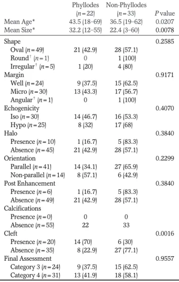
Table 1. Ultrasonographic Features of Phyllodes Tumors and Non-phyllodes Tumors
Phyllodes Non-Phyllodes (n=22) (n=33) P value Mean Age* 43.5 (18-69) 36.5 (19-62) 0.0207 Mean Size* 32.2 (12-55) 22.4 (3-60)0 0.0078 Shape 0.2585
Oval (n=49) 21 (42.9) 28 (57.1)
Round�(n=1) 0 .1 (100)
Irregular�(n=5) 1 (20) 4 (80).
Margin 0.9171
Well (n=24) 09 (37.5) 15 (62.5) Micro (n=30) 13 (43.3) 17 (56.7) Angular�(n=1) 0 .1 (100)
Echogenicity 0.4070
Iso (n=30) 14 (46.7) 16 (53.3) Hypo (n=25) 8 (32) 17 (68)0.
Halo 0.3840 Presence (n=10) 01 (16.7) 05 (83.3)
Absence (n=45) 21 (42.9) 28 (57.1)
Orientation 0.2299
Parallel (n=41) 14 (34.1) 27 (65.9) Non-parallel (n=14) 08 (57.1) 06 (42.9)
Post Enhancement 0.3840
Presence (n=6) 01 (16.7) 05 (83.3) Absence (n=49) 21 (42.9) 28 (57.1) Calcifications
Presence (n=0) 0 0
Absence (n=55) 22 33
Cleft 0.0016
Presence (n=20) 14 (70).0 6 (30).
Absence (n=35) 08 (22.9) 27 (77.1)
Final Assessment 0.9557
Category 3 (n=24) 09 (37.5) 15 (62.5) Category 4 (n=31) 13 (41.9) 18 (58.1) Note.─*; Student t test
�Fisher’s exact test
었다. 엽상종양 22명의 환자의 평균나이는 43.5 ± 11.60세 (18~69세)였으며 종양의 평균 크기는 32.2 ± 14.07 mm (12~55 mm)인데 반하여 엽상종양이 아닌 33명의 평균 나이 는 36.5 ± 10.25세(19~62세)로, 병변의 평균 크기는 22.4
± 13.64 mm (12~55 mm)로, 평균 크기와 평균 나이 모두 통계적으로 유의한 차이를 보였다(p=0.0078, p=0.0207) (Table 1).
초음파 소견은 엽상종양과 엽상종양이 아닌 종양이 서로 유 사하였으나 틈새는 엽상종양에서 통계적으로 유의하게 더 많 이 보였다 (p=0.0016) (Table 1).
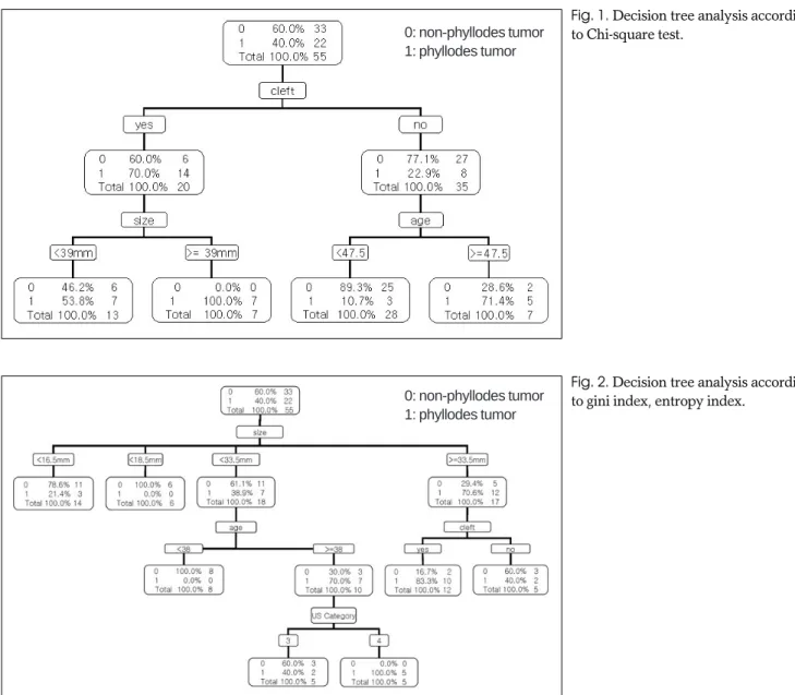
의사결정 나무모형을 이용한 분석에서 나무의 분리기준을 카이 제곱 검정법(Fig. 1)과 gini index, entropy index(Fig.
2)로 나눌 수 있으며 분리기준을 카이 제곱 검정법을 이용해 분석하였을 때 초음파 소견에서 틈새가 있으면서 크기가 39 mm이상인 모형은 7예 중 7예(100%) 모두 엽상 종양으로 분 류되었고(Figs. 3, 4) 틈새가 없으면서 나이가 47세 이하인 28예 중 25예(89.3%)가 엽상 종양이 아닌 종양으로 분류되었
다(Figs. 5, 6).
Gini index, entropy index를 기준으로 분리하였을 때 크 기가 33.5 mm 미만이지만 환자의 나이가 38세 이상이고 초음 파 검사의 최종범주가 범주 4에 해당하는 5예는 모두 엽상종 양이었으며 크기가 33.5 mm 이상이면서 틈새가 있는 12예 중 10예(83.3%)가 엽상종양이었다(Figs. 3, 4). 엽상종양이 아 닌 종양을 가장 잘 분류하는 모형은 크기가 33.5 mm 미만이 고 환자의 나이가 38세 미만인 경우(8예, 100%)였다.
고 찰
임상적으로 유방의 수술 전 조직검사에서 FELCS로 진단되 면 추가적인 수술적 절제가 권유된다(5). 이는 FELCS가 엽 상종양일 가능성이 있으며 엽상종양은 재발을 잘하는 국소침 습(locally invasive) 병변으로 음성변연(negative margin) 을 확보하기 위해 광범위절제술(wide surgical excision)로 치료되어야 하기 때문이다(2, 11, 12). 그러나 엽상종양은 단
Fig. 2. Decision tree analysis according to gini index, entropy index.
Fig. 1. Decision tree analysis according to Chi-square test.
0: non-phyllodes tumor 1: phyllodes tumor
0: non-phyllodes tumor 1: phyllodes tumor
순절제(enucleation) 혹은 추적관찰(observation)이 가능한 섬유선종과 임상, 영상, 조직 소견이 유사해 감별진단이 어려 운 것으로 잘 알려져 있다(2).
엽상종양의 수술 전 진단에는 중심부바늘생검에 의한 조직 검사가 가장 정확한 것으로 알려져 있다(13). 엽상종양에 대한 중심부바늘생검의 진단율은 64.7~83.3% 정도였으며(4, 5, 8), 유방의 수술 전 중심부바늘생검에서 진단된 FELCS를 수 술하였을 때 22~44% 정도가 엽상종양인 것으로 확진되었다 (1, 6-8). 본 연구에서는 55예의 FELCS 중 22예(40%)가 엽 상종양이었으며 이는 이전 보고들과 비슷한 수치이다. 현재 수 술 전 진단된 FELCS에 대해서 일괄적인 수술절제가 이루어 지고 있어 엽상종양을 불충분하게 제거하여 재발의 원인이 되 기도 하고 섬유선종을 불필요하게 수술하기도 한다. 외과의가 수술 전 엽상종양이나 혹은 섬유선종의 가능성을 염두에 둔다 면 좀더 적합한 수술적 방법을 고려할 수 있으며, 또한 젊은 여 성에서 보이는 섬유선종의 가능성이 많은 FELCS에 대해서는 수술적 절제보다는 추적관찰과 같은 방법을 적용할 수 있으리 라 여겨진다. Jacobs 등(1)은 중심부바늘생검에서 진단된
Fig. 4. 32-year-old woman with a palpable mass diagnosed as fibroepithelial lesion with cellular stroma at ultrasonography guided core biopsy ultrasonogram shows a 34 mm sized, oval, not-circumscribed and hypoechoic mass with clefts was cate- gorized as Category 4. It was confirmed as phyllodes tumor at surgical excision.
A B
C
Fig. 3. 18-year-old woman with a palpable mass diagnosed as fi- broepithelial lesion with cellular stroma at ultrasonography guided core biopsy.
A. Ultrasonography shows a 41 mm sized, irregular, not-circum- scribed and hypoechoic mass with clefts categorized as Category 4. It was confirmed as benign phyllodes tumor at sur- gical excision.
B. Photomicrograph of core needle biopsy specimen shows in- creased stromal cellularity (arrows) and normal ducts (arrow- heads) (H and E, ×40).
C. Photomicrograph of surgical excision specimen shows ep- ithelial lining stroma with significantly increased cellularity, which is more compatible with phyllodes tumor than fibroade- noma (H and E, ×40).
FELCS의 병리학적 소견을 연구하였는데 그들은 경한 간질세 포증가(mildly increased stromal cellularity)를 보이고, 유 사 분열이 없으며(no mitoses), 낮은 증식 지수(low proliferation indices)를 가지는 경우 엽상종양의 가능성이 적어 수술적 절제가 필요하지 않다고 제시하였다.
이에 본 연구에서는 중심부바늘생검에서 진단된 FELCS의 임상소견과 더불어 영상소견을 분석하여 엽상종양을 예측할 수 있는 인자가 있는지 알아보고자 하였다. 이제까지 엽상종양 과 섬유선종의 영상소견은 매우 유사해 감별하기 어려운 것으 로 알려져 있으며 최근 110개의 엽상종양과 2,204개의 섬유선 종 의 초 음 파 소 견 을 비 교 분 석 한 연 구 에 서 엽 상 형 (lobulation) 모양, 비균일한 내부에코, 석회화가 없는 점이 엽상종양에서 통계적으로 우세하게 많이 보이는 소견이라고 하였으나(14) 이 또한 비특이적인 소견이라고 할 수 있다. 저 자들은 초음파 소견을 분석하기 위해 이전 연구와 같이 BIRADS-lexicon을 사용하였으며(15) Stravros(10)가 제시 한 틈 새 유 무 여 부 를 추 가 하 였 다 . 틈 새 는 소 내 관 형 (intracanalicular type)의 섬유선종에서도 보일 수 있지만 (10) 이에 대한 연구가 없었기 때문에 엽상종양을 진단하는 데 있어서 어느 정도의 통계적 유의성이 있는지 알려져 있지 않았 다. 본 연구에서 중심부바늘생검에서 진단된 FELCS 중 엽상 종양과 엽상종양이 아닌 병변을 비교하였을 때 환자의 나이, 병변의 크기, 초음파 소견 중 틈새가 각각 통계적으로 유의한 차이를 보였다(p=0207, p=0.0078, p=0.0016). 엽상종양 에서 환자의 나이가 더 많았고, 병변의 크기가 더 컸으며, 틈새 가 더 많이 보였다(43.5세/36.5세, 32.2 mm/22.4 mm, 70%/30%). 또한, 여러 변수를 한꺼번에 고려하여 얻는 의사 결정나무 모형에 의하면 엽상종양을 가장 잘 예측하는 모형은 초음파 소견에서 틈새가 있으면서 크기가 39 mm 이상인 모형 (7예 중 7예, 100%)이거나 크기가 33.5 mm 미만이지만 환 자의 나이가 38세 이상이고 초음파 검사의 최종 평가(final
assessment)가 범주 4에 해당하는 모형(5예 중 5예, 100%) 이었으며, 또한 크기가 33.5 mm 이상이면서 틈새가 있는 모 형 12예 중 10예(83.3%)가 엽상종양이었다 주로 섬유선종으 로 이루어진 엽상종양이 아닌 병변을 가장 잘 예측하는 모형은 크기가 33.5 mm 미만이고 환자의 나이가 38세 미만인 경우 (8예 중 8예, 100%)였으며 틈새가 없으면서 나이가 47세 이 하인 28예 중 25예(89.3%)가 엽상 종양이 아닌 병변으로 분 류되었다.
본 연구의 제한점은 첫째, 대상의 수가 적은 점이다. 그러나 FELCS에 대한 지금까지의 연구 중에서는 가장 많은 수를 대 상으로 한 것으로 알고 있다. 둘째, 중심부바늘생검에서 FELCS 혹은 섬유선종, 엽상종양의 진단에서 객관적인 기준 이 모호하다는 것이다. 따라서 병리 조직을 판독하는 데 있어 서 판독자간의 불일치(inter-observer disagreement)가 있 을 수 있다. 셋째, 본 연구에서 이용된 통계 방법 중 의사결정 나무모형은 원래 대용량 데이터에 쓰이는 비모수적인 방법으 로 본 연구에서는 총 데이터가 50여 개뿐이어서 이 모형을 일 반화하는 데는 한계가 있을 수 있다.
결론적으로 중심부바늘생검에서 유방의 FELCS로 진단된 병변 중 엽상종양을 예측하는 데 있어서 환자의 나이, 병변의 크기와 틈새의 초음파 소견이 도움이 될 수 있고 틈새가 있으 면서 크기가 39 mm 이상인 모형과 크기가 33.5 mm 미만이 지만 환자의 나이가 38세 이상이고 초음파 검사의 최종범주가 범주 4에 해당하는 모형이 엽상종양을 가장 잘 예측할 수 있었 다.
Acknowledgement
This research was supported by the Basic Science Research Program through the National Research Foundation of Korea (NRF) funded by the Ministry of Education, Science and Technology (2009-0067048).
Fig. 5. 19-year-old woman with a palpable mass diagnosed as fibroepithelial lesion with cellular stroma at ultrasonography guided core biopsy Ultrasonogram shows a 60 mm sized, oval, circumscribed and hypoechoic mass categorized as Category 3. It was confirmed as fibroadenoma at surgical excision.
Fig. 6. 42-year-old woman with a palpable mass diagnosed as fibroepithelial lesion with cellular stroma at ultrasonography guided core biopsy Ultrasonogram shows a 16 mm sized, oval, circumscribed and hypoechoic mass categorized as Category 3. It was confirmed as fibroadenoma at surgical excision.
참 고 문 헌
1. Jacobs TW, Chen YY, Guinee DG, Holden JA, Cha I, Bauermeister DE, et al. Fibroepithelial lesions with cellular stroma on breast core needle biopsy: are there predictors of outcome on surgical ex- cision? Am J Clin Patho 2005;124:342-354
2. Rosen PP. Fibroepithelial lesions. In Rosen’s breast pathology, 2nd ed. Philadelphia: Lipincott Williams & Williams, 2001:163-200 3. Shousha S. Issues in the interpretation of breast core biopsies. Int J
Surg Pathol 2003;11:167-176
4. Bode MK, Rissanen T, Apaja-Sarkkinen M. Ultrasonography and core needle biopsy in the differential diagnosis of fibroadenoma and tumor phyllodes. Acta Radiol 2007;48:708-713
5. Jacobs TW, Connolly JL, Schnitt SJ. Nonmalignant lesions in breast core needle biopsies to excise or not to excise? Am J Surg Pathol 2002;26:1095-1110
6. Dershaw DD, Morris Ea, Liberman L, Abramson AF.
Nondiagnostic stereotaxic core breast biopsy: results of rebiopsy.
Radiology 1996;198:323-325
7. Meyer JE, Smith DN, Lester SC, DiPiro PJ, Denison CM, Harvey SC, et al. Large-needle core biopsy: nonmalignant breast abnormal- ities evaluated with surgical excision or repeat core biopsy.
Radiology 1998;206:717-720
8. Komenaka IK, EL-Tammer M, Pile-Spellman E, Hibshoosh H.
Core needle biopsy as a diagnostic tool to differentiate phyllodes tumor from fibroadenoma. Arch Surg 2003;138:987-990
9. Jung HK, Kim EK, Go KH, Rho JY. Phyllodes tumors or fibroep- ithelial lesions with cellular stroma of breast diagnosed at sono- graphically guided core needle biopsy: comparison between re- sults on excision biopsy and sonographic findings. J Korean Soc Ultrasound Med 2011;30:45-53
10. Stravros AT. Atypical, high-risk, premalignant, and locally aggressive lesions. In: Stavros AT. Breast Utrasound. Philadelphia: Lippincott, Williams & Wilkins, 2001:695-701
11. Reinfuss M, Mitus J, Duda K, Stelmach A, Rys J, Smolak K. The treatment and prognosis of patients with phyllodes tumor of the breast: an analysis of 170 cases. Cancer 1996;77:910-916
12. Salvadori B, Cusumano F, Del Bo R, Delledonne V, Grassi M, Rovini D, et al. Surgical treatment of phyllodes tumors of the breast. Cancer 1989;63:2532-2536
13. Jacklin RK, Ridgway PF, Ziprin P, Healy V, Hadjiminas D, Darzi A. Optimising preoperative diagnosis in phyllodes tumour of the breast. J Clin Pathol 2006;59:454-459
14. Chao Tc, Lo YF, Chen SC, Chen MF. Sonographic features of phyl- lodes tumors of the berast. Ultrasound Obstet Gynecol 2002;20:64- 71
15. American College of Radiology. Breast imaging reporting and data system (BI-RADS), 4th ed. Reston, VA: American College of Radiology, 2003
J Korean Soc Radiol 2011;64:603-609
Address reprint requests to : Eun-Kyung Kim, M.D., Department of Radiology, Yonsei University College of Medicine 250 Seongsanno, Seodaemun-gu, Seoul 120-752, Korea.
Tel. 82-2-2228-7400 Fax. 82-2-393-3035 E-mail: [email protected]
Can We Predict Phyllodes Tumor among Fibroepithelial Lesions with Cellular Stroma Diagnosed at Breast Core Needle Biopsy?
1Hae Kyoung Jung, M.D., Kyung Hee Ko, M.D., Ji Young Rho, M.D., Hee Jung Moon, M.D.2, Eun-Kyung Kim, M.D.2, Min Jung Kim, M.D.2, Byeong-Woo Park, M.D.2
1Department of Radiology, CHA University College of Medicine
2Department of Radiology, Yonsei University College of Medicine
Purpose: To evaluate the surgical outcomes of fibroepithelial lesion with cellular stroma (FELCS) diagnosed at sonography guided core needle biopsy of breast masses, and to determine whether the clinical and imaging features of this lesion could predict the presence of a phyllodes tumor.
Materials and Methods: We retrospectively reviewed the pathologic results of sonography guided core needle biopsy of solid breast masses. A total of 55 FELCS diagnosed with this procedure that underwent subsequent surgical excision were included in this study; their medical records and radiologic images were retrospectively reviewed.
Results: The results of the surgical excision revealed 22 (40%) phyllodes tumors and 33 (60%) non-phyllodes tumors: 30 (54.6%) fibroadenomas, 1 (1.8%) adenosis, 1 (1.8%) fibrocystic changes and 1 (1.8%) fibroadenoma- tous hyperplasia. Lesion size and patient age were significantly different between phyllodes tumors and non- phyllodes tumors groups (32.2 ± 14.07 mm/22.4 ± 13.64 mm, p=0.0078, 43.5 ± 11.60 years/36.5 ± 10.25 years, p=0.0207). Among the sonographic features, only cleft was significantly more visible in phyllodes tu- mors than in non-phyllodes tumors (n=14 (70%)/n=6 (30%), p=0.0016).
Conclusion: The size of the lesions, the age of the patients, and the sonographic features of cleft were the sig- nificant helpful variables to predict phyllodes tumors among FELCS diagnosed at breast core biopsy.
Index words :Breast
Phyllodes tumor Ultrasonography Biopsy