유방의 국소 병변에 대한 병리학적 진단을 얻기 위한 방법 으로 영상 유도하 경피적 생검법이 수술적 생검을 대치하고 있으며 무증상 여성을 대상으로 한 검진이 증가하면서 우연히 발견되는 비촉지성 병변의 조직학적 진단에 널리 쓰이고 있다 (1-4). 비촉지성 병변에 고식적으로 사용되던 위치결정술 (localization)을 통한 생검의 경우 흉터와 유방 변형 등을 초 래하고 양성으로 진단되는 경우가 70% 이상으로 불필요한 수 술이 문제로 지적되어 왔으며, 악성으로 진단되는 경우 또 한 번의 수술을 해야 하는 단점이 있다 (1, 2, 5, 6). 이에 비해 초음파 유도하 핵 생검은 초음파 검사 도중 생검의 적응이 되 는 병변을 선별하여 바로 시행할 수 있고 기구가 간단하며 검 사 시간이 짧아 최근 국내에서 활발히 시행되고있다 (7, 8).
그러나 수술적 생검에 비해 위 음성률이 높고 조직학적 저급 평가(underestimation)의 가능성이 있기 때문에 (9-11) 생검 결과를 확인한 후에도 지속적인 환자 추적과 정기적인 의학적 감사(medical audit)를 실시하여 초음파검사와 핵생검의 효과 를 평가하는 것이 필요하다. 국내에서도 이러한 시도가 있었 으나 증례 숫자가 적고 추적검사 기간이 짧았으며 18게이지 생검침을 사용한 제한적인 보고였다 (7, 8).
저자들은 1996년부터 본 기관에서 유방의 비촉지성 병변에 대해 시행한 초음파 유도하 핵 생검의 추적 및 결과분석을 통 하여 유방의 비촉지성 병변의 진단에 초음파 유도하 핵 생검 의 유용성을 알아보고자 하였다.
대상과 방법
1996년 4월 부터 2000년 12월 까지 본원에서 시행한 초음 파 유도하 핵 생검 932예(901명) 중 비촉지성 유방 병변 440
유방의 비촉지성 병변에 대한 초음파 유도하 핵 생검의 유용성
1유재경・김은경・김미혜2・곽진영・오기근・박병우3・이경식3
목적: 비촉지성 유방병변에 대한 초음파 유도하 핵 생검의 유용성을 알아보고자 하였다.
대상과 방법: 1996년 4월부터 2000년 12월까지 본원에서 시행한 초음파 유도하 핵 생검 932
예 중 비촉지성 유방병변 440예(428명)를 대상으로 핵생검 결과와 수술결과를 비교, 고찰하 고 수술을 시행하지 않은 경우 외래 추적 검사기록이나 유방촬영술, 유방 초음파 소견을 분석 하여 핵생검 결과와 일치하는지 여부를 알아보았다. 환자는 모두 여자였고 평균 연령은 43.9 세였다. 병변의 크기는 0.3-3 cm으로 평균 0.9 cm이었다. 440예의 생검중 197예에서 16게 이지 생검침을 사용하였고 243예에서 14게이지 생검침을 사용하였다.
결과: 초음파 유도하 핵생검 결과 침윤성 유방암이 53예, 상피내암이 7예, 비전형상피증식이 4 예, 기타 악성이 4예(전이성 유방암 2예, 림프종 2예)였고 나머지 372예(84.5%)는 양성 병변 으로 진단되었다. 생검결과 침윤성 유방암으로 판명된 53예 중 45예가 수술하여 44예는 침윤 성 유방암이였고 1예는 잔류암이 발견되지 않았다. 상피내암과 비전형 상피증식으로 진단된 11예는 모두 수술을 하여 상피내암의 4예와 비전형상피세포증식증의 2예가 침윤성 유방암으 로 최종 진단되었다. 핵생검결과 양성으로 판명된 372예중 49예에서 수술을 시행하였고 수술 결과 1예의 상피내암과 5예의 침윤성 유방암이 진단되는 위음성 생검을 보였는데 (8.3%) 6 예 모두 영상 소견에서 악성이 의심된 병변이었다. 6예의 위음성 생검중 16게이지를 사용한 경우가4예로 위음성율 12%, 14게이지를 사용한 경우가 2예로 위음성율 5.1%였으며 통계학 적으로 의미있는 차이는 없었다 (p=0.26).
결론: 유방의 비촉지성 병변에 대한 초음파 유도하 핵생검은 수술적 생검을 대치할 수 있는 유 용한 방법이며 위음성 생검을 극복하기 위해서 영상 소견과의 밀접한 연관이 필요하다.
1연세대학교 의과대학 진단방사선과학교실
2미즈메디병원 진단방사선과
3연세대학교 의과대학 외과학교실
이 논문은 2001년 11월 12일 접수하여 2002년 2월 21일에 채택되었음.
예(428명)를 대상으로 하였다. 모두 여자로 평균 연령은 43.9 세(21-74세), 병변의 크기는 0.3 cm에서 3 cm (평균 0.9 cm) 이었다. 자동 생검용 총(automated biopsy gun. Promac 2.2, Manan, U.S.A.)과 2.2 cm long through needle을 사용하였는 데 2000년 2월까지 시행한 197예는 16게이지 생검침을, 2000 년 3월 이후의 243예는 14게이지 생검침을 사용하였다. 생검 횟수는 5회를 원칙으로 하였고 크기가 작거나 액체가 포함되 어 생검 도중 병변이 소실되는 경우는 3-4회, 미세 석회화와 같이 병변이 산재되어 있는 경우는 7-8회 시행하였다. 초음 파 기기는 HDI 3000(ATL, Bothell, U.S.A.)과 Diasonic spec- tra(GE medical systems, Wisconsin, U.S.A.)를 사용 하였고 초음파와 핵생검은 유방영상을 전공한 4명의 방사선과 전문의 에 의해 시행되었다. 428명 중 311명에서 유방촬영술을 시행 하였다. 환자의 추적 기간은 8개월에서 5년 4개월로 평균 21 개월 이었다.
총 440예의 핵생검 결과와 수술결과를 비교하고 수술을 시 행하지 않은 경우 외래 추적검사 기록과 추적 초음파, 유방촬 영 소견을 분석하였다. 또한14게이지 생검침과 16게이지 생검 침을 이용했을 때 위음성 생검의 비율에 차이가 있는지를 알 아보기 위해서 Fisher’s exact test를 사용하여 통계학적인 분 석을 하였다.
결 과
총 440예의 비촉지성 유방병변에 대한 초음파 유도하 핵 생 검 결과와 수술 결과는 표 1과 같다.
핵생검에서 침윤성 유방암으로 나온 예는 53예로 이중 45 예가 수술 하였고 잔류암이 발견되지 않은 1예를 제외하고 모 두 수술결과 침윤성 유방암이었다. 수술을 시행하지 않은 8예 중 4예는 다른 장기에 전이가 있었고 4예는 수술을 거부하고 타 병원으로 가서 추적되지 않은 경우였다. 핵생검에서 상피 내암으로 진단된 예가 7예 있었고 모두 수술하여 상피내암 4
예, 침윤성 유방암 3예로 43%(3/7)의 상피내암 저급평가 (DCIS underestimation)가 있었다. 핵생검에서 비전형상피증 식증으로 나온 예가 4예 있었고 모두 수술을 시행하여 2예는 최종적으로 비전형상피증식증으로 진단되었고 나머지 2예는 침윤성 유방암으로 진단되어 50%의 비전형상피증식증 저급평 가(ADH underestimation)를 보였다. 그 외 기타 악성으로 림 프종 2예, 타장기에서 유방으로의 전이암 2예가 있었고 이들 은 모두 수술을 시행하지 않았다.
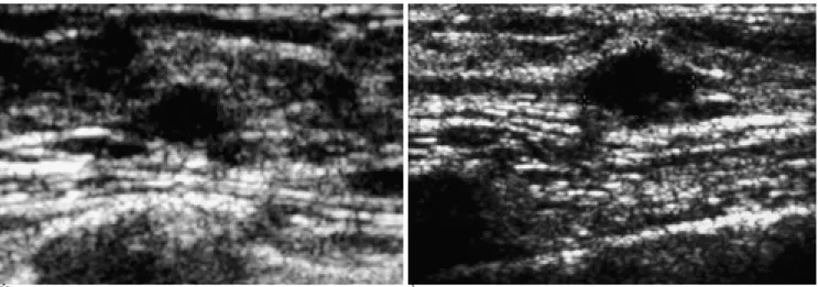
생검결과 양성 병변으로 진단된 경우는 372예 였고 (Fig.1), 이중 49명이 수술을 시행하였다. 수술을 시행한 이유는 방사 선학적 소견과의 불일치(29예), 환자나 주치의의 불안(16예), 이전에 유방암의 병력(4예) 등 이였다. 수술을 시행한 49명 중 43명은 생검결과와 일치하는 양성 병변으로 진단되었으나 나
A B
Fig. 1. 41-year-old woman without clinical symptom. US shows slightly hypoechoic, microlobulated, ovoid shaped nodule (arrows heads). US-guided core biopsy before (A) and after (B) firing shows that echogenic needle(arrows) was passed within nodule and di- agnosed as fibrosis, which was considered as concordant result. On US after six-month(not shown), the nodule decreased in size.
Table 1. Comparison of Core Biopsy Result and Operation Result
Core Biopsy Operation
Result No. of Case(%) Result No. of Case(%) Benign 43
Benign 372(84.5%) DCIS 1 49
IDC 5
ADH 4(0.9%) ADH 2 4
IDC 2
DCIS 7(1.6%) DCIS 4 7
IDC 3
IDC 53(12%) IDC 44 45
NR 1
Other malignancy 4(0.9%)
Total 440 105
ADH: atypical ductal hyperplasia DCIS: ductal carcinoma in situ IDC: invasive ductal carcinoma NR: no residual tumor
Other malignancy: lymphoma(n=2), metastasis(n=2)
머지 6예는 최종적으로 악성으로 진단되어 8.3% (6/72)의 위 음성율을 보였다. 이들 6예의 핵생검 결과는 기질 섬유화 (stromal fibrosis) 3예, 상피증식증(ductal epithelial hyper- plasia) 2예, 선종성증식증(adenomatous hyperplasia) 각 1예 였다. 위음성 생검은 모두 방사선학적 소견상 악성이 의심되 는 category 4 병변 이었다. 이들의 평균 크기는 1.2 cm으로 핵생검을 시행한 전체 병변의 평균크기 0.9 cm과 비교하여 의 미있는 차이는 없었다. 6예 중 4예는 생검 직후 수술을 시행 하였고 (Fig. 2) 나머지 2예는 6개월 후 추적검사에서 크기 변 화가 있어 수술을 시행하였다 (Fig. 3). 위음성 생검 결과를 보 인 6예 중 14게이지 생검침을 사용한 경우가 2예(5.1%), 16
게이지 생검침을 사용한 경우가 4예(12%) 였으나 통계적으로 의미있는 차이는 없었다 (Fisher’s exact test, p-value=0.26).
핵생검상 양성으로 진단되어 수술을 시행하지 않은 323예 는 평균 21개월(8개월-5년4개월)의 추적검사 기간동안 임상 적이나 방사선학적으로 악성 소견을 보이지 않았다.
고 찰
영상 유도하 경피적 생검법은 유방 병변의 초기 접근법으로 점차 증가하는 추세이며 특히 비촉지성 병변의 조직학적 진단 에 있어 수술적 생검을 대치하고 있다 (1-4). 영상 유도하 경
A B
Fig. 3. 53-year-old woman with incidentally found breast nodule was diagnosed as fibrosis on core biopsy (A) and it was regarded as concordant result. But after 6 months, the nodule increased in size (B) and repeated biopsy revealed invasive ductal carcinoma.
A B C
Fig. 2. 50-year-old woman was found to have suspicious microcalcification on screening mammogram (A). On sonogram, only spotty calcifications (arrows) were visible without definite mass and core biopsy was done targeting on calcification up to 8 times (B). Ductal epithelial hyperplasia was diagnosed on pathology. So mammogram-guided localization biopsy was done and it proved to be ductal carcinoma in situ (C).
피적 생검법으로는 초음파 유도하 핵 생검과 유방촬영술을 이 용한 정위(stereotactic) 생검법이 주로 쓰이고 있으나 (12, 13) 국내에서는 정위 생검에 필요한 기구의 보급이 미흡하여 초음파 유도하 핵 생검이 주종을 이루고 있다 (7, 8). 초음파 유도하 핵 생검은 정위 생검법에 비해 기구가 간단하고 방사 선 노출이 없으며 액와를 포함한 유방 전체 병변에 손쉽게 접 근할 수 있는 등 많은 장점을 가지고 있으나 (12, 14, 15) 초 음파에서 잘 보이지 않는 병변은 검사에 어려움이 있다는 단 점이 있다. 본 연구에서도 초음파상 뚜렷한 종괴 없이 의심스 러운 미세 석회화만 관찰되었던 예에서 위음성 생검이 있었다 (Fig. 2). 이러한 병변의 경우 유방촬영술 유도 생검이 효과적 이며 시술 후 반드시 검체 유방촬영술(specimen mammo- gram)을 촬영하여 확인하는 과정이 필요하다 (16).
초음파 유도 핵 생검에 14게이지 생검침이 보편적으로 사 용되고 있는데 이는 초기에 사용되었던 16게이지에 비하여 생 검의 정확도는 의미있게 높이면서 시술에 따르는 합병증을 최 소화 할 수 있기 때문이다 (17). 본 연구에서는 14게이지를 사용했을 때 16게이지에 비해 낮은 위음성 생검률을 보였으 나 악성의 증례 수가 작아서 통계학적 의의는 없었다. 영상 유 도하 경피적 생검법은 수술적 생검에 비해 간단하게 조직학적 진단을 내릴 수 있고 암으로 진단되는 경우 미리 수술 계획을 세울 수 있어 더 만족스러운 수술 결과를 얻을 수 있다 (18).
또한 이러한 영상 유도하 경피적 생검법의 도입으로 양성으로 진단된 경우 불필요한 수술 횟수를 줄일 수 있고 결과적으로 의료비 절감 효과를 얻을 수 있다는 연구도 보고되고 있다 (19, 20). 그렇지만 수술적 생검과 비교해 피할 수 없는 한계로 지 적되는 것이 바로 위음성 생검과 조직학적 저급평가(under- estimation) 이다. 14게이지 핵생검의 경우 위음성 생검률이 평균 2.8%(0.3-8.2%)로 보고되고 있는데 이 중 추적 검사 도 중 발견되는 지연 위 음성률은 30% 가량이고 나머지 70%는 생검 직후 수술로 발견되는 경우라고 한다 (21, 22). 본 연구 에서는 14게이지를 사용 했을 때 5%(2예)의 위 음성률을 보 였는데 2예 모두 생검 직후 영상 소견과 조직검사 결과의 불 일치로 수술을 시행 하였다. 16게이지 핵생검까지 포함한 총 6예의 위음성 생검중 2예(33.3%)의 지연 위음성 생검이 나타 났다. 이들은 모두 핵생검 결과에서는 양성으로 나왔으나 6개 월 후 추적검사에서 크기 변화가 관찰되어 수술을 시행한 경 우였다. 위음성 생검을 감소시키기 위해 숙련된 기술뿐만 아 니라 생검침의 크기나 생검 횟수 등이 중요하며 또한 병변의 크기나 위치도 고려 대상이 된다 (23). 본 연구에서는 생검침 의 크기나 병변의 크기가 위음성 생검여부와 의미있는 연관이 없는 것으로 나왔지만 이는 증례 수가 작기 때문일 것으로 사 료된다. 위음성 생검을 피하기 위하여 앞서 언급한 대로 미세 석회화를 대상으로 한 경우 반드시 검체 유방 촬영술로 확인 해야 하며 (16) 무엇보다도 방사선 소견과의 연관에 주의를 두어 의심되는 경우 바로 수술을 비롯한 재 생검을 권해야 한 다. 또한 양성 생검결과가 나온 경우에도 환자에게 추적 검사 의 필요성을 강조하는 것이 중요하다.
조직학적 저급평가도 핵생검의 또다른 중요한 한계로 (24)
14게이지 핵생검의 경우 침윤성암을 상피내암으로 진단하는 상피내암 저급평가가 16-35%(5,25-28) 유방암을 비전형상 피증식증으로 진단하는 비전형상피증식 저급평가가 20-56%
로 보고되고 있다(5,11,25, 29-32). 본 연구에서 16게이지를 포함해서 비전형상피증식증 저급평가가 2예(50%), 상피내암 저급평가가 3예(43%)였다. 최근 14게이지 자동총 핵생검 대 신 11게이지 진공 보조 생검(vacuum assisted biopsy)이 더 많은 조직을 얻을 수 있어 정확한 조직학적 진단을 얻을 수 있 다고 보고되나 역시 0-38%의 비전형상피증식증 저급평가(25, 30-33)와 16-35%의 상피내암 저급평가가 있어 (13, 25, 27, 28, 34) 수술적 생검에 대한 한계는 여전히 남아있다.
결론적으로 비촉지성 유방병변에 대한 초음파 유도하 핵 생 검은 조직학적 진단에 유용한 방법으로 수술적 생검과 비교할 때 위음성 생검의 한계를 가지고 있지만 방사선 소견과의 주 의 깊은 연관, 밀접한 추적검사 등으로 이를 극복하는 것이 중 요하다.
참 고 문 헌
1. Elvecrog EL, Lechner MC, Nelson MT. Nonpalpable breast le- sions: Correlation of stereotaxic large-core needle biopsy and surgi- cal biopsy results. Radiology 1993;188:453-455
2. Gisvold JJ, Goellner JR, Grant CS, et al. Breast biopsy: A compara- tive study of stereotaxically guided core and excisional techniques.
AJR Am J Roentgenol 1994;162:815-820
3. Parker SH, Burbank F, Jackman RJ, et al. Percutaneous large-core breast biopsy: A multi-institutional study. Radiology 1994;193:359- 364
4. Parker SH, Jobe WE, Dennis MA, et al. US-guided automated large-core breast biopsy. Radiology 1993;187:507-511
5. Jackman RJ, Nowels KW, Shepard MJ, Finkelstein SI, Marzoni FA.
Stereotaxic large-core needle biopsy of 450 nonpalpable breast le- sions with surgical correlation in lesions with cancer or atypical hyperplasia. Radiology 1994;193:91-95
6. Parker SH, Lovin JD, Jobe WE, Burke BJ, Hopper KD, Yakes WF.
Nonpalpable breast lesions: stereotactic automated large-core biop- sies. Radiology 1991;180:403-407
7. 곽민숙, 김학수, 이한경 외. 비촉진 유방병변에 대한 초음파 유도 자 동총생검의 유용성. 대한방사선의학회지 1997;37:943-947 8. 이청근, 김충현, 신경숙 외. 유방질환에 있어서 초음파를 이용한 자
동총생검의 이용가치 및 그 결과. 대한초음파의학회지 1994;13:
211-216
9. Burbank F, Parker SH. Methods for evaluating the quality of an imag- ing-guided breast biopsy program. In: Parker SH, ed. Interventional breast procedures. In: Feig SA, ed. Seminars in breast disease, vol. 1, no. 2. Philadelphia: Saunders, 1998:1:71-83
10. Liberman L, Cohen MA, Dershaw DD, Abramson AF, Hann LE, Rosen PP. Atypical ductal hyperplasia diagnosed at stereotaxic core biopsy of breast lesions: an indication for surgical biopsy. AJR Am J Roentgenol 1995;164:1111-1113
11. Liberman L, Dershaw DD, Glassman J, et al. Analysis of cancers not diagnosed at stereotactic core breast biopsy. Radiology 1997;
203:151-157
12. Rubin E, Dempsey PJ, Pile NS, et al. Needle-localization biopsy of the breast: impact of a selective core needle biopsy program on yield. Radiology 1995;195:627-631
13. Meyer JE, Smith DN, Lester SC, et al. Large core needle biopsy of
nonpalpable breast lesions. JAMA 1999;281:1638-1641
14. Parker SH, Jobe WE, Dennis MA, et al. US-guided automated large-core breast biopsy. Radiology 1993;187:507-511
15. Liberman L, Feng TL, Dershaw DD, Morris EA, Abramson AF.
Ultrasound-guided core breast biopsy: utility and cost-effective- ness. Radiology 1998;208:717-723
16. Liberman L, Evans WP, Dershaw DD, et al. Specimen radiography of microcalcifications in stereotaxic mammary core biopsy speci- mens. Radiology 1994;190:223-225
17. Helbich TH, Rudas M, Haitel A, et al. Evaluation of needle size for breast biopsy: Comparison of 14-,16-,18-gauge biopsy needles. AJR Am J Roentgenol 1998;171:59-63
18. Liberman L, LaTrenta LR, Dershaw DD. Impact of core biopsy on the surgical management of impalpable breast cancer: Another look at margins (letter). AJR Am J Roentgenol 1997;169:1464-1465 19. Jackman RJ, Marzoni FA, Finkelstein SI, Shepard MJ. Benefits of
diagnosing nonpalpable breast cancer with stereotactic large-core needle biopsy: Lower costs and fewer operations (abstr). Radiology 1996;201(P):311
20. Lindfors KK, Rosenquist CJ. Needle core biopsy guided with mam- mography: A study of cost effectiveness. Radiology 1994;190:217- 222
21. Jackman RJ, Nowels KW, Rodriguez-Soto J, Marzoni FA, Finkelstein SI, Shepard MJ. Stereotactic, automated, large-core needle biopsy of nonpalpable breast lesions: False-negative and histologic underestimation rates after long-term follow-up.
Radiology 1999;210:799-805
22. Lee CH, Philpotts LE, Horvath LJ, Tocino I. Follow-up of breast le- sions diagnosed as benign with stereotactic core-needle biopsy:
Frequency of mammographic change and false-negative rate.
Radiology 1999;212:189-194
23. Liberman L, Dershaw DD, Glassman J, et al. Analysis of cancers not diagnosed at stereotactic core breast biopsy. Radiology 1997;
203:151-157
24. Burbank F, Parker SH. Methods for evaluating the quality of an imag- ing-guided breast biopsy program. In: Parker SH, ed. Interventional breast procedures. In: Feig SA, ed. Seminars in breast disease, vol. 1, no. 2. Philadelphia: Saunders, 1998;1:71-83
25. Burbank F. Stereotactic breast biopsy of atypical ductal hyperpla-
sia and ductal carcinoma in situ lesions: improved accuracy with a directional, vacuum-assisted biopsy instrument. Radiology 1997;202:843-847
26. Liberman L, Dershaw DD, Rosen PP, et al. Stereotaxic core biopsy of breast carcinoma: Accuracy at predicting invasion. Radiology 1995;194:379-381
27. Jackman RJ, Burbank FH, Parker SH, et al. Accuracy of sampling ductal carcinoma in situ by three stereotactic breast biopsy meth- ods (abstr). Radiology 1998;209(P):197-198
28. Won B, Reynolds HE, Lazaridis CL, Jackson VP. Stereotactic biop- sy of ductal carcinoma in situ of the breast using an 11-gauge vacu- um-assisted device: Persistent underestimation of disease. AJR Am J Roentgenol 1999;173:227-229
29. Liberman L, Cohen MA, Dershaw DD, Abramson AF, Hann LE, Rosen PP. Atypical ductal hyperplasia diagnosed at stereotaxic core biopsy of breast lesions: an indication for surgical biopsy. AJR Am J Roentgenol 1995;164:1111-1113
30. Jackman RJ, Burbank F, Parker SH, et al. Atypical ductal hyperpla- sia diagnosed at stereotactic breast biopsy: Improved reliability with 14-gauge, directional, vacuum-assisted biopsy. Radiology 1997;204:485-488
31. Jackman RJ, Burbank FH, Parker SH, et al. Atypical ductal hyper- plasia diagnosed by 11-gauge, directional, vacuum-assisted breast biopsy: How often is carcinoma found at surgery? (abstr).
Radiology 1997;205(P):325
32. Philpotts LE, Shaheen NA, Carter D, Lange RC, Lee CH. Compar- ison of rebiopsy rates after stereotactic core needle biopsy of the breast with 11-gauge vacuum suction probe versus 14-gauge nee- dle and automatic gun. AJR Am J Roentgenol 1999;172:683-687 33. Brem RF, Behrndt VS, Sanow L, Gatewood OMB. Atypical ductal
hyperplasia: Histologic underestimation of carcinoma in tissue har- vested from impalpable breast lesions using 11-gauge stereotacti- cally guided directional vacuum-assisted biopsy. AJR Am J Roentgenol 1999;172:1405-1407
34. Liberman L, Vuolo M, Dershaw DD, et al. Epithelial displacement after stereotactic 11-gauge directional vacuum-assisted breast biop- sy. AJR Am J Roentgenol 1999;172:677- 681
J Korean Radiol Soc 2002;46:601-606
Address reprint requests to : Eun-Kyung Kim, M.D., Department of Diagnostic Radiology, Yonsei University College of Medicine, 134, Shinchon-dong, Seodaemun-gu, Seoul 120-752, Korea.
Tel. 82-2-361-6687 Fax. 82-2-393-3035 E-mail: [email protected]
The Usefulness of Ultrasound-Guided Core Needle Biopsy for Non-Palpable Breast Lesion
1Jai Kyung You, M.D., Eun-Kyung Kim, M.D., Mi Hye Kim, M.D.2, Jin-Young Kwak, M.D., Ki Keun Oh, M.D., Byung Woo Park, M.D.3, Kyong Sik Lee, M.D.3
1Department of Diagnostic Radiology, Yonsei University College of Medicine
2Department of Diagnostic Radiology, Miz Medi Hospital
3Department of General Surgery, Yonsei University College of Medicine
Purpose: To determine the usefulness of ultrasound-guided core biopsy for the diagnosis of non-palpable beast lesions.
Materials and Methods: Between April 1996 and December 2000, 932 lesions in 901 patients were the object of ultrasound-guided core biopsy. Of these, 440 non-palpable lesions ranging in size from 0.3 to 3.0 (average, 0.9)cm, and found in 428 patients (all women aged, on average, 43.9 years), were included in this study. The pathologic results of core biopsy were compared with the available surgical data, and clinical and radiologic follow-up data were also reviewed. A 16-gauge needle was used in 197 lesions, and a 14-gauge neadle in the other 243.
Results: At core biopsy, 53 lesions were diagnosed as invasive carcinoma, and 45 of these were excised. Forth- four were confirmed as invasive carcinoma, and in one case there was no residual tumor. Seven lesions, diag- nosed as ductal carcinoma in situ at core biopsy, were surgically removed, and the final diagnosis was ductal carcinoma in four cases and invasive carcinoma in two. Two of four cases initially diagnosed as atypical ductal hyperplasia were finally diagnosed as invasive carcinoma after surgery. Six lesions diagnosed at core biopsy as benign were later found to be malignant (false-negative rate, 8.3%). Radiologic imaging suggested that all six le- sions-for two of which, a 14-gauge needle was used, and for four, a 16-gauge needle-were malignant. The false -negative rate was 5.1% and 12%, respectively, whithout statistical significance (p=0.26).
Conclusion: Ultrasound-guided core needle biopsy for non-palpable breast lesions is useful and can replace surgical excision. To avoid false-negative assessment, however, strict radiologic-histopathologic correlation is required.
Index words :Breast, biopsy
Breast neoplasms, diagnosis Breast, US
Breast, diseases