Veterinary Science
Phacoemulsification and acryl foldable intraocular lens implantation in dogs: 32 cases
Na-Young Yi, Shin-Ae Park, Man-Bok Jeong, Won-Tae Kim, Se-Eun Kim, Je-Min Chae, Kang-Moon Seo*
Department of Veterinary Ophthalmology, College of Veterinary Medicine, Seoul National University, Seoul 151-742, Korea
This study evaluated the surgical outcome and complications of phacoemulsification and the implantation of an acryl foldable intraocular lens (IOL) with a squared edge in dogs with cataracts. Thirty-two eyes from 26 dogs were examined. The mean follow up period was 75.9 days ranging from 23 to 226 days. The complications after phacoemulsification were posterior capsular opacity (PCO) around the IOL (n = 11), ocular hypertension (n = 4), focal posterior synechia (n = 4), hyphema (n = 3) and corneal ulcer (n = 2). The complications associated with the IOL were decenteration of the optic (n = 2) and ventral haptic displacement (n = 1). Most cases of PCO were found only around the margin of the IOL, and all eyes had vision during the observation period. In conclusion, the implantation of an acryl-foldable lens with a squared edge at the time of phacoemulsification is an effective method for preserving the central visual field of dogs with cataract.
Key words: cataract, dog, foldable, intraocular lens, pha- coemulsification
Introduction
Cataract extraction is a type of ophthalmic surgery frequently performed in veterinary medicine. Various surgical techniques have been used but phacoemulsification with the implantation of an intraocular lens (IOL) is currently the treatment of choice. IOL implantation improves the optics of the aphakic eye and reduces the formation of posterior capsular opacity (PCO) after surgery [1,15]. However, IOL decenteration or luxation can cause visual alterations or increase the risk of postoperative complications [1,15]. Another potential disadvantage of IOL implantation is transient intraocular inflammation [11].
In order to achieve postoperative emmetropia, many
researchers have documented the diopter (D) of IOL optic in dogs [3,17] with an IOL of approximately 40 D being mainly used in canine cataract surgery. The most widely used IOL material in veterinary practice is polymethylmethacrylate (PMMA) [12]. However, the use of various IOL materials such as silicone and hydroxyethylmethacrylate (HEMA) optic IOL have also been examined [6,10]. Because acrylic and silicone lenses are flexible, they can be implanted through a small corneal incision (2.5-3.5 mm) [5]. This small incision reduces the surgically induced astigmatism associated the use of a PMMA optic IOL, which requires a corneal incision large enough to accommodate an 8-9 mm implant [5,8]. Recently, 41 D foldable IOL made from silicone was reported in dogs [6]. However, it is a posterior chamber type IOL, which carries a high risk of pupil capture, chronic iridocyclitis and posterior synechia [8].
An acryl IOL with a squared edge is associated with a lower incidence of induced PCO than other lens materials [5,14]. This is because acryl lenses have strong adhesion to the posterior capsule and the specific optical design (squared) can inhibit the migration of lens epithelial cells into the optic area [5]. Flexible acryl IOLs are commonly implanted in dogs but the IOL optic is not specifically designed to prevent PCO [7]. This study evaluated the surgical outcomes and complications of phacoemulsification with the implantation of a flexible acryl IOL in dogs with cataracts. The study also examined the effects of the squared edge design of the IOL on PCO formation.
Materials and Methods
The medical records of dogs undergoing phacoemulsification with the implantation of an IOL at the Veterinary Medical Teaching Hospital of Seoul National University between October 2004 and January 2006 were reviewed. The following information was analyzed: signalment (age, gender and breed), stages of cataract, surgical outcomes and postoperative complications.
Postoperative ocular hypertension was defined as an intraocular pressure (IOP) >25 mmHg within 72 h after surgery, and >30 mmHg after 72 h after surgery. Aqueous
*Corresponding author
Tel: +82-2-880-1258; Fax: +82-2-884-8651
E-mail: [email protected]
flare was graded on a scale of 0 to 3+ (0 = normal anterior chamber; 1+ = mild flare; 2+ = moderate flare, the slit lamp beam in the anterior chamber is equally opaque as the beam passing through the normal lens; and 3+ = severe flare, the slit beam in the anterior chamber is more opaque than that passing through the lens) [10]. Corneal opacity was rated from 0 to 4+ (0=normal cornea; 1+=mild loss of transparency;
2+ = moderate loss of transparency; 3+ = severe loss of transparency; 4+ = opaque cornea) [10]. PCO was graded from 0 to 3+ (0 = none; 1+ = decreased transparency but the fundic examination is unobstructed; 2+=decreased transparency, and the fundic examination is partially obstructed; and 3+ = decreased transparency, and the fundic examination is completely obstructed) [18]. The complications relevant to the implantation of the IOL were recorded.
Preoperatively, a complete ophthalmic examination including a Schirmer tear test (STT), slit lamp biomicroscopy, applanation tonometry, fluorescein staining test and indirect ophthalmoscopy were performed in all dogs. Topical corticosteroids/antibiotics (dexamethasone/neomycin/polymyxin B) were administered if a dog had lens-induced uveitis (LIU).
Prior to surgery, electroretinography and ocular ultrasonography were performed in all eyes to exclude retinal diseases and to evaluate the posterior segment of the eye.
During pre-surgical preparation, all the dogs received topical corticosteroids/antibiotics (dexamethasone/neomycin/
polymyxin B), topical non-steroidal anti-inflammatory agents (0.03% flurbiprofen) and mydriatics (1% atropine sulfate and 1% tropicamide). Also, systemic non-steroidal anti- inflammatory agents (flunixin meglumine, 0.5 mg/kg, IV), corticosterids (dexamethasone 2 mg/kg, IV) and antibiotics (cefazoline, 30 mg/kg, IV) were administered.
General anesthesia was induced using routine procedures, and maintained with isoflurane inhalation. One-handed phacoemulsification with a modification of the “crater divide and conquer” method was performed through a 3 mm corneal incision [8,15]. The procedure used a single piece of IOL of 40 D, which was a flexible, injectable and consisted of an acryl polymer optic and polypropylene haptics (Tekia, USA). The IOL optic had a square edge. The IOL optic was folded using IOL-holding forceps and inserted into the IOL cartridge. After filling the capsular bag and anterior chamber with a sodium hyaluronate (Hyaltech, UK), the IOL in the cartridge was inserted into the capsular bag with the IOL inserter without enlarging the corneal incision (Fig. 1-a).
The corneal incision was closed with interrupted 8-0 polyglactin 910 sutures (Johnson & Johnson, Belgium).
Postoperatively, the intraocular pressure, aqueous flare, corneal edema, fibrin formation and PCO were evaluated at each ophthalmic examination. Where necessary, a non- steroidal anti-inflammatory agent (0.03% flurbiprofen), corticosteroids/antibiotics (dexamethasone/neomycin/polymyxin B), antibiotics (0.3% ciprofloxacin) and mydriatics (1%
atropine sulfate) were administered topically to control the intraocular inflammation and maintain mydriasis. Corticosteroids (prednisolone 1 mg/kg) and antibiotics (amoxicillin/clavunic acid 12.5 mg/kg) were administered orally every 12 h for 7 days. The medical treatment for dogs that developed ocular hypertension after surgery included the administration of mannitol (1 g/kg, IV), topical β -adrenergic receptor blocker (0.5% timolol maleate) and carbonic anhydrase inhibitors (2% dorzolamide hydrochloride). During the follow-up period, the surgical outcomes were divided into four categories according to the specific criteria reported by Davidson et al . [4]. The Fischer’s exact test was used to compare the percentage of fibrin formation according to the cataract stage. All statistical analysis was performed using the SPSS 12.0 analysis program. A p value < 0.05 was considered significant.
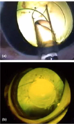
Fig. 1. Photograph of the IOL placement and pseudophakic eye
after phacoemulsification. (a) A flexible IOL in the cartridge was
inserted into the capsular bag through a 3 mm corneal incision
with an IOL inserter. (b) Four weeks after surgery, a properly
positioned IOL in the capsular bag can be seen in an 8-year-old,
male Cocker spaniel. Vacuole-type posterior capsular opacity are
formed around the margin of the optic, and there is no PCO
behind the IOL optic.
Results
During the observation period, the phacoemulsification and implantation of IOLs was performed on 32 eyes in 26 dogs (6 bilateral and 20 unilateral). The age of the dogs ranged from 9 months to 14 years, with a mean age of 5.4 years. The study consisted of 2 males, 6 castrated males, 16 females and 2 spayed females. The most prevalent breed was the Cocker spaniel (n = 7). Other affected breeds were the toy Poodle (n = 5), miniature Schnauzer (n = 3), Shih Tzu (n = 3), Siberian Husky (n=2), Yorkshire terrier (n = 2), Maltese terrier (n = 2) and mixed breed dog (n = 2). The postoperative follow-up periods were as follows: 14-30 days in 32 eyes, 30-60 days in 28 eyes, 60- 90 days in 18 eyes, and >90 days in 10 eyes. The surgically removed cataracts were immature (n = 9, 28.1%), mature (n = 18, 56.3%) and hypermature (n = 5, 15.6%). The preoperative LIU was detected in 11 (34.4%) eyes from 9 dogs.
After surgery, a return of vision was reported in all eyes.
The mean IOP (mmHg) decreased below the normal IOP range one day after surgery, and returned to the normal range within two weeks after surgery (Table 1). One day after surgery, the mean flare grade increased but had resolved in all eyes within 14 days (Table 1). During the two weeks after surgery, 8 eyes showed mild fibrin formation in the anterior chamber or surface of the IOL, and 3 eyes showed moderate fibrin formation. Intra-cameral injections of a tissue plasminogen activator (tPA, 25 µ g) were performed in two dogs (4 and 16 days after surgery, respectively) with moderate fibrin formation in the anterior chamber, which was resolved within one day after the injection. Mild fibrin in the other cases had dissolved
spontaneously within 3 weeks. All the dogs developed 1+ to 2+ corneal edema adjacent to the corneal incision that was caused by mechanical trauma during surgery. The corneal edema in all eyes regressed spontaneously within one week even though the opacity had remained around the corneal incisions. The postoperative results were evaluated (Table 2). The rate of excellent or good postoperative outcomes was 93.8%. The hypermature cataracts showed significantly more fibrin formation than the other stages of cataracts ( p <
0.05).
Table 3 lists the postoperative complications after surgery.
The most common complication was PCO formation around the optic of the IOL (11 eyes). However, the central vision still remained because no PCO was observed behind the optic of the IOL. Most PCO was mainly noted around the margin of the IOL (Fig. 1-b). The incidence of PCO increased over time, and more PCO accumulated in dogs that had already had PCO. During the 72 h postoperative period, postoperative ocular hypertension was developed two Cocker spaniels but was resolved after medical treatments for glaucoma. Ocular hypertension also developed in a Cocker spaniel and a toy Poodle at 9 and 11 days after surgery, respectively. Focal posterior synechia was observed in four eyes within 30 days. Mild hyphema was noted one day after surgery in three cases. Within one day spontaneous resorption occurred in two eyes, and irrigation and aspiration (I/A) was performed in one eye. No dogs showed a recurrence of hyphema during the follow-up period.
Linear shaped, superficial corneal ulcers in the center of the cornea were found in both eyes of one dog. Complications associated with the implantation of the IOL were optic decenteration (n = 2) and ventral haptic displacement from
Table 1. Intraocular pressure (IOP) and flare grade of the dogs that underwent surgery according to the follow-up periods (mean ± SD) Periods
1 day before
surgery <3 days 3-14 days 14-30 days 30-60 days 60-90 days >90 days IOP (mm Hg) 16.16 ± 4.04 13.42 ± 3.16 15.21 ± 5.30 16.26 ± 5.35 16.05 ± 4.71 15.30 ± 4.42 16.13 ± 5.14
Flare grade* 00.00 ± 0.00 01.50 ± 0.79 00.21 ± 0.05 00.00 ± 0.00 00.00 ± 0.00 00.00 ± 0.00 00.00 ± 0.00
*Flare was graded on a scale of 0 to 3+ following Gilger et al. [10].
Table 2. Postoperative results in 32 eyes according to the cataract stage during the follow-up period*
Category
†Immature (n = 9) Stage of cataract Mature (n = 18) Hypermature (n = 5) Total (n = 32)
Excellent 3/9 12/18 4/5 19/32
Good 4/9 6/18 1/5 11/32
Fair 2/9 0/18 0/5 2/32
Poor 0/9 0/18 0/5 0/32
*Mean: 75.9 days.
†
Excellent = Clear visual axis and no intraocular lens (IOL) decenteration; Good = 1+ posterior capsular opacity (decreased transparency , but the
fundus was visual with no hindrance), focal posterior synechia or mild IOL decenteration; Fair = 2+ or greater posterior capsular opacity (decreased
transparency, the fundus was partially visual or transparency was decreased or the fundus was completely not visible), multifocal posterior synechia,
IOL haptic dislocation or iris capture; Poor = Visual loss from complications.
the capsular bag (n = 1). There were no other complications such as persistent uveitis, endophthalmitis or retinal detachment during the observation period.
Discussion
The surgical outcome in this study was similar to those reported in previous study of phacoemulsification with an IOL [4]. Hypermature cataracts were associated with more fibrin formation than the other cataract stages. That may be because a hypermature cataract is the type of cataract that most commonly increases the risk of lens-induced uveitis if cataract extraction is performed [19]. The frequent problems during the placement of a PMMA optic IOL are miosis, limited diameter anterior capsulectomy and irregular margins of the anterior capsulectomy [8]. However, a small anterior capsulectomy was not a factor in this study because a flexible IOL could be inserted into the capsular bag through an IOL cartridge opening. No dog showed miosis as a result of iris irritation during implantation. PCO was the most common complication associated with lens extraction [15].
The residual lens epithelial cells proliferated to form a lentoid undergoing fibrous pseudometaplasia [2,15]. PCO is also derived from the bonding between the capsulectomy margin and the posterior capsule [16]. Histologically, a lens fibrous membrane was observed in the adhesion that had formed between the capsulectomy margin and the posterior capsule, and the adhesion caused severe wrinkling of the posterior capsule [16]. An intraocular lens might reduce the incidence of PCO because it prevents wrinkling of the posterior capsule and limits the space for lens epithelial cells to migrate by contacting the posterior capsule [15,16].
Previous studies showed that an acryl optic IOL had a significantly lower incidence of PCO than a PMMA optic IOL [13,14]. It may be associated with the biophysical properties of acryl polymer to increase the contact between the optic and posterior capsule [5,14]. The squared edge design of the optic provides a mechanical barrier by
inhibiting the migration of lens epithelial cells to the optic, which can preserve the central visual field [5]. In this study, most PCO was formed around the optic, and the central vision was preserved in all cases. It is believed that the appearance of PCO was caused by the high level of adhesion of the optic to the posterior capsule and the squared edge design of the optic. Therefore, the squared edged, acryl IOL described in this paper appears to be the most effective IOL in preventing PCO. However, a long- term follow-up will be needed because PCO may develop several years after cataract surgery.
Ocular hypertension after cataract surgery may often occur in some breeds, such as Shih Tzu, Cocker spaniel and Boston terrier [18]. Similar to a previous study, the Cocker spaniel was the most common breed that developed glaucoma in this study. Transient ocular hypertension generally occurs within 72 h in dogs that have undergone phacoemulsification [15]. However, aphakic glaucoma can occur within one month after surgery when the dogs have persistent anterior uveitis or a fully dilated pupil [8]. Two dogs that developed ocular hypertension 7 days after surgery received persistent topical mydriatics therapy with no flare in anterior chamber being noted in these dogs. This might be related to the moderately to fully dilated pupil resulting from the continuous application of mydriatics, which may cause the occlusion of the outflow channels. The incidence of other complications were similar to previous reports [4,18]. One dog that developed a bilateral corneal ulcer was a brachycephalic breed and the condition was associated with a macropalpebral fissure. It has been reported that infrequent blinking rates, thin central precorneal tear film and an exposed cornea may predispose the dog to corneal ulcers after cataract surgery, particularly in brachycephalic breeds [8]. The use of an ocular lubricant after surgery may relieve the problems associated with dryness in these breeds such as corneal ulcers and/or erosion. TPA has been used to dissolve the hyphema or fibrin, which is formed from surgery or inflammation [9,15], and is effective in fibrinolysis in eyes with moderate fibrin
Table 3. Prevalence of postoperative complications after surgery during the various follow-up periods in 32 eyes
Complications Follow-up period
< 3 days
(32 eyes) 3-14 days
(32 eyes) 14-30 days
(32 eyes) 30-60 days
(28 eyes) 60-90 days
(18 eyes) > 90 days (10 eyes)
PCO (1+) 0 (0)* 3 (9.4) 4 (12.5) 6 (18.8) 4 (22.2) 3 (42.9)
POH 2 (6.3) 2 (6.3) 0 (0) 0 (0) 0 (0) 0 (0)
Posterior synechia 0 (0) 3 (9.4) 1 (3.1) 0 (0) 0 (0) 0 (0)
Hyphema 3 (9.4) 0 (0) 0 (0) 0 (0) 0 (0) 0 (0)
Corneal ulcer 2 (6.3) 0 (0) 0 (0) 0 (0) 0 (0) 0 (0)
IOL problems
†2 (6.3) 0 (0) 0 (0) 0 (0) 0 (0) 0 (0)
*Number of eye (percentage of eyes with the complication).
†