Type of tooth movement during en masse retraction of the maxillary anterior teeth using labial versus lingual biocreative therapy in adults: A randomized clinical trial Mais M
12
0
0
전체 글
(2) Sadek et al • Labial versus lingual biocreative therapy. INTRODUCTION Dentoalveolar protrusion is commonly treated by premolar extraction followed by anterior retraction to achieve a harmonious facial profile and an adequate incisor relationship. The applied biomechanical system should ensure adequate retraction of the anterior teeth for proper function, esthetics, and stability. In severe protrusion cases, maximum anchorage is often required. For anterior retraction, miniscrew anchorage was reported to be more effective than conventional methods for anchorage reinforcement.1 En masse anterior retraction is often performed using sliding mechanics with the retraction force applied from a hook crimped or soldered to the archwire and interdental miniscrews. In the presence of a rigid continuous archwire, the line of action of the retraction force relative to the center of resistance of the entire arch will dictate the displacement pattern of the dentition.2 The retraction force at the archwire level will result in rotation of the entire arch around the center of resistance, producing a tendency toward posterior open bite, anterior deep overbite with lingual inclination of the anterior segment, and steepening of the occlusal plane.3 To avoid this problem, the "biocreative therapy technique" was introduced by Chung et al.4 In this technique, brackets are bonded only on the six anterior teeth, and temporary anchorage devices (TADs) are used as the only source of anchorage. To provide bodily tooth movement during retraction, anterior torque moment is generated on the anterior teeth by using gable bends in labial biocreative therapy type I,4 and an overlay intrusion arch in type II.5 No change in the posterior occlusion is expected, since the forces generated during anterior retraction are not against the posterior teeth, but rather applied against the mini-implants. This approach can also minimize iatrogenic effects on the periodontium, since the posterior segments are not engaged during anterior retraction. After en masse retraction, shortterm fixed appliances or clear aligners can be used as the finishing stage. However, according to Mo et al., 6 this protocol requires adequate stability of the mini-implant against the moment applied to secure torque and vertical control over the anterior segment during retraction. They recommended using the C-implant (sandblasted, largegrit, acid-etched mini-implant; C-implant, Seoul, Korea), which can resist rotational moments. In biocreative therapy type I, gable bends on 0.016 × 0.022-inch (in) stainless steel utility archwires are used for anterior torque control during retraction.4 The archwire is placed into the hole of the C-implant. Changes in the length of the retraction hook and the degree of the gable bend will affect anterior torque and vertical. 382. control during retraction. The biocreative therapy type II technique can be considered as an improved method of applying Burstone’s segmented intrusion arch technique.7 An intrusion overlay archwire is inserted posteriorly into the hole of the mini-implant and ligated anteriorly to the archwire, between the two central incisors. This produces forces that control both the torque and the vertical position of the incisor segment. On the other hand, lingual biocreative therapy was introduced8 to overcome many of the disadvantages of conventional lingual orthodontics, such as excessive chair time, patient discomfort, and expensive lab procedures. Lingual biocreative therapy uses a bonded lingual retractor and a palatal plate for en masse anterior retraction. A soldered hook on the lingual retractor carries the point of application of the retraction force close to the center of resistance of the anterior segment to provide torque control during retraction. Biocreative therapy can thus offer significant advantages, including controlled tooth movement, no need for complex appliances, and skeletal anchorage with minimal reliance on patient compliance. Several case reports were published showing successful anterior retraction using labial,4,5,9 and lingual biocreative therapy.8,10 However, only a few studies have analyzed the results of anterior retraction using these techniques. In 2009, Kim et al.11 conducted two retrospective studies; one to evaluate labial biocreative therapy in 200911 and the other to evaluate lingual biocreative therapy in 2011.12 They found that significant anterior retraction was achieved with maximum anchorage using TADs as the only source of anchorage. Using finite element analysis, factors that affect effective torque control during en masse anterior retraction by using labial biocreative therapy type I and type II techniques6,13 and the lingual biocreative technique were evaluated by Mo et al.14 in 2013. Torque control was found to vary depending on the height of the anterior retraction hook in both techniques as well as the amount of intrusion force used in labial biocreative technique type II. Jee et al.15 compared the effects of a preformed C-wire with those of a conventional C-wire for en masse retraction, with TADs as the only source of anchorage. Full anterior retraction with controlled tipping was found without alteration of the posterior occlusion in both groups. However, they reported that a preformed C-wire can allow for simultaneous leveling and space closure, and thus ensure faster treatment. The existing body of research on the biocreative technique mainly consists of case reports,4,5,9,10 threedimensional finite element analyses,6,13,14,16 and retrospective studies.11,12,17 No randomized controlled trial has been published comparing the treatment effects using labial versus lingual biocreative techniques. Accordingly,. https://doi.org/10.4041/kjod.2019.49.6.381. www.e-kjo.org.
(3) Sadek et al • Labial versus lingual biocreative therapy. this study was conducted to compare the type of tooth movement during en masse anterior retraction using labial versus lingual biocreative therapy.. MATERIALS AND METHODS This study was a two-arm randomized clinical trial with a 1 : 1 allocation ratio. The trial was performed in the outpatient clinic at the Department of Orthodontic, Ain Shams University. The trial was registered at ClinicalTrials.gov with the identifier NCT03239275. The protocol of this study was approved by the Ethical Committee of the Faculty of Dentistry, Ain Shams University (approval number: FDASU-RECID091408). Before treatment, all participants signed a detailed written consent form. Participants for the study were adult female patients showing maxillary dentoalveolar protrusion in need for extraction of the maxillary first premolars and anterior retraction with maximum anchorage. Participants were judged to have maxillary dentoalveolar protrusion when they had a convex profile, upper incisor to A-Pog linear measurement more than 5 mm, and procumbent lips. Subjects were excluded if they showed severe crowding in the maxillary anterior segment, previous orthodontic treatment, obvious periodontal disease, and signs of bone loss or systemic diseases (such as bleeding disorders, bisphosphonate therapy, chemotherapy, and radiotherapy). A computerized random sequence table was generated, and the randomization was made in blocks to ensure a 1 : 1 allocation ratio. Patients were randomly assigned to either group A or B and allocated into the two groups using sequentially numbered, opaque, sealed envelopes. A person not involved in the trial was responsible for implementing the randomization and opening of the envelopes. Patients in group A (14 patients, 20.5 ± 2.1 years) were treated with labial biocreative therapy type II, while those in group B (14 patients, 21.1 ± 2.5 years) were treated with the lingual biocreative therapy technique. Blinding of the patients and the operator to the type of intervention was impossible. Blinding the outcome assessor was not possible during analysis of cone beam. computed tomography (CBCT) scans because of the presence of the brackets in the labial group. Blinding regarding the time point of the scans was also not possible because of the presence of the extraction space in the pre-treatment scans. Interventions In group A (labial biocreative therapy group), preadjusted straight wire brackets with a 0.018 × 0.025-in slot were bonded to the maxillary six anterior teeth. Leveling and alignment were then performed till a 0.017 × 0.025in stainless steel wire was obtained. To ensure that the wire was passive, it was left in place for 4 weeks before starting retraction. During that time, extraction of the right and left first premolars was performed and en masse retraction of the six anterior teeth was started. Two AbsoAnchor (Dentos Inc., Daegu, Korea) self-drilling bracket head mini-implants (right and left-handed screws, 1.6 mm diameter and 8 mm length) were placed buccally in the mucogingival junction between the maxillary second premolar and first molar. Next, 10-mm-long hooks were crimped distal to the lateral incisors. An overlay reverse curve of 0.016 × 0.022-in nickel-titanium (NiTi) wire was inserted posteriorly into the hole of the mini-implant. Figure 2. Initial photographs after final set-up, lingual biocreative therapy.. Figure 1. Initial photographs after final set-up, labial biocreative therapy. www.e-kjo.org. https://doi.org/10.4041/kjod.2019.49.6.381. 383.
(4) Sadek et al • Labial versus lingual biocreative therapy. (hole diameter, 0.75-mm) and ligated anteriorly (onepoint contact) onto the wire at the midline between the two central incisors. Closed NiTi coil springs (G4TM NiTi closed coil springs; G&H Wire Company, Franklin, IN, USA) were used to provide a consistent force of 200 g per side for en masse retraction of the anterior teeth (Figure 1). In group B (lingual biocreative therapy group), the lingual retractor was fabricated from a chrome cobalt alloy and sandblasted to provide adequate bond strength. It was bonded to the lingual surface of the six anterior teeth so that they were rigidly splinted together and had 10-mm retraction hooks that were located between the central and lateral incisors, as recommended by Kim et al.12 and Mo et al.14 A cross-type miniplate (C palatal plate; Gebrüder Martin GmbH, Tuttlingen, Germany) was fixed mid-palatally to provide skeletal anchorage for en masse retraction of the anterior segment. Its main arm had three holes for inserting the microscrews and two horizontal arms with holes for the NiTi closing coil springs. A retraction force of 200 g per side was used (Figure 2). In both groups, participants underwent follow-up examinations every 6 weeks. Retraction was stopped when a Class I canine relationship was achieved and an adequate incisor relationship was obtained (Figures 3 and 4). The main outcome of the study was the type of tooth movement during anterior retraction. Secondary outcomes were molar anchorage loss, vertical movement of the crown of the six maxillary anterior teeth, inclination of the maxillary occlusal plane, and time required to complete the retraction phase. CBCT scans were obtained for every subject before and after retraction by using the iCAT scanner (Model 17/19 series; Imaging Sciences International, Hatfield, PA, USA). The CBCT scanner parameters were set to 120 kVp at 5 mA for a. total scan time of 8.9 seconds, and the field of view was 17- × 23-mm. The Digital Imaging and Communications in Medicine (DICOM) files were processed into volumetric images using InVivo 5 ver. 5.2 software (Anatomage, San Jose, CA, USA). CBCT analysis involved orienting the CBCT unit, defining the desired landmarks, creating the reference lines and reference planes, and then obtaining linear and angular measurements. The landmarks, reference lines, and planes are shown in Tables 1 and 2. Table 3 shows the linear and angular measurements used (Figure 5). Sample size calculation Sample size calculation was based on the studies by Kim et al.11 and Kim et al.12 G*power software (Universität Düsseldorf, Düsseldorf, Germany) showed that group sample sizes of 12 each would achieve 80.09% power to reject the null hypothesis of equal means with a significance level (alpha) of 0.05 using a two-sided twosample equal variance t- test. To compensate for possible dropouts, a sample size of 28 patients was selected and 14 patients were included in each group. Statistical analysis The statistical analysis was performed using IBM SPSS ver. 20 (IBM Corp., Armonk, NY, USA). The statistician was blinded and data from the patients were presented with no indication of which treatment the patients received. All variables were measured for both sides, and averages were then taken for the right and left sides. Shapiro–Wilk test of normality was used to test the normality hypothesis of all quantitative variables. Paired ttest was performed to compare the pre- and post-treatment CBCT measurements within the labial and lingual groups. An independent-sample t -test was performed for comparing the mean treatment changes between the. Figure 3. Retraction finished, labial biocreative therapy.. Figure 4. Retraction finished, lingual biocreative therapy. 384. https://doi.org/10.4041/kjod.2019.49.6.381. www.e-kjo.org.
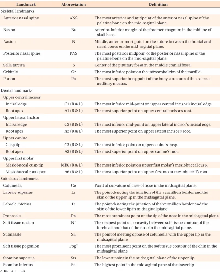
(5) Sadek et al • Labial versus lingual biocreative therapy. Table 1. Landmarks used in the three-dimensional cone beam computed tomography analysis Landmark. Abbreviation. Definition. Skeletal landmarks Anterior nasal spine. ANS. The most anterior and midpoint of the anterior nasal spine of the palatine bone on the mid-sagittal plane.. Basion. Ba. Anterior-inferior margin of the foramen magnum in the midline of skull base.. Nasion. N. Middle, anterior-most point on the suture between the frontal and nasal bones on the mid-sagittal plane.. Posterior nasal spine Sella turcica. PNS S. The most posterior midpoint of the posterior nasal spine of the palatine bone on the mid-sagittal plane. Center of the pituitary fossa in the middle cranial fossa.. Orbitale. Or. The most inferior point on the infraorbital rim of the maxilla.. Porion. Po. The most superior bony point of the bony structure of the external auditory meatus.. Incisal edge. C1 (R & L). The most inferior mid-point on upper central incisor’s incisal edge.. Root apex. A1 (R & L). The most superior point on upper central incisor’s root.. Incisal edge. C2 (R & L). The most inferior mid-point on upper lateral incisor’s incisal edge.. Root apex. A2 (R & L). The most superior point on upper lateral incisor’s root.. Cusp tip. C3 (R & L). The most inferior point on upper canine’s cusp.. Root apex. A3 (R & L). The most superior point on upper canine’s root.. Dental landmarks Upper central incisor. Upper lateral incisor. Upper canine. Upper first molar Mesiobuccal cusp tip Mesiobuccal root apex. MB6 (R & L). The most inferior point on upper first molar’s mesiobuccal cusp.. A6 (R & L). The most superior point on upper first molar mesiobuccal’s root.. Soft tissue landmarks Columella. Co. Point of curvature of base of nose in the midsagittal plane.. Labrale superius. Ls. The point denoting the junction of the vermillion border and the skin of the upper lip in the midsagittal plane.. Labrale inferius. Li. The point denoting the junction of the vermillion border and the skin of the lower lip in midsagittal plane.. Pronasale. Pn. The most prominent point on the tip of the nose in the midsagittal plane.. Soft tissue nasion. N”. The deepest point of concavity between soft tissue contour of the forehead and that of the nose in the midsagittal plane.. Subnasale. Sn. The point of meeting of base of columella with the upper lip in the midsagittal plane.. Soft tissue pogonion. Pog”. The most prominent point on the soft tissue contour of the chin in the midsagittal plane.. Stomion superius. Sts. The lowest point in the midsagittal plane of the upper lip.. Stomion inferius. Sti. The highest point in the midsagittal pane of the lower lip.. R, Right; L, left.. www.e-kjo.org. https://doi.org/10.4041/kjod.2019.49.6.381. 385.
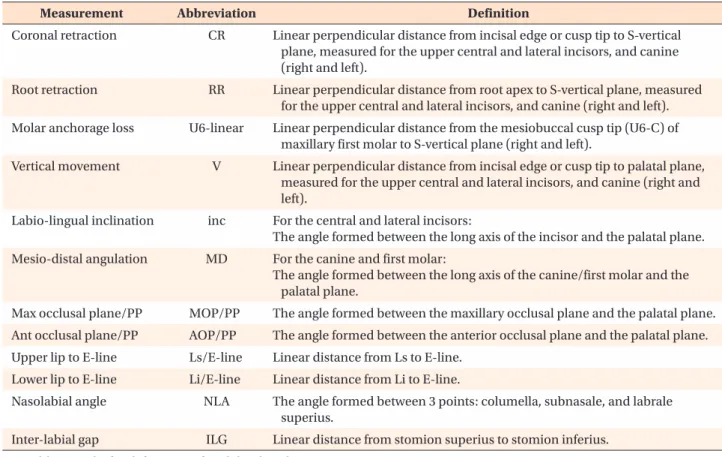
(6) Sadek et al • Labial versus lingual biocreative therapy. Table 2. Reference lines and planes used in the three-dimensional cone beam computed tomography analysis Variable. Definition. Reference line Upper central incisor long axis (R and L) Line connecting incisal edge and root apex of the upper central incisor. Upper lateral incisor long axis (R and L). Line connecting incisal edge and root apex of the upper lateral incisor.. Upper canine long axis (R and L). Line connecting cusp tip and root apex of the upper canine.. Upper first molar long axis (R and L). Line connecting mesiobuccal cusp tip and mesiobuccal root apex of the upper first molar.. E-line. Line connecting pronasale and soft tissue pogonion.. Reference plane Frankfurt horizontal plane (FHP). Plane defined by 3 landmarks: right orbitale, right porion and left porion.. Mid-sagittal plane (MSP). Plane defined by 3 landmarks: nasion, anterior nasal spine, and basion.. Palatal plane (PP). Plane defined by two landmarks: ANS and PNS points and perpendicular on the midsagittal plane.. S vertical plane (S Ver). Plane through Sella and perpendicular to the Frankfort horizontal plane and midsagittal planes.. Maxillary occlusal plane (MOP). Plane defined by 3 landmarks: incisal edge of the upper left central incisor, and mesiobuccal cusp tips of the upper right and left first molar.. Anterior occlusal plane (AOP). Plane defined by 3 landmarks: incisal edge of the upper left central incisor, and cusp tip of upper right and left canines.. See Table 1 for definitions of each landmark or measurement.. Table 3. Linear and angular measurements used in the three-dimensional cone beam computed tomography analysis Measurement. Abbreviation. Definition. Coronal retraction. CR. Linear perpendicular distance from incisal edge or cusp tip to S-vertical plane, measured for the upper central and lateral incisors, and canine (right and left).. Root retraction. RR. Linear perpendicular distance from root apex to S-vertical plane, measured for the upper central and lateral incisors, and canine (right and left).. Molar anchorage loss Vertical movement. U6-linear. Linear perpendicular distance from the mesiobuccal cusp tip (U6-C) of maxillary first molar to S-vertical plane (right and left).. V. Linear perpendicular distance from incisal edge or cusp tip to palatal plane, measured for the upper central and lateral incisors, and canine (right and left).. Labio-lingual inclination. inc. For the central and lateral incisors: The angle formed between the long axis of the incisor and the palatal plane.. Mesio-distal angulation. MD. For the canine and first molar: The angle formed between the long axis of the canine/first molar and the palatal plane.. Max occlusal plane/PP. MOP/PP. The angle formed between the maxillary occlusal plane and the palatal plane.. Ant occlusal plane/PP. AOP/PP. The angle formed between the anterior occlusal plane and the palatal plane.. Upper lip to E-line. Ls/E-line. Linear distance from Ls to E-line.. Lower lip to E-line. Li/E-line. Linear distance from Li to E-line.. Nasolabial angle. NLA. The angle formed between 3 points: columella, subnasale, and labrale superius.. Inter-labial gap. ILG. Linear distance from stomion superius to stomion inferius.. See Tables 1 and 2 for definitions of each landmark or measurement.. 386. https://doi.org/10.4041/kjod.2019.49.6.381. www.e-kjo.org.
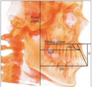
(7) Sadek et al • Labial versus lingual biocreative therapy. two groups. The significance level was set at p < 0.05. Intra-examiner and inter-examiner error analysis for the CBCT measurements were performed using concordance correlation coefficients (CCC). The closer the CCC was to 1.0, higher was the reliability of the measurement.. RESULTS Twenty-eight patients were enrolled in the trial. Four patients were lost to follow-up. The details are provided in the CONSORT flow diagram (Figure 6). The baseline demographic characteristics are presented in Table 4. Participants’ recruitment and follow-up was carried out over 20 months from January 2016 till September 2017. In the error analysis for the CBCT measurements, the CCC values ranged between 0.796 and 0.997, indicating good to excellent agreement. Shapiro–Wilk test of normality showed that variables were normally distributed; therefore, parametric tests were used for analyzing the data. Retraction durations for both groups are shown in Table 5. No statistically significant differences were found between the two groups. Table 6 shows descriptive sta-. Table 4. Baseline data for both treatment groups Variable Age (yr). Figure 5. Cone beam computed tomography measurements for the upper right central incisor. S ver, S-vertical plane; CR, crown retraction; RR, root retraction; V, vertical movement; inc, labio-lingual inclination.. Labial group (n = 14). Lingual group (n = 14). 20.5 ± 2.1. 21.1 ± 2.5. Overjet (mm). 7.2 ± 1.1. 7.5 ± 0.8. Overbite (mm). 5.9 ± 0.9. 5.6 ± 1.2. 116.7 ± 1.5. 117.1 ± 1.8. Upper central incisor inclination to palatal plane (o). Values are presented as mean ± standard deviation.. Figure 6. CONSORT 2010 flow diagram. www.e-kjo.org. https://doi.org/10.4041/kjod.2019.49.6.381. 387.
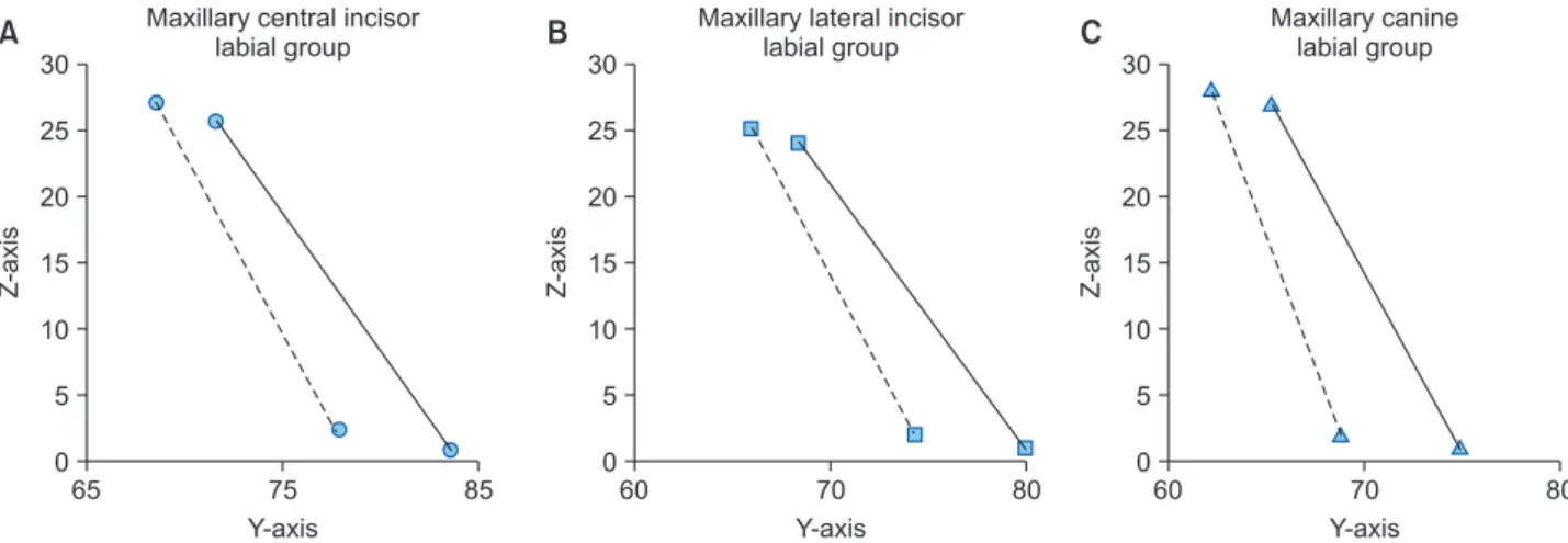
(8) Sadek et al • Labial versus lingual biocreative therapy. Table 5. Comparison of the duration of retraction between the two groups Measurement Duration of retraction (mo). Group. Mean Diff. Mean ± SD. Labial. 12.30 ± 2.72. Lingual. 11.93 ± 3.17. 0.37. 95% CI of the difference Maxilla. Mandible. −2.79. 3.54. p -value 0.80312. SD, Standard deviation; Mean Diff, mean difference; CI, confidence interval.. tistics for the intragroup and intergroup comparisons for all variables measured. The mean change in the maxillary central incisor inclination was –4.41 ± 2.33o in the labial group and –10.26 ± 4.70o in the lingual group. When comparing both groups, significant differences were found. The inclination of the central incisor was reduced more in the lingual group (i.e., more lingual tipping; mean difference, 5.85 ± 1.85o). The canine showed significant distal tipping in the lingual group (mean difference, 6.98 ± 1.25o). Figures 7 and 8 show tooth axis graphs for the maxillary central and lateral incisors and canine before and after retraction. No significant differences were found between the two groups in relation to the amount of anterior retraction, except for coronal retraction of the maxillary central incisor (1.35 mm more retraction in the lingual group) and root retraction of the canine (1.8 mm more root retraction in the labial group). The maxillary central incisor showed a mean intrusion of 1.46 ± 0.54 mm in the labial group and 1.45 ± 1.02 mm in the lingual group, with no significant difference between the two groups. However, significant differences in intrusion of the lateral incisor and canine were found between the two groups. The canine was significantly more intruded (mean difference, 1.67 ± 0.49 mm) in the lingual group. Good anchorage control was achieved in both groups. The mean molar mesial movement was 0.63 ± 0.51 mm in the labial group and 0.47 ± 0.26 mm in the lingual group. Significant improvement in the facial profile was achieved in both groups. Adverse effects No serious adverse effects were observed other than gingivitis associated with plaque accumulation. One miniscrew failed in the labial group due to poor oral hygiene. Retraction was stopped, oral hygiene measures were given to the patient, and the miniscrew was reinserted after 6 weeks. No other problems occurred.. DISCUSSION The aim of this study was to compare the treatment effects observed when using labial versus lingual bio-. 388. creative therapy for en masse retraction of the maxillary anterior teeth. Regarding the mean retraction period, no statistically significant differences in the duration of the retraction phase were found between the two groups. Jee et al.15 reported the duration to be 13.44 ± 4.30 months in the conventional C-wire group, and Kim et al.11 reported an average duration of 13.94 ± 5.61 months, which is close to our results. In labial biocreative therapy type II, various patterns of tooth movement could be obtained through the combination of intrusion force, retraction force, and length of the retraction hook. In the labial group, 10-mm-long crimpable hooks were used to achieve bodily retraction of the anterior segment.6,18 An overlay reverse-curved 0.016 × 0.022-in NiTi wire would produce an intrusion force of 70 g6 and a counterclockwise moment that was used for anterior vertical and torque control during retraction. Two AbsoAnchor bracket head (right and left) mini-implants were used instead of the partially osseointegrated C-implants recommended by Chung et al.5 They were self-drilling and could be loaded immediately. The right bracket head mini-implant was inserted into the right side and turned clockwise during insertion. The left bracket head mini-implant was inserted into the left side and turned counterclockwise during insertion. The tip-back moment created by the overlay reverse curve wire on the mini-implant would serve to stabilize rather than loosen the screw, tightening it even further. The primary outcome of our study was to assess the type of tooth movement during retraction. Kim et al.11 reported a mean change of 15.33 ± 6.85o for the central incisor and Jee et al.15 observed uprighting of the maxillary anterior teeth with a 13.77o change in central incisor inclination. Accordingly, in comparison with other studies, our results show better torque control for the maxillary incisors with only a mean change of 4.41 ± 2.33o. Sung et al.19 suggested that en masse bodily retraction of the anterior teeth seems to be difficult with conventional sliding mechanics by using orthodontic miniimplants. We completely agree with this. Even though the line of action of force was applied close to the center of resistance for the six anterior teeth and a counterclockwise moment was added via the reverse curve,. https://doi.org/10.4041/kjod.2019.49.6.381. www.e-kjo.org.
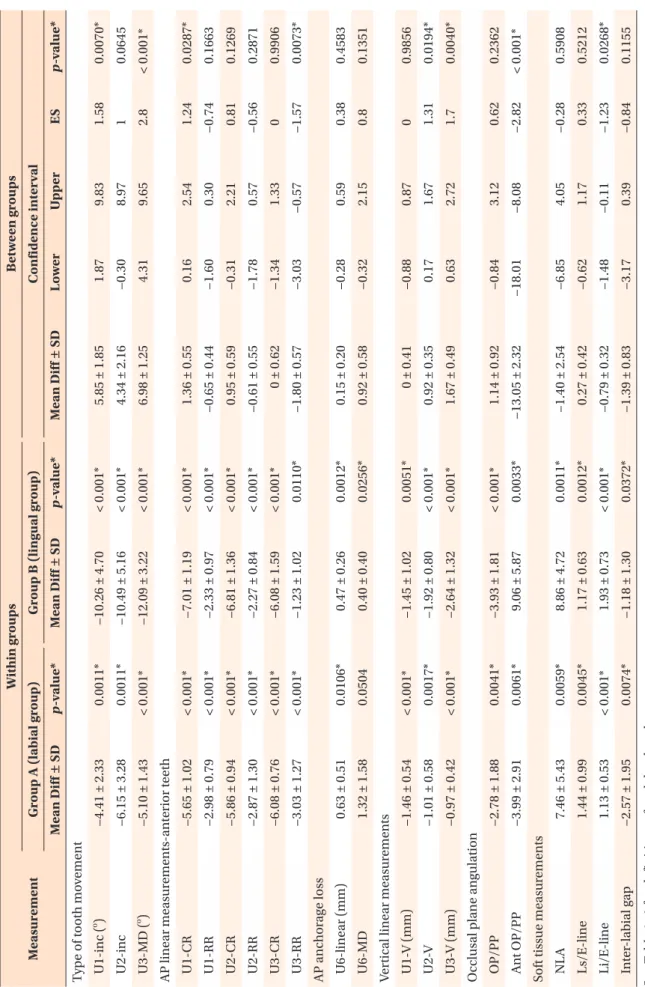
(9) www.e-kjo.org. U3-MD ( ). −2.98 ± 0.79. −5.86 ± 0.94. −2.87 ± 1.30. −6.08 ± 0.76. −3.03 ± 1.27. U1-RR. U2-CR. U2-RR. U3-CR. U3-RR. https://doi.org/10.4041/kjod.2019.49.6.381. 1.32 ± 1.58. U6-MD. −1.01 ± 0.58. −0.97 ± 0.42. U2-V. U3-V (mm). −3.99 ± 2.91. Ant OP/PP. 1.13 ± 0.53. Li/E-line 0.0074*. < 0.001*. 0.0045*. 0.0059*. 0.0061*. 0.0041*. < 0.001*. 0.0017*. < 0.001*. 0.0504. 0.0106*. < 0.001*. < 0.001*. < 0.001*. < 0.001*. < 0.001*. < 0.001*. < 0.001*. 0.0011*. 0.0011*. p -value*. −1.18 ± 1.30. 1.93 ± 0.73. 1.17 ± 0.63. 8.86 ± 4.72. 9.06 ± 5.87. −3.93 ± 1.81. −2.64 ± 1.32. −1.92 ± 0.80. −1.45 ± 1.02. 0.40 ± 0.40. 0.47 ± 0.26. −1.23 ± 1.02. −6.08 ± 1.59. −2.27 ± 0.84. −6.81 ± 1.36. −2.33 ± 0.97. −7.01 ± 1.19. −12.09 ± 3.22. −10.49 ± 5.16. −10.26 ± 4.70. Mean Diff ± SD. See Table 1–3 for definitions of each landmark or measurement. Mean Diff ± SD, Mean ± standard deviation; AP, anteroposterior; ES, effect size. *p < 0.05.. −2.57 ± 1.95. 1.44 ± 0.99. Ls/E-line. Inter-labial gap. 7.46 ± 5.43. NLA. Soft tissue measurements. −2.78 ± 1.88. OP/PP. Occlusal plane angulation. −1.46 ± 0.54. U1-V (mm). Vertical linear measurements. 0.63 ± 0.51. U6-linear (mm). AP anchorage loss. −5.65 ± 1.02. U1-CR. AP linear measurements-anterior teeth. −5.10 ± 1.43. −6.15 ± 3.28. U2-inc. o. −4.41 ± 2.33. Mean Diff ± SD. 0.0372*. < 0.001*. 0.0012*. 0.0011*. 0.0033*. < 0.001*. < 0.001*. < 0.001*. 0.0051*. 0.0256*. 0.0012*. 0.0110*. < 0.001*. < 0.001*. < 0.001*. < 0.001*. < 0.001*. < 0.001*. < 0.001*. < 0.001*. p -value*. Group B (lingual group). Within groups. Group A (labial group). U1-inc (o). Type of tooth movement. Measurement. Table 6. Descriptive statistics and intragroup and intergroup comparisons. −1.39 ± 0.83. −0.79 ± 0.32. 0.27 ± 0.42. −1.40 ± 2.54. −13.05 ± 2.32. 1.14 ± 0.92. 1.67 ± 0.49. 0.92 ± 0.35. 0 ± 0.41. 0.92 ± 0.58. 0.15 ± 0.20. −1.80 ± 0.57. 0 ± 0.62. −0.61 ± 0.55. 0.95 ± 0.59. −0.65 ± 0.44. 1.36 ± 0.55. 6.98 ± 1.25. 4.34 ± 2.16. 5.85 ± 1.85. Mean Diff ± SD. Between groups. −3.17. −1.48. −0.62. −6.85. −18.01. −0.84. 0.63. 0.17. −0.88. −0.32. −0.28. −3.03. −1.34. −1.78. −0.31. −1.60. 0.16. 4.31. −0.30. 1.87. Lower. 0.39. −0.11. 1.17. 4.05. −8.08. 3.12. 2.72. 1.67. 0.87. 2.15. 0.59. −0.57. 1.33. 0.57. 2.21. 0.30. 2.54. 9.65. 8.97. 9.83. Upper. Confidence interval. −0.84. −1.23. 0.33. −0.28. −2.82. 0.62. 1.7. 1.31. 0. 0.8. 0.38. −1.57. 0. −0.56. 0.81. −0.74. 1.24. 2.8. 1. 1.58. ES. 0.1155. 0.0268*. 0.5212. 0.5908. < 0.001*. 0.2362. 0.0040*. 0.0194*. 0.9856. 0.1351. 0.4583. 0.0073*. 0.9906. 0.2871. 0.1269. 0.1663. 0.0287*. < 0.001*. 0.0645. 0.0070*. p -value*. Sadek et al • Labial versus lingual biocreative therapy. 389.
(10) Sadek et al • Labial versus lingual biocreative therapy. Maxillary central incisor labial group. 30. B. Maxillary lateral incisor labial group. 30. C. 25. 20. 20. 20. Z-axis. 25. 15. 15. 15. 10. 10. 10. 5. 5. 5. 0. 0 65. 75 Y-axis. 85. Maxillary canine labial group. 30. 25. Z-axis. Z-axis. A. 0 60. 70 Y-axis. 80. 60. 70 Y-axis. 80. Figure 7. Changes in the axes of the maxillary anterior teeth after retraction in the labial group. Tooth axis graph. A, Upper central incisor; B, upper lateral incisor; C, upper canine (solid line, before retraction; dotted line, after retraction). Incisor, Midpoint of incisal edge to root apex; canine, cusp tip to root apex. Maxillary central incisor lingual group. 30. B. Maxillary lateral incisor lingual group. 30. C. 25. 20. 20. 20. Z-axis. 25. 15. 15. 15. 10. 10. 10. 5. 5. 5. 0. 0 60. 70. 80. 90. Maxillary canine lingual group. 30. 25. Z-axis. Z-axis. A. 0 60. 70. Y-axis. 80. 90. 60. 70. Y-axis. 80. 90. Y-axis. Figure 8. Changes of the axes of the maxillary anterior teeth after retraction in the lingual group. Tooth axis graph. A, Upper central incisor; B, upper lateral incisor; C, upper canine (solid line, before retraction; dotted line, after retraction). Incisor, Midpoint of incisal edge to root apex; canine, cusp tip to root apex. exactly equal amount of crown and root retraction for the central and lateral incisors was not achieved. Despite the many advantages of conducting a finite element study, the accuracy of such studies might be affected by major factors such that the results can only represent a theoretical scenario that cannot be achieved clinically; a clinical study is essential for confirmation. Remarkable tipping of the anterior segment during retraction was frequently reported by clinicians despite using forces and moments that were adjusted to achieve pure retraction of the anterior teeth. Furthermore, it was previously reported that the desired bodily movement of the anterior segment requires further correction of the force system, even though the biomechanical design employed should theoretically provide bodily movement of the segment.20 This is quite close to our experience in this study. Our. 390. results are based on a clinical study rather than a theoretical model and can be well applied in clinical practice, resulting in anterior retraction with good torque control. As for the lingual biocreative therapy group, the “vertical bowing effect” occurred during anterior retraction, with lingual tipping of the incisors and distal tipping and intrusion of the canines. The maxillary central and lateral incisors showed controlled tipping movement. The mean change in central incisor inclination was similar to that reported by Seo et al.,17 where a 10.65 ± 5.17o change in the labiolingual inclination of the central incisor occurred. Kim et al.12 found a decrease of 7.8o in the SN-U1 angle in the group where 10-mm long retraction hooks were used, while the group with shorter retraction hooks showed a decrease of 12.1o. Therefore, it can be recommended that for cases with flared incisors that will. https://doi.org/10.4041/kjod.2019.49.6.381. www.e-kjo.org.
(11) Sadek et al • Labial versus lingual biocreative therapy. need controlled lingual tipping, a retraction hook length of 10 mm should be used. On the other hand, a longer hook should be used if bodily tooth movement is desired. One important consideration is the morphology of the palatal vault. Furthermore, the exact location of the center of resistance of the anterior segment is affected by the morphology of alveolar bone and roots.21 In some challenging cases, we might need to resort to additional TADs or other complex biomechanics. This may warrant further study. As for anterior vertical control, similar to our results, Kim et al.11 reported a 0.63-mm intrusion of the maxillary central incisor using the labial technique and Seo et al.17 reported a 1.18 ± 2.04 mm intrusion for the central incisor using the lingual biocreative technique. The often-reported adverse effect of increasing the incisor display and creating a gummy smile, which is associated with anterior retraction in extraction, did not occur with these mechanics. Significant differences in the intrusion of the lateral incisor and canine were found between the two groups. The canine was significantly more intruded in the lingual group than in the labial group. Different measures could be applied for proper vertical control of the canine during retraction using the lingual biocreative technique. The use of anterior triangular elastics from buttons bonded to the maxillary canine and the lower arch was recommended.14 Jang et al.16 recommended using torqueing springs for canine vertical control. However, they reported that the length of the retraction hook and the resultant line of action of force were more important factors than the incorporation of the torqueing springs. Mo et al.14 recommended sectioning the canine from the anterior segment to allow individual control of the canine. At the end of the retraction phase, full fixed appliances had to be bonded to the maxillary and mandibual arches to continue the levelling and alignment phases, followed by finishing and settling of the occlusion. Similar to previous studies,11,12 good anchorage control was achieved in both groups. This is of paramount importance to maximize anterior retraction in patients with dentoalveolar protrusion and protrusive lips. Furthermore, both groups showed significant soft tissue changes following anterior retraction, with a significant increase in nasolabial angle and reduction of the interlabial gap and the linear distance from Labrale Superius and Labrale Inferius to E-Line. Kim et al.11 reported significant soft tissue changes using the labial technique, where the upper and lower lips to the E-line moved posteriorly 1.87 ± 0.91 mm and 2.75 ± 1.80 mm, respectively. Limitations Blinding of the patients and the operator to the type. www.e-kjo.org. https://doi.org/10.4041/kjod.2019.49.6.381. of intervention was impossible. Blinding of the outcome assessor was not possible during analysis of the CBCT scans because of the presence of the brackets in the labial group. Two patients were lost to follow-up in each group. However, the final sample size was equal to the sample size calculated and the number of patients analyzed was the same in both groups. The vertical positions of the miniscrews are different in the two techniques, and this might have had an impact on the final inclination of the incisors. This should be considered while interpreting the results. Generalizability This randomized clinical trial was performed at one center; thus, the generalizability of these findings is limited. However, this strengthens the standardization for all steps.. CONCLUSION Significant differences were found between the two groups regarding the type of tooth movement. The inclination of the incisors was reduced more in the lingual group (i.e., more lingual tipping). The canine showed significant distal tipping and intrusion in the lingual group. Using the labial biocreative therapy with a 10-mm anterior retraction hook and a reverse curve overlay, anterior retraction with good torque control, vertical control, and anchorage control were achieved. Using the lingual biocreative therapy with a 10-mm anterior retraction hook, anterior retraction occurred with controlled tipping movement. Clockwise rotation of the anterior segment occurred with significant distal tipping and intrusion of the canine. No statistically significant differences in the duration of the retraction phase were found between the two groups.. CONFLICTS OF INTEREST No potential conflict of interest relevant to this article was reported.. REFERENCES 1. Antoszewska-Smith J, Sarul M, Łyczek J, Konopka T, Kawala B. Effectiveness of orthodontic miniscrew implants in anchorage reinforcement during en-masse retraction: a systematic review and meta-analysis. Am J Orthod Dentofacial Orthop 2017;151:440-55. 2. Sung EH, Kim SJ, Chun YS, Park YC, Yu HS, Lee KJ. Distalization pattern of whole maxillary dentition. 391.
(12) Sadek et al • Labial versus lingual biocreative therapy. according to force application points. Korean J Orthod 2015;45:20-8. 3. Lee KJ, Park YC. The biomechanics of miniscrews: from single-tooth control to total-arch movement. In: Burstone CJ, Choy K, eds. The biomechanical foundation of clinical orthodontics. Chicago: Quintessence Publishing; 2015. p. 435. 4. Chung KR, Kim SH, Kook YA, Son JH. Anterior torque control using partial-osseointegrated miniimplants: biocreative therapy type I technique. World J Orthod 2008;9:95-104. 5. Chung KR, Kim SH, Kook YA, Choo H. Anterior torque control using partial-osseointegrated miniimplants: biocreative therapy type II technique. World J Orthod 2008;9:105-13. 6. Mo SS, Kim SH, Sung SJ, Chung KR, Chun YS, Kook YA, et al. Factors controlling anterior torque with C-implants depend on en-masse retraction without posterior appliances: biocreative therapy type II technique. Am J Orthod Dentofacial Orthop 2011;139:e183-99. 7. Burstone CJ. The segmented arch approach to space closure. Am J Orthod 1982;82:361-78. 8. Chung KR, Kook YA, Kim SH, Mo SS, Jung JA. Class II malocclusion treated by combining a lingual retractor and a palatal plate. Am J Orthod Dentofacial Orthop 2008;133:112-23. 9. Chung KR, Cho JH, Kim SH, Kook YA, Cozzani M. Unusual extraction treatment in Class II division 1 using C-orthodontic mini-implants. Angle Orthod 2007;77:155-66. 10. Chung KR, Jeong DM, Park HJ, Kim SH, Nelson G. Severe bidentoalveolar protrusion treated with lingual Biocreative therapy using palatal miniplate. Korean J Orthod 2010;40:276-87. 11. Kim SH, Hwang YS, Ferreira A, Chung KR. Analysis of temporary skeletal anchorage devices used for en-masse retraction: a preliminary study. Am J Orthod Dentofacial Orthop 2009;136:268-76. 12. Kim JS, Kim SH, Kook YA, Chung KR, Nelson G. Analysis of lingual en masse retraction combining a C-lingual retractor and a palatal plate. Angle Orthod 2011;81:662-9.. 392. 13. Mo SS, Kim SH, Sung SJ, Chung KR, Chun YS, Kook YA, et al. Factors controlling anterior torque during C-implant-dependent en-masse retraction without posterior appliances. Am J Orthod Dentofacial Orthop 2011;140:72-80. 14. Mo SS, Kim SH, Sung SJ, Chung KR, Chun YS, Kook YA, et al. Torque control during lingual anterior retraction without posterior appliances. Korean J Orthod 2013;43:3-14. 15. Jee JH, Ahn HW, Seo KW, Kim SH, Kook YA, Chung KR, et al. En-masse retraction with a preformed nickel-titanium and stainless steel archwire assembly and temporary skeletal anchorage devices without posterior bonding. Korean J Orthod 2014;44:23645. 16. Jang HJ, Roh WJ, Joo BH, Park KH, Kim SJ, Park YG. Locating the center of resistance of maxillary anterior teeth retracted by Double J Retractor with palatal miniscrews. Angle Orthod 2010;80:1023-8. 17. Seo KW, Kwon SY, Kim KA, Park KH, Kim SH, Ahn HW, et al. Displacement pattern of the anterior segment using antero-posterior lingual retractor combined with a palatal plate. Korean J Orthod 2015;45:289-98. 18. Tominaga JY, Ozaki H, Chiang PC, Sumi M, Tanaka M, Koga Y, et al. Effect of bracket slot and archwire dimensions on anterior tooth movement during space closure in sliding mechanics: a 3-dimensional finite element study. Am J Orthod Dentofacial Orthop 2014;146:166-74. 19. Sung SJ, Jang GW, Chun YS, Moon YS. Effective en-masse retraction design with orthodontic miniimplant anchorage: a finite element analysis. Am J Orthod Dentofacial Orthop 2010;137:648-57. 20. Bourauel C, Drescher D. Retraktion der oberen Schneidezähne mit pseudoelastischen Behandlungselementen- Rechnerische und biomechanische Prüfung sowie klinische Anwendung. J Orofac Orthop 1994;55:36-44. 21. Melsen B, Fotis V, Burstone CJ. Vertical force considerations in differential space closure. J Clin Orthod 1990;14:678-83.. https://doi.org/10.4041/kjod.2019.49.6.381. www.e-kjo.org.
(13)
수치


+4
관련 문서
The meeting was attended by Assistant Foreign Minister for GCC Affairs, Ambassador, Nasser Al-Muzayyen, and Deputy Assistant Foreign Minister for the Office of the
“ Sheikh Nasser has a written message from HH the Amir, Sheikh Sabah Al-Ahmad Al-Jaber Al-Sabah to the Chinese President, Chi Gen Beng related to enhancing mutual
On his part, CEO of Express Roads Authority, Saud Al-Naqqi said that the heavy rains of the previous day led to clogging parts of the express
Kuwait will celebrate on Sunday the fourth anniversary of the UN honoring and proclamation of His Highness the Amir, Sheikh Sabah Al-Ahmad Al-Jaber Al-Sabah as
The Joseon government designed and promulgated the Taegeukgi as a national flag for diplomatic and political purposes, but it was the independence movement that made it
• 이명의 치료에 대한 매커니즘과 디지털 음향 기술에 대한 상업적으로의 급속한 발전으로 인해 치료 옵션은 증가했 지만, 선택 가이드 라인은 거의 없음.. •
The proposal of the cell theory as the birth of contemporary cell biology Microscopic studies of plant tissues by Schleiden and of animal tissues by Microscopic studies of
Department of Internal Medicine, Novosibirsk State University, Russia 1 , Department of Cardiology, Surgut State University, Russia 2 , Department Fundamental
