http://e-nrp.org
Protective role of oligonol from oxidative stress-induced inflammation in C6 glial cell
Jae Hyun Ahn
1, Ji Won Choi
1, Ji Myung Choi
1, Takahiro Maeda
2, Hajime Fujii
2, Takako Yokozawa
3and Eun Ju Cho
1§1Department of Food Science and Nutrition, Pusan National University, Busandaehak-ro 63 beon-gil, Geumjeong-gu, Busan 609-735, Korea
2Amino Up Chemical Co., Ltd, Sapporo 004-0839, Japan
3Institute of Natural Medicine, University of Toyama, Toyama 930-0194, Japan
BACKGROUND/OBJECTIVES: Natural products or active components with a protective effect against oxidative stress have attracted significant attention for prevention and treatment of degenerative disease. Oligonol is a low molecular weight polyphenol containing catechin-type monomers and oligomers derived from Litchi chinensis Sonn. We investigated the protective effect and its related mechanism of oligonol against oxidative stress.
MATERIALS/METHODS: Oxidative stress in C6 glial cells was induced by hydrogen peroxide (H
2O
2) and the protective effects of oligonol on cell viability, nitric oxide (NO) and reactive oxygen species (ROS) synthesis, and mRNA expression related to oxidative stress were determined.
RESULTS: Treatment with oligonol inhibited NO and ROS formation under cellular oxidative stress in C6 glial cells. In addition, it recovered cell viability in a dose dependent-manner. Treatment with oligonol also resulted in down-regulated mRNA expression related to oxidative stress, nuclear factor kappa-B (NF-κB) p65, cyclooxygenase-2 (COX-2), and inducible nitric oxide synthase (iNOS), compared with the control group treated with H
2O
2.In particular, expression of NF-κB p65, COX-2, and iNOS was effectively reduced to the normal level by treatment with 10 μg/mL and 25 μg/mL of oligonol.
CONCLUSIONS: These results indicate that oligonol has protective activity against oxidative stress-induced inflammation. Oligonol might be a promising agent for treatment of degenerative diseases through inhibition of ROS formation and NF-κB pathway gene expression.
Nutrition Research and Practice 2015;9(2):123-128; doi:10.4162/nrp.2015.9.2.123; pISSN 1976-1457 eISSN 2005-6168
Keywords: Oligonol, C6cell, inflammation, nitric oxide, oxidative stress
INTRODUCTION
3)Oxidative stress and inflammatory process are mainly attributed to neurodegenerative diseases, including dementia, which has recently attracted significant attention. As brain cells die, interaction between neurons would be inhibited, resulting in impairment of memory and cognitive function, known as dementia [1,2]. Overproduction of free radicals leads to oxidative stress, which induces cells to go under an inflammation mechanism and lowers cell viability. In particular, nitric oxide (NO) reacts with membrane lipids to induce lipid peroxidation [3]. NO and superoxide (O
2-) combine to form a highly reactive intermediate, peroxynitrite (ONOO
-). ONOO
-induces DNA strand breaks, lipid peroxidation, and protein nitration. By overpro- duction of NO and generation of ONOO
-, glial cells would be damaged [4,5]. Injury of glial cells produces a variety of pro-inflammatory and neurotoxic factors, including nuclear factor kappa B (NF-κB), inducible nitric oxide synthases (iNOS), cycloxygenase-2 (COX-2), and several cytokines [3]. As above, this process can surely occur in C6 glial cells, in which the
oxidative damage can lead to dementia. Therefore, attenuation of oxidative stress and inhibition of inflammatory process in glial cells are crucial for prevention of neurodegenerative diseases.
Natural extract products have recently attracted significant attention for prevention and treatment of degenerative diseases. Litchi chinensis Sonn., a subtropical fruit, is widely cultivated in Africa, South-East Asia, such as China, Taiwan, Vietnam, and Japan etc. [6,7]. The fresh fruit has a sweet flavor and contains sugars, citric acid, vitamin C, and polyphenols [8].
It has traditionally been used as a tonic for the function of heart and liver and it has been reported to exhibit strong antioxidant activities [9,10]. It contains polyphenolic compounds such as condensed tannins, epicatechin, procyanidin A2, anthocyanin, quercetin 3-rutinoside (rutin), and quercetin glucoside [10,11].
Oligonol is a phenolic product derived from Litchi chinensis Sonn. extract containing catechin-type monomers and oligomers of proanthocyanidins [12]. Emerging evidence indicates that oligonol has some biological effects [13], including anticancer, antioxidant, and anti-inflammatory effects, as well as beneficial
§Corresponding Author: Eun Ju Cho, Tel. 82-51-510-2837, Fax. 82-51-583-3648, Email. [email protected] Received: May 26, 2014, Revised: August 21, 2014, Accepted: August 27, 2014
This is an Open Access article distributed under the terms of the Creative Commons Attribution Non-Commercial License (http://creativecommons.org/licenses/by-nc/3.0/) which permits unrestricted non-commercial use, distribution, and reproduction in any medium, provided the original work is properly cited.
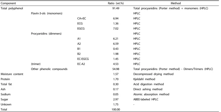
Component Ratio (wt.%) Method
Total polyphenol 91.49 Total procyanidins (Porter method) + monomers (HPLC)
Flavin-3-ols (monomers) HPLC
CA+EC 6.94 HPLC
ECG 1.36 HPLC
EGCG 7.02 HPLC
Procyanidins (dimmers) HPLC
A1 6.21 HPLC
A2 6.59 HPLC
B1 0.43 HPLC
B2 1.98 HPLC
EC-EGCG 1.45 HPLC
(trimer) EC-A2 4.53 HPLC
Other phenolic compounds 54.98 Total procyanidins (Porter method) - Dimers/Trimers (HPLC)
Moisture content 1.57 Decompressed drying method
Protein 1.70 Kjeldahl method
Total fat 0.30 Acid digestion method
Ash 0.17 Direct ashing method
Sodium 0.05 Atomic absorption method
Sugar 2.97 ABEE-labeled HPLC
Unkown 1.75 -
Total 100.00
CA: catechin, EC: epicatechin, EGC: epicatechin gallate, EGCG: epigallocatechin gallate, A1: procyanidinA1, A2: procyanidinA2, B1: procyanidinB1, B2: procyanidin B2.
Table 1. Oligonol components
activity for NO bioavailability and regulatory effect on lipid metabolism. However, study on the protective role of oligonol against oxidative stress in C6 glial cells has not yet been conducted. In the current study, we investigated the protective effect and its related mechanisms of oligonol against oxidative stress-induced inflammation in C6 glial cells.
MATERIALS AND METHODS Oligonol
Oligonol was provided by Amino Up Chemical Co., Ltd.
(Sapporo, Japan). Oligonol was produced from lychee fruit (Litchi chinensis Sonn.) extract using a patented technology process (international patent WO 2004/103988 AI) at Amino Up Chemical Co., Ltd. (Sapporo, Japan) [5]. Briefly, dried Litchi chinensis Sonn. was extracted with 50% [volume to volume (v/v)] ethanol. The filtrate was evaporated and passed through a DIAION HP-20 column, and eluted with ethanol. The eluate was then evaporated to dryness, yielding a dark brown powder.
It was heated at 60°C for 16 h, filtered through a DIAION HP-20 column, washed with water and eluted with 40% (v/v) ethanol.
Evaporation of the eluate yielded a reddish brown powder containing the monomeric and oligomeric proanthocyanidin mixture, oligonol. The characteristics of the oligonol preparation used in this study (Batch No. OLF1202S) are shown in Table 1 [12]. Oligonol was dissolved in dimethyl sulfoxide (DMSO) and diluted to an appropriate concentration in culture media.
Cell culture
C6 glial cells (Korean Cell Line Bank, Seoul, Korea) were maintained in a culture flask containing 10% fetal bovine serum supplemented with Dulbecco’s Modified Eagle’s Medium
(Welgene, Daegu, Korea) (pH 7.2) at 37°C in a humidified atmosphere of 5% CO
2in air. All subsequent procedures were performed under these conditions. The cells were subcultured with 0.05% trypsin-EDTA in phosphate buffer saline.
Induction of oxidative stress
After confluence had been reached, the cells were seeded into 96-well plates at 5 × 10
4cells/mL and allowed to adhere for 2 h. Next, cells were treated with sodium nitroprusside (SNP) (500 μM) or hydrogen peroxide (H
2O
2) (500 μM). After 24 h of incubation, samples were treated in the test wells at various concentrations for 24 h.
Cell viability
Cell viability was assessed using the MTT colorimetric assay.
MTT solution (5 mg/mL) was added to each 96-well culture plate, followed by incubation for 4 h at 37°C; the medium containing MTT was then removed. The incorporated formazan crystals in the viable cells were solubilized with 200 μL of DMSO, and, after 30 min, the absorbance of each well was read at 540 nm using a microplate reader (Model 680, Bio-Rad, Hercules, CA, USA).
NO level
The amount of NO production was assayed by measuring the
accumulation of nitrite, using a microplate assay method based
on the Griess reaction. Briefly, 100 μL of culture supernatant
was allowed to react with 100 μL of Griess reagent (Sigma-
Aldrich Co. St. Louis, MO, USA), followed by incubation at room
temperature for 15 min. The optical density of the samples was
measured at 540 nm using a microplate reader.
Fig. 1. NO formation of C6 cells treated with SNP. After treatment of C6 cells with SNP (500 μM) for 24 h, oligonol (5, 10, and 25 μg/mL) was added for 24 h, and NO formation was then measured by Griess reaction. Values are expressed as mean ± SD.
a-cMeans with different letters are significantly different (P< 0.05) by Duncan's multiple range test. Normal, SNP- and oligonol-non-treated cells; Control, SNP-treated cells.
Fig. 2. Effect of oligonol on viability of C6 glial cells treated with SNP. After treatment of C6 cells with SNP (500 μM) for 24 h, oligonol (5, 10, and 25 μg/mL) was added for 24 h, and cell viability was then measured by MTT assay. Values are expressed as mean ± SD. a-cMeans with different letters are significantly different (P< 0.05) by Duncan's multiple range test. Normal, SNP- and oligonol-non-treated cells; Control, SNP-treated cells.
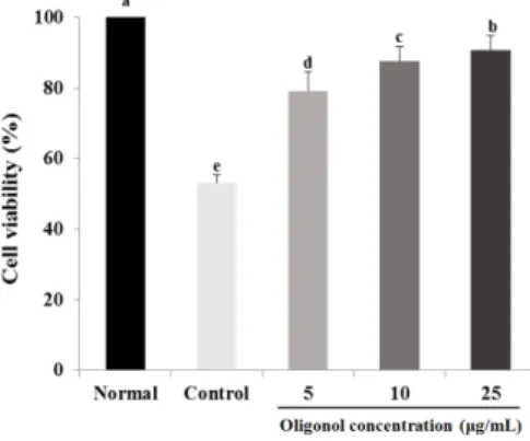
Fig. 3. Effect of oligonol on viability of C6 glial cells treated with H2O2. After treatment of C6 cells with H2O2 (500 μM) for 24 h, oligonol (5, 10, and 25 μg/mL) was added for 24 h, and cell viability was then measured by MTT assay. Values are expressed as mean ± SD. a-eMeans with different letters are significantly different (P< 0.05) by Duncan's multiple range test. Normal, H2O2- and oligonol-non-treated cells; Control, H2O2-treated cells.
Reactive oxygen species (ROS) generation
After 24 h of sample treatment, 2’,7’-dichlorofluorescein diacetate (DCFDA) (Sigma-Aldrich Co. St. Louis, MO, USA) (80 μM) was added, and the cells were incubated for 30 min at 37°C. Then, fluorescence intensity was measured at 485 nm excitation and 535 nm emission using a fluorescence spectrophotometer (FLUOstar OPTIMA, BMG Labtech, Ortenberg, Germany).
RNA extraction and Reverse transcription-polymerase chain reaction (RT-PCR)
Total RNA was isolated using Trizol reagent (Invitrogen Co., Carlsbad, CA, USA) by following the manufacturer’s methods.
The total RNA was digested with RNAse-free DNase (Roche, Indianapolis, IN, USA) for 15 min at 37°C and re-purified using the RNase kit according to the manufacturer’s protocol (Qiagen, La Jolla, CA, USA). cDNA was synthesized from 2 μg of total RNA by incubation at 37°C for 1 h with AM reverse transcriptase (Amersham, Arlington, USA) and random hexanucleotides according to the manufacturer’s instructions. The following primers were used to amplify the specific genes of interest:
forward, 5’-TTC-AAA-TCA-GAT-TCT-GGG-AAA-AT-3’ and reverse, 5’-AGA-TCA-TCT-CTG-CCT-GAG-TAT-CTT-3’ for the COX-2 gene;
forward, 5’-AGA-GAG-ATC-CGG-TTC-ACA-3’ and reverse, 5’-CAC- AGA-GCT-GAG-GGT-ACA-3’ for the iNOS gene; and forward, 5’-GCA-GCC-TAT-CAC-CAA-CTC-3’ and reverse, 5’-TAC-TCC-TTC- TTC-TCC-ACC-3’ for the NF-κB p65 gene.
Statistical analysis
The measurement data are expressed as mean ± SD. The data were examined using a one-way analysis of variance. Signi- ficance was verified by performing Duncan’s multiple test using SAS software (version 6.0, SAS Institute, Cary, NC, U.S.A.).
RESULTS
Protective effect of oligonol against oxidative stress induced by SNP
SNP was applied for induction of NO in C6 glial cells and the protective effects of oligonol were investigated. Fig. 1 shows that the control produced 10.34 μM of NO by SNP, two times
more than the normal group. However treatment with oligonol at a concentration of 5 and 10 μg/mL led to a decrease in formation of NO to 9 μM and 4.5 μM, respectively. This result indicated that SNP induced overproduction of NO, whereas oligonol significantly inhibited the production of NO. As shown in Fig. 2, the cell viability of the control group, which was only treated with SNP, was 28.23%, indicating that cells died as a result of oxidative stress induced by SNP. Treatment with oligonol resulted in a significant increase of cell viability in a concentration dependent-manner.
Protective effect of oligonol against oxidative stress induced by H
2O
2Fig. 3 shows the protective effects of oligonol on oxidative stress induced by H
2O
2. The cell viability of the control group showed a significant decrease to 43.2% compared to the normal group, with 100%. C6 glial cells were damaged by treatment with H
2O
2. However, treatment with 5 and 10 μg/mL concen- trations of oligonol led to an increase of cell viability to 79.2%
and 87.5%, respectively. In particular, at the concentration of
25 μg/mL, the cell viability was recovered to almost normal level.
Fig. 4. ROS formation of C6 cells treated with H2O2. After treatment of C6 cells with H2O2 (500 μM) for 24 h, oligonol (5, 10, and 25 μg/mL) was added for 24 h, and intracellular ROS was then measured by monitoring the fluorescence increase during 60 min (A) and presented fluorescence intensity at 60 min (B). Values are expressed as mean ± SD. a-bMeans with different letters are significantly different (P< 0.05) by Duncan's multiple range test. Normal, H2O2- and oligonol-non-treated cells; Control, H2O2-treated cells.
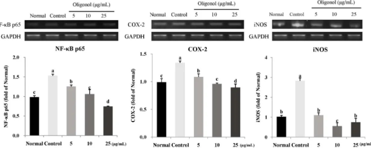
Fig. 5. Effect of oligonol on mRNA expression of NF-κB (A), COX-2 (B), and iNOS (C) under H2O2-induced oxidative stress in C6 glial cells. After treatment of C6 cells with H2O2 (500 μM) for 24 h, oligonol (5, 10, and 25 μg/mL) was added for 24 h. Total RNA was isolated and RT-PCR was performed using indicated primers. The amplified PCR products were run on a 1% agarose gel and visualized by EtBr staining. GAPDH was a house-keeping control gene. Fold ratio = mRNA expression/Normal group. a-dMeans with different letters are significantly different (P< 0.05) by Duncan's multiple range test. Normal, H2O2- and oligonol-non-treated cells; Control, H2O2-treated cells.
Fig. 4 shows the scavenging effect of ROS of oligonol determined by DCFDA assay. The control group showed a steady increase of ROS formation compared to the normal group. Based on these data we can predict that oxidative stress is induced by increases of ROS formation. However, treatment with oligonol at a concentration of 5, 10, and 25 μg/mL led to a decrease in ROS formation to 89.7%, 87.1%, and 86.4%, respectively. This result indicates that oligonol inhibited ROS formation in a concentration-dependent manner.
Protective mechanism of oligonol against oxidative stress To investigate the protective mechanism against oxidative stress and inflammation, we observed the effect of oligonol on mRNA expression of NF-κB p65, COX-2, and iNOS in H
2O
2treated C6 glial cells (Fig. 5). Control group treated with H
2O
2showed higher expression of mRNA in NF-κB p65 compared with the normal group. However, treatment with 5 and 10 μg/mL oligonol led to decreased expression of NF-κB p65. At a concentration higher than 10 μg/mL, NF-κB p65 mRNA expression was down-regulated to almost the normal level.
COX-2 mRNA expression showed a pattern form similar to that
of NF-κB p65. Treatment of glial cells also increased the expression of COX-2 mRNA. However, oligonol had a down- regulatory effect against overexpression of COX-2 mRNA. In addition, marked up-regulation of iNOS was observed by H
2O
2treatment, whereas oligonol induced a significant decrease in iNOS expression. Combining all results together, oligonol regulated mRNA expression related to oxidative stress and inflammation such as NF-κB p65, COX-2, and iNOS.
DISCUSSION
Oligonol is a low molecular weight polyphenol in Litchi chinensis Sonn. Oligonol contains catechin-type monomers and oligomers derived by a novel manufacturing process of the Litchi chinensis Sonn. [14]. Several studies have reported on the antioxidative and anti-inflammatory potential of oligonol [13,15].
These results support the capacity of oligonol in regulating the
pathological condition of chronic disease, such as cancer,
diabetes, cardiovascular disease, and neurodegenerative disorders
as well as aging progression. It has already been shown that
oligonol is absorbed from the intestine, and has a strong
antioxidative activity and anticancer effects. Based on these evidences, we could expect that oligonol might have protective effects against neurodegenerative diseases induced by oxidative stress. However, study on the protective effect of oligonol against oxidative stress in glial cells related to cognitive and memory impairment has not yet been conducted.
The number of people suffering from neurodegenerative disease is increasing. The search for functional food or bioactive compounds against neurodegenerative disease is crucial for prevention and treatment of neurodegenerative disease. The oxidative stress and inflammatory process induced by free radicals are the main cause of neurodegenerative disease. In particular, overproduction of NO causes damage to cells and is very important factor in neurodegenerative diseases. NO has several potential roles in the brain, as a neurotransmitter, a potential source of free radicals, and a potential mediator of inflammation [16,17]. Overproduction of NO can lead to react with membrane lipids to induce lipid peroxidation. Indirectly the combination of NO and O
2-can form highly reactive intermediates, such as ONOO
-, which can induce DNA strand breaks, lipid peroxidation, and protein nitration [18]. Injury of glial cells also occurred by NO through activation of microglia.
Activated microglia produce a variety of pro-inflammatory and neurotoxic factors, including cytokines, such as tumor necrosis factor-α (TNF-α) and interleukin-1β (IL-1β) [3,5,19,20]. In the current study, NO was induced by SNP in C6 glial cells and the effect of oligonol was investigated. C6 glial cells were used and oxidative stress was induced by SNP. As shown in Fig. 1, oligonol inhibited the production of NO, suggesting a protective role against NO-induced oxidative stress. As the concentration of oligonol increases, production of NO shows a progressive decrease, almost to the normal level. In addition, as shown in Fig. 2, treatment with oligonol resulted in elevation of cell viability compared with the control group. In this experiment we confirmed that SNP elevated NO production and inhibited cell growth. However, oligonol had a protective role against inflammation by inhibiting production of NO, resulting in elevation of cell viability.
H
2O
2is one of the reactive oxygen species (ROS) involved in oxidative stress. By the high accumulation of oxygen radicals and H
2O
2, protein changes occur in intracellular and intercellular signaling pathways, so that proteins become inactivated or damaged, resulting in cellular degeneration and death [21]. The current results also support that treatment of C6 glial cells with H
2O
2resulted in increased ROS production and decreased cell viability. However, oligonol inhibits ROS and it can protect brain cells from death induced by overproduction of ROS. By inhibiting inflammatory factors, oligonol can protect against glial cell death from oxidative stress and it would be related to improvement of memory and cognitive function. H
2O
2regulates expression of inflammatory factor genes such as NF-κ B, iNOS, and COX-2. NF-κB activation is essential for iNOS expression and NO regulates NF-κB at various points in its activation cascade. NO has a positive effect on NF-κB activation by affecting signaling cascades [22]. Activation of NF-κB culminates in translocation of NF-κB to the nucleus and an increase in gene expression such as iNOS and COX-2 [23-25].
iNOS has the ability to produce NO used during host defense.
However, overproduction of NO is also injurious to host cells, leading to neurotoxicity and disease. iNOS gene expression is regulated through transcriptional controls [26]. Chemical stimuli that regulate NF-κB activity can also modulate iNOS induction [27,28]. COX-2 is activated by inflammation inducible factor such as IL-1, TNF, LPS, and reactive oxygen intermediates [29]. COX-2 induces inflammation by increasing the formation of eicosanoids.
COX-2 increases the conversion of arachidonic acid into prosta- glandin E2 (PGE2), which sets up a negative feedback loop because PGE2 acts through cyclic AMP to inhibit transcription [17]. Which inactivates proteins or cells leading to death. In this study, we suggested the theory that oligonol might have an effect on inflammatory factor gene which is regulated by H
2O
2. Oxidative stress was induced in C6 glial cells by H
2O
2; however, oligonol had a protective effect on inflammatory factor genes.
Oligonol decreased expression of mRNA of NF-κB, iNOS, and COX-2. Several reports have also demonstrated the anti-inflam- matory effect of oligonol [30,31]. Oligonol decreased pro-inflam- matory cytokines (IL-1β, IL-6, TNF-α), COX-2 and iNOS in LPS-stimulated RAW 264.7 cells [30]. In addition, in the kidney of type 2 diabetic mice, inflammation-related genes and NF-κB transcription-stimulated AGE-RAGE interaction were down- regulated [31]. Together with this evidence, we suggest that oligonol regulates oxidative stress-induced inflammation.
The current results indicated that oligonol blocked the oxidative stress induced by H
2O
2and also caused a decrease in expression mRNA of inflammatory factor, which has a connection with H
2O
2mechanism. These results indicated that oligonol regulated the mechanisms related to oxidative stress and inflammation, eventually leading to elevation of cell viability.
Although further study on the protective mechanism of oligonol from oxidative stress is needed in order to support these findings, the current study suggests the promising role of oligonol as an antioxidant against oxidative stress in glial cells related to cognitive impairment.
REFERENCES
1. Coyle JT, Puttfarcken P. Oxidative stress, glutamate, and neurode- generative disorders. Science 1993;262:689-95.
2. Tuppo EE, Arias HR. The role of inflammation in Alzheimer's disease.
Int J Biochem Cell Biol 2005;37:289-305.
3. Liu B, Hong JS. Role of microglia in inflammation-mediated neuro- degenerative diseases: mechanisms and strategies for therapeutic intervention. J Pharmacol Exp Ther 2003;304:1-7.
4. Cirino G, Distrutti E, Wallace JL. Nitric oxide and inflammation.
Inflamm Allergy Drug Targets 2006;5:115-9.
5. Pahan K, Raymond JR, Singh I. Inhibition of phosphatidylinositol 3-kinase induces nitric-oxide synthase in lipopolysaccharide- or cytokine-stimulated C6 glial cells. J Biol Chem 1999;274:7528-36.
6. Jiang G, Lin S, Wen L, Jiang Y, Zhao M, Chen F, Prasad KN, Duan X, Yang B. Identification of a novel phenolic compound in litchi (Litchi chinensis Sonn.) pericarp and bioactivity evaluation. Food Chem 2013;136:563-8.
7. Wang L, Lou G, Ma Z, Liu X. Chemical constituents with antioxidant activities from litchi (Litchi chinensis Sonn.) seeds. Food Chem 2011;126:1081-7.
8. Ong PK, Acree TE. Similarities in the aroma chemistry of Gewür-
ztraminer variety wines and lychee (Litchi chinesis sonn.) fruit. J Agric Food Chem 1999;47:665-70.
9. Yang B, Wang J, Zhao M, Liu Y, Wang W, Jiang Y. Identification of polysaccharides from pericarp tissues of litchi (Litchi chinensis Sonn.) fruit in relation to their antioxidant activities. Carbohydr Res 2006;341:634-8.
10. Bhoopat L, Srichairatanakool S, Kanjanapothi D, Taesotikul T, Thananchai H, Bhoopat T. Hepatoprotective effects of lychee (Litchi chinensis Sonn.): a combination of antioxidant and anti-apoptotic activities. J Ethnopharmacol 2011;136:55-66.
11. Sarni-Manchado P, Le Roux E, Le Guernevé C, Lozano Y, Cheynier V. Phenolic composition of litchi fruit pericarp. J Agric Food Chem 2000;48:5995-6002.
12. Fujii H, Nishioka H, Wakame K, Magnuson BA, Roberts A. Acute, subchronic and genotoxicity studies conducted with Oligonol, an oligomerized polyphenol formulated from lychee and green tea extracts. Food Chem Toxicol 2008;46:3553-62.
13. Noh JS, Park CH, Yokozawa T. Treatment with oligonol, a low-molecular polyphenol derived from lychee fruit, attenuates diabetes-induced hepatic damage through regulation of oxidative stress and lipid metabolism. Br J Nutr 2011;106:1013-22.
14. Kundu JK, Chang EJ, Fujii H, Sun B, Surh YJ. Oligonol inhibits UVB-induced COX-2 expression in HR-1 hairless mouse skin--AP-1 and C/EBP as potential upstream targets. Photochem Photobiol 2008;84:399-406.
15. Zhang XH, Yokoo H, Nishioka H, Fujii H, Matsuda N, Hayashi T, Hattori Y. Beneficial effect of the oligomerized polyphenol oligonol on high glucose-induced changes in eNOS phosphorylation and dephosphorylation in endothelial cells. Br J Pharmacol 2010;159:
928-38.
16. Ialenti A, Ianaro A, Moncada S, Di Rosa M. Modulation of acute inflammation by endogenous nitric oxide. Eur J Pharmacol 1992;
211:177-82.
17. Surh YJ, Chun KS, Cha HH, Han SS, Keum YS, Park KK, Lee SS.
Molecular mechanisms underlying chemopreventive activities of anti-inflammatory phytochemicals: down-regulation of COX-2 and iNOS through suppression of NF-κB activation. Mutat Res 2001;480- 481:243-68.
18. Dobashi K, Pahan K, Chahal A, Singh I. Modulation of endogenous antioxidant enzymes by nitric oxide in rat C6 glial cells. J
Neurochem 1997;68:1896-903.
19. Garcion E, Sindji L, Montero-Menei C, Andre C, Brachet P, Darcy F. Expression of inducible nitric oxide synthase during rat brain inflammation: regulation by 1,25-dihydroxyvitamin D3. Glia 1998;22:
282-94.
20. Syapin PJ, Militante JD, Garrett DK, Ren L. Cytokine-induced iNOS expression in C6 glial cells: transcriptional inhibition by ethanol.
J Pharmacol Exp Ther 2001;298:744-52.
21. Peterson LJ, Flood PM. Oxidative stress and microglial cells in Parkinson's disease. Mediators Inflamm 2012;2012:401264.
22. McCafferty DM, Mudgett JS, Swain MG, Kubes P. Inducible nitric oxide synthase plays a critical role in resolving intestinal inflam- mation. Gastroenterology 1997;112:1022-7.
23. Aktan F. iNOS-mediated nitric oxide production and its regulation.
Life Sci 2004;75:639-53.
24. Grandison L, Nolan GP, Pfaff DW. Activation of the transcription factor NF-κB in GH3 pituitary cells. Mol Cell Endocrinol 1994;106:
9-15.
25. O'Neill LA, Kaltschmidt C. NF-κB: a crucial transcription factor for glial and neuronal cell function. Trends Neurosci 1997;20:252-8.
26. Gookin JL, Chiang S, Allen J, Armstrong MU, Stauffer SH, Finnegan C, Murtaugh MP. NF-κB-mediated expression of iNOS promotes epithelial defense against infection by Cryptosporidium parvum in neonatal piglets. Am J Physiol Gastrointest Liver Physiol 2006;290:
G164-74.
27. Korhonen R, Lahti A, Kankaanranta H, Moilanen E. Nitric oxide production and signaling in inflammation. Curr Drug Targets Inflamm Allergy 2005;4:471-9.
28. Kubes P, McCafferty DM. Nitric oxide and intestinal inflammation.
Am J Med 2000;109:150-8.
29. Kidd BL, Urban LA. Mechanisms of inflammatory pain. Br J Anaesth 2001;87:3-11.
30. Yum HW, Zhong X, Park J, Na HK, Kim N, Lee HS, Surh YJ. Oligonol inhibits dextran sulfate sodium-induced colitis and colonic adenoma formation in mice. Antioxid Redox Signal 2013;19:102-14.
31. Noh JS, Kim HY, Park CH, Fujii H, Yokozawa T. Hypolipidaemic and antioxidative effects of oligonol, a low-molecular-weight polyp- henol derived from lychee fruit, on renal damage in type 2 diabetic mice. Br J Nutr 2010;104:1120-8.