9 DOI: 10.4196/kjpp.2011.15.1.9
ABBREVIATIONS: DC, 1-docosanoyl cafferate; LPS, lipopolysaccha- ride; TNF-α, tumor necrosis factor α; IL-1β, interleukin 1β;
NF-kB, nuclear factor kappa B; IKB, inhibitor of NF-kB; iNOS, inducible nitric oxide synthase; NO, nitric oxide; CAPE, caffeic acid phenylethyl ester.
Received November 19, 2010, Revised January 17, 2011, Accepted January 17, 2011
Corresponding to: Wanjoo Chun, Department of Pharmacology, College of Medicine, Kangwon National University, Hyoja 2-dong, Chuncheon 200-701, Korea. (Tel) 82-33-250-8853, (Fax) 82-33- 255-8809, (E-mail) [email protected]
#These authors contributed equally to this work and should be considered co-first authors.
Anti-inflammatory Activity of 1-docosanoyl Cafferate Isolated from Rhus verniciflua in LPS-stimulated BV2 Microglial Cells
Jae-Won Lee1,#, Il-Young Cheong2,#, Hae-Sung Kim1, Jae Jun Lee1, Yong-Suk Lee2,5, Yong-Soo Kwon3, Myong-Jo Kim4, Hee Jae Lee1, Sung-Soo Kim1, and Wanjoo Chun1
Departments of 1Pharmacology, 2Anesthesiology, College of Medicine, 3College of Pharmacy, 4Division of Bio-resources Technology, Kangwon National University, Chuncheon 200-701, 5Korea Institute of Radiological and Medical Sciences, Seoul 139-706, Korea
Although various derivatives of caffeic acid have been reported to possess a wide variety of biological activities such as protection of neuronal cells against excitotoxicity, the biological activity of 1-docosanoyl cafferate (DC) has not been examined. The objective of the present study was to evaluate the anti-inflammatory effects of DC, isolated from the stem bark of Rhus verniciflua, on lipopoly- saccharide (LPS)-stimulated BV2 microglial cells. Pretreatment of cells with DC significantly attenuated LPS-induced NO production, and mRNA and protein expression of iNOS in a concentration- dependent manner. DC also significantly suppressed LPS-induced release of cytokines such as TNF-α and IL-1β . Consistent with the decrease in cytokine release, DC dose-dependently and significantly attenuated LPS-induced mRNA expression of these cytokines. Furthermore, DC significantly sup- pressed LPS-induced degradation of IKB, which retains NF-kB in the cytoplasm. Therefore, nuclear translocation of NF-kB induced by LPS stimulation was significantly suppressed with DC pretreat- ment. Taken together, the present study suggests that DC exerts its anti-inflammatory activity through the suppression of NF-kB translocation to the nucleus.
Key Words: 1-Docosanoyl cafferate (DC), BV2 microglial cells, Lipopolysaccharide, Cytokines, iNOS, NF-kB
INTRODUCTION
Microglia, resident macrophages and immune surveil- lance cells in the central nervous system (CNS), have been reported to play a critical role in host defense and tissue repair in the CNS [1]. However, microglia have also been proposed to play a pathogenic role in neuroinflammatory conditions such as ischemia [2], multiple sclerosis [3], Parkinson’s disease [4], Alzheimer’s disease [5] and HIV-as- sociated dementia [6]. It has been suggested that during these immunologically mediated CNS diseases, activated microglia contribute to neurodegenerative process by pro- ducing excessive amounts of proinflammatory molecules such as nitric oxide (NO) and several cytokines [7-10].
Therefore, novel pharmacological agents that can suppress microglia-induced overproduction of inflammatory media- tors might provide a promising strategy for treating in- flammation-related CNS diseases.
Rhus verniciflua, a deciduous tree in the anacardiaceae family, exhibits a variety of biological activities such as an- tioxidant [11,12], anti-microbial [13], anti-cancer [14,15],
and anti-platelet properties [16]. However, the chemical components of Rhus verniciflua and their biological proper- ties have not been fully elucidated. Previously, we reported that stigmasterols, which were isolated from Rhus vernici- flua, were neuroprotective against kainic acid-induced ex- citotoxicity [17]. To further isolate and identify biologically active compounds from Rhus verniciflua, 1-docosanoyl caff- erate (DC) was purified from the plant's stem bark.
DC is an ester derivative of caffeic acid, a simple phenyl- propanoid that possesses a wide variety of biological activ- ities: it has antioxidant and anti-inflammatory properties [18] and is neuroprotective against Aβ-induced neuro- toxicity [19]. Ester derivatives of caffeic acid also exhibit various biological activities [20]. For example, caffeic acid phenylethyl ester (CAPE), an antioxidant component of propolis, protects against glutamate-induced neurotoxicity [21] and cerebral ischemia [22]. Although DC was pre- viously identified in the root of Sohora subprostrata [23], its biological activity was never demonstrated.
In the present study, we investigated the anti-inflam- matory activity of DC isolated from the stem bark of Rhus verniciflua. We studied a possible underlying mechanism by which DC exerts its anti-inflammatory action in LPS-stimulated BV2 microglial cells.
Fig. 1. Chemical structure of 1-docosanoyl cafferate (DC).
METHODS Reagents and cell culture
Bacterial lipopolysaccharide (LPS, E. coli serotype 055:
B5) was purchased from Sigma-Aldrich (St. Louis, MO, USA). DC was isolated and identified from the stem bark of Rhus verniciflua (Fig. 1). The compound was dissolved in ethanol and added to the cell culture at the desired concentrations. The BV2 microglia cell line was maintained in Dulbecco's modified Eagle's medium (DMEM; Gibco BRL, Grand Island, NY, USA) supplemented with 5% heat-in- activated fetal bovine serum (FBS; Gibco BRL) and 50μg/
ml gentamicin (Sigma, St. Louis, MO, USA). Cells were maintained in a humidified incubator at 37oC with 5% CO2. In all experiments, cells were incubated in the presence of the indicated concentration of DC before the addition of LPS (200 ng/ml).
Cell viability
Cell viability was determined by 3-(4,5-dimethylthiazol- 2-yl)-2,5-diphenyltetrazolium bromide (MTT) assay. BV2 microglial cells were plated into 96-well plates at a density of 5×10⁴ cells per well for 24 hr. BV2 microglial cells were incubated with various concentrations of DC for 24 hr. MTT (0.5 mg/ml in PBS) was added to each well and the cells were incubated for 3 hr at 37oC. The resulting formazan crystals were dissolved in dimethyl sulfoxide (DMSO). The optical density was measured at 540 nm. The results were expressed as a percentage of surviving cells over control cells.
Nitrite quantification assay
The production of NO was estimated by measuring the amount of nitrite, a major stable metabolite of NO, using the Griess reagent (0.1% naphthylethylenediamine dihy- drochloride and 1% sulfanilamide in 5% phosphoric acid).
DC-pretreated BV2 microglial cells were stimulated with LPS in 12-well plates for 24 hr, and then 100μl of each culture medium was mixed with an equal volume of Griess reagent. The absorbance at 540 nm was measured on a mi- croplate reader. The results were expressed as a percentage of released NO from LPS-stimulated BV2 cells. To prepare a standard curve, sodium nitrite was diluted in culture me- dium to the desired concentrations.
TNF-α and IL-1β ELISA
BV2 microglia cells were treated with DC in the absence or presence of LPS. After 24 hr incubation, TNF-α and IL- 1β levels in culture media were quantified using mono- clonal anti-IL-1β or anti-TNF-α antibody according to the manufacturer’s instruction (R & D Systems).
Western blot analysis
BV2 microglial cells were washed twice with PBS and lysed with lysis buffer [50 mM Tris-HCl, pH 8.0, 150 mM NaCl, 0.02% sodium azide, 0.1% sodium dodecyl sulfate (SDS), 1% NP-40, 0.5% sodium deoxycholate, 1 mM phenyl methylsulfonyl fluoride]. Equal amounts of protein were separated on 10% SDS-polyacrylamide gel. Proteins were transferred to Hypond PVDF membranes (Amersham Biosciences, Piscataway, NJ, USA). The membranes were blocked using 5% skim milk in TBST for 1 hr at room tem- perature and sequentially incubated with an appropriate antibody: anti-iNOS (BD Bioscience, Franklin Lakes, NJ), anti-IkB-α (Santa Cruz Biotechnology Inc., Eugene, OR, USA), or anti-β-actin (Sigma, St. Louis, MO, USA). After thoroughly washing with TBST, the membranes were washed three times with TBST and incubated with HRP- conjugated goat anti-rabbit IgG (Jackson ImmunoResearch) or HRP-conjugated goat anti-mouse IgG (Jackson Immuno- Research) for 2 hr at room temperature. The blots were de- veloped using enhanced chemiluminescence detection kits (ECL Amersham Biosciences, Piscataway, NJ, USA).
Immunofluorescence assay
The effect of DC on the translocation to the nucleus of the p65 subunit of NF-kB was examined by immuno- fluorescence assay using confocal microscopy. BV2 micro- glial cells were cultured on? under? sterile coverslips, pre- treated with DC for 1 hr, and stimulated with LPS. At 30 min after the LPS treatment, the cells were fixed in 4%
paraformaldehyde for 20 min at room temperature. The fixed cells were then permeabilized with 0.1% Triton X-100 in PBS and blocked with 3% BSA. Afterwards, the cells were sequentially incubated with rabbit p65 antibody (Santa Cruz Biotechnology Inc., Eugene, OR, USA) at room temperature and Alexa 546-labeled goat anti-rabbit IgG (Molecular Probes, Eugene, OR, USA) at room temperature for 1 hr. Cells were stained with 0.5μg/ml of Hoechst stain- ing solution (Hoechst 33258) for 10 min at room tempera- ture. After washing with PBS, the samples were mounted and observed by confocal microscopy.
RNA isolation and quantitative real time RT-PCR Total RNA was isolated using the Total RNA Extraction Kit (iNtRON Biotechnology, Inc, USA) according to the manufacturer's instruction. Total RNA (2μg) obtained from cells was reverse-transcribed using oligo-(dT) 15 primers (Promega, Madison, WI, USA). The cDNA products were used immediately for SYBR (Takara, JAPAN) real time RT-PCR using primers specific for iNOS, TNF-α, IL-1β and β-actin (BIO NEER, KOREA). Quantitative changes in mRNA levels were estimated by real time PCR using the following cycling conditions: 35 cycles of denaturation at 94oC for 10 sec; annealing at 61oC for 15 sec; and ex-
A B
C D
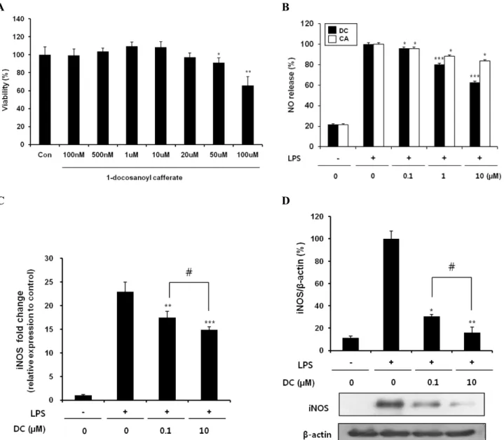
Fig. 2. Effects of DC on NO production and iNOS expression in LPS-stimulated BV2 microglial cells. (A) Effect of DC on the viability of BV2 microglial cells. No noticeable cell death was observed up to 20μM. (B) Concentration-dependent suppression of LPS-induced NO production by DC and CA (caffeic acid). (C) Inhibitory effect of DC on LPS-induced upregulation of iNOS mRNA expression. (D) Suppression of LPS-induced iNOS protein expression by DC: top, quantitative analysis of immunoblots; bottom, representative immunoblot of iNOS.
β-Actin was used as an internal control. Quantitative data represent three independent experiments and are expressed as mean±SD.
*p<0.05, **p<0.01, and ***p<0.001 indicate statistically significant differences compared to LPS alone. #p<0.05 indicate statistically significant differences between the indicated groups.
tension at 72oC for 20 sec; followed by 2 min at 72oC, in the presence of SYBR Green (1:10,000 dilution of a stock solution from Molecular Probes) carried out in a 20μL re- action volume.
Statistical analysis
All values shown in the figures are expressed as mean±
SD for at least three independent experiments. Statistical analysis was carried out by one-way analysis of variance (ANOVA) with Tukey’s post-hoc test using SPSS software 12K (SPSS, Chicago, IL, USA). A value of p<0.05 was con- sidered statistically significant.
RESULTS
1-Docosanoyl cafferate (DC) inhibits NO production and iNOS expression in LPS-stimulated BV2 cells The effects of DC on NO production and iNOS expression were examined in LPS-stimulated BV2 microglial cells. No noticeable cell death was observed in the concentration range of DC used, although higher concentrations of DC appeared to be cytotoxic (Fig. 2A). Treatment of LPS re- sulted in excessive production of NO and up-regulation of iNOS as indicated by increases in both mRNA and protein levels (Fig. 2B∼D). Pretreatment of cells with DC sig-
A B
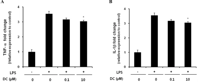
Fig. 3. Inhibitory effects of DC on LPS-induced release of TNF-α (A) and IL-1β (B) in BV2 microglial cells. BV2 microglia cells were incubated with 200 ng/ml of LPS in the presence or absence of the indicated concentrations of DC for 24 hr. Cell culture media were collected and subjected to TNF-α and IL-1β sandwich ELISAs. Data represent three independent experiments, each run in triplicate, and are expressed as mean±SD. *p<0.05 and **p<0.01 indicate statistically significant differences compared to LPS alone. ##p<0.01 indicates a statistically significant difference between the indicated groups.
A B
Fig. 4. Effects of DC on the gene expression of TNF-α (A) and IL-1β (B) in LPS-stimulated BV2 microglial cells. Cells were incubated with DC for 1 hr prior to exposure to 200 ng/ml LPS. Total RNA was isolated 6 hr after LPS treatment. TNF-α and IL-1β mRNA levels were determined by real time PCR. Data represent three independent experiments, each done in triplicate, and are expressed as mean±SD.
*p<0.05 indicates a statistically significant difference compared to LPS alone.
nificantly suppressed LPS-induced NO production in a con- centration-dependent manner (Fig. 2B). Caffeic acid also caused a significant suppression of LPS-induced NO production. However, DC elicited a more robust attenuation compared to caffeic acid, presumably due to the increased lipophilicity of DC. Consistent with the decrease in NO pro- duction, DC also significantly attenuated LPS-induced up-regulation of iNOS expression - including both iNOS mRNA (Fig. 2C) and iNOS protein levels (Fig. 2D). DC in- hibited LPS-induced NO production and iNOS expression without causing cell toxicity.
DC attenuates the expression of pro-inflammatory cytokines in LPS-stimulated BV2 cells
To determine the effect of DC on the expression of pro-in- flammatory cytokines such as TNF-α and IL-1β, extra- cellular release and mRNA expression of these cytokines were examined using an ELISA assay and quantitative RT-PCR, respectively. DC significantly suppressed LPS-in- duced extracellular release of TNF-α and IL-1β (Fig. 3).
Extracellular release of IL-1β was almost completely sup-
Fig. 5. Inhibitory effects of DC on LPS-induced IkB-α degradation in BV2 microglial cells. The intracellular level of IKB-α was deter- mined using immunoblotting analysis: top, quantitative analysis of immunoblots; bottom, representative immunoblot of IKB-α. β -Actin was used as an internal control. Quantitative data represent three independent experiments and are expressed as mean±SD. *p
<0.05 and **p<0.01 indicate statistically significant differences compared to LPS alone. ##p<0.01 indicates a statistically signifi- cant difference between the indicated groups.
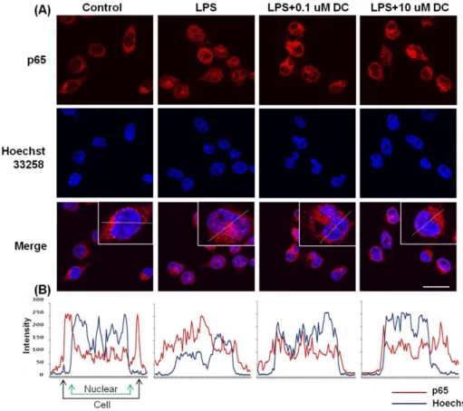
Fig. 6. Blockade by DC of nuclear translocation of the p65 subunit of NF-kB in LPS-stimulated BV2 microglial cells. (A) Localization of the NF-kB p65 subunit was deter- mined using a p65 antibody and an Alexa 546-labeled goat anti-rabbit IgG antibody. Nuclei were visualized by Hoechst staining (Hoechst 33258).
In basal conditions, immunostaining of p65 subunit was diffuse through- out the cytoplasm. LPS stimulation resulted in the translocation of p65 subunits into the nucleus. Pretreat- ment with DC attenuates LPS- induced nuclear translocation of the p65 subunit. (B) Line scanning analysis of confocal images further visualizes the intracellular locali- zation of the p65. Scale bar, 20μm.
pressed at a DC concentration of 10μM. The inhibition by DC was attributable to the attenuated expression of these genes as DC decreased mRNA levels of these cytokines (Fig.
4).
DC suppresses LPS-induced degradation of IKB and nuclear translocation of NF-kB
Given the previous report that the transcription factor NF-kB is the predominant regulator of numerous in- flammatory cytokine genes and that IKB inhibits the nu- clear translocation of NF-kB by retaining it in the cyto- plasm [24,25], we examined the effects of DC on LPS-in- duced nuclear translocation of NF-kB and degradation of cytosolic IKB. LPS significantly decreased intracellular lev- els of IKB; pretreatment with DC significantly attenuated this effect of LPS (Fig. 5). To further determine whether the change in the level of IKB affects the nuclear trans- location of NF-kB, we used confocal microscopy. Immuno- staining of the RelA (p65) subunit of NF-kB was present predominantly in the cytoplasm under basal conditions (Fig. 6). LPS increased nuclear translocation of the p65 sub- unit of NF-kB. Pretreatment with DC attenuated the effect of LPS (Fig. 6). Line scanning analysis of confocal images clearly demonstrated the intensity of the intracellular local- ization of the p65 subunit (Fig. 6B).
DISCUSSION
In the present study, we demonstrated that DC isolated from the stem bark of Rhus verniciflua possesses anti-in-
flammatory activity in LPS-stimulated BV2 microglial cells. DC significantly suppressed LPS-induced increases in the production of NO, upregulation of iNOS expression, and release of cytokines such as TNF-α and IL-1β. Further- more, DC significantly attenuated LPS-induced IKB degra- dation and subsequent nuclear translocation of NF-kB.
Numerous ester derivatives of caffeic acid such as CAPE [21], chlorogenic acid [26], pedicularioside A [27], forsytho- side B [28], and echinachoside [29] exhibit a wide range of biological activities including antioxidant and anti-in- flammatory properties, inhibition of caspase-3, and main- tenance of mitochondrial function. However, ester deriva- tives of caffeic acid with high molecular weight aliphatic alcohols such as DC and 1-eicosanoyl cafferate have rarely been isolated. DC was isolated from sophora subprostrata [23] and 1-eicosanoyl cafferate from Echinosophora kore- ensis [30]. Furthermore, their biological activities have not been demonstrated. High molecular fatty alcohols in their chemical structures are thought to increase the lipid sol- ubility of the compounds, leading to enhanced penetration of the compounds through the plasma membrane and into cells. In the present study, DC exhibited a significant an- ti-inflammatory activity in the micromolar range, suggest- ing that DC might efficiently enter cells due to its high lipid solubility. Although DC was not first isolated from Rhus verniciflua, the present study demonstrated its anti-in- flammatory activity for the first time.
Aberrant activation of microglia contributes to neuronal damage in several pathologic conditions by releasing proin- flammatory cytokines and oxidants such as TNF-α, inter- leukin (IL)-6, IL-lβ, IL-6, IL-10, IL-12, interferon-γ, and NO [8,31]. Not surprisingly, then, suppression of microglial activation and subsequent release of cytokines protected neuronal damage in several inflammation-related CNS dis- eases [2,32-34]. In the present study, DC significantly atte- nuated LPS-induced NO production through the sup- pression of iNOS expression. In addition, DC suppressed the LPS-induced increases in the release of TNF-α and IL- lβ and in their gene expression. To our knowledge, effects of DC on other proinflammatory cytokines have not been reported. Therefore, further studies will be necessary to ex- amine the effect of DC on the release and gene expression of other cytokines.
The inappropriate regulation of NF-kB and its down- stream genes has been associated with various pathological conditions including cancer and autoimmune diseases [35,36]. For example, NF-kB-dependent microglial activa- tion was a crucial contributor to ischemia [37]. Consistent with these reports, DC significantly attenuated LPS-in- duced nuclear translocation of NF-kB and subsequent re- lease of proinflammatory cytokines in the present study.
However, it has also been suggested that NF-kB is an- ti-apoptotic [38] and that activation of NF-kB can provide neuroprotection against amyloid β-induced toxicity [39]
and against excitotoxic or oxidative stress [40,41]. Overall, these studies suggest diverse functions for NF-kB in the nervous system depending on the cellular context.
In conclusion, our results demonstrate that DC exerts an- ti-inflammatory activity such as suppression of NO pro- duction and cytokine release by inhibiting nuclear trans- location of NF-kB in LPS-stimulated BV2 microglial cells.
This suggests that DC might be a valuable therapeutic agent in the treatment of inflammation-related brain path- ologies such as ischemia and neurodegenerative diseases.
ACKNOWLEDGEMENTS
This work was supported by the Korea Research Founda- tion Grant funded by the Korean Government (MOEHRD, Basic Research Promotion Fund) (KRF-2008-E00028).
REFERENCES
1. Perry VH, Gordon S. Macrophages and microglia in the nervous system. Trends Neurosci. 1988;11:273-277.
2. Hailer NP. Immunosuppression after traumatic or ischemic CNS damage: it is neuroprotective and illuminates the role of microglial cells. Prog Neurobiol. 2008;84:211-233.
3. Matsumoto H. Some markers reflecting the pathology and disease activity of multiple sclerosis. No To Shinkei. 1992;44:
95-102.
4. McGeer PL, McGeer EG. Glial cell reactions in neurodegene- rative diseases: pathophysiology and therapeutic interventions.
Alzheimer Dis Assoc Disord. 1998;12 Suppl 2:S1-6.
5. Itagaki S, McGeer PL, Akiyama H, Zhu S, Selkoe D. Relation- ship of microglia and astrocytes to amyloid deposits of Alzhei- mer disease. J Neuroimmunol. 1989;24:173-182.
6. Merrill JE, Chen IS. HIV-1, macrophages, glial cells, and cytokines in AIDS nervous system disease. FASEB J. 1991;5:
2391-2397.
7. Chao CC, Hu S, Close K, Choi CS, Molitor TW, Novick WJ, Peterson PK. Cytokine release from microglia: differential inhibition by pentoxifylline and dexamethasone. J Infect Dis.
1992;166:847-853.
8. Graeber MB, Streit WJ. Microglia: biology and pathology. Acta Neuropathol. 2010;119:89-105.
9. Merrill JE, Benveniste EN. Cytokines in inflammatory brain lesions: helpful and harmful. Trends Neurosci. 1996;19:331-338.
10. Tuttolomondo A, Di Raimondo D, di Sciacca R, Pinto A, Licata G. Inflammatory cytokines in acute ischemic stroke. Curr Pharm Des. 2008;14:3574-3589.
11. Kim MJ, Choi WC, Barshinikov AM, Koba-yashi A. Anticancer and antioxidant activity of allergen-removed extract in Rhus verniciflua stokes. Korean J Med Crop Sci. 2002;10:288- 293.
12. Kim ML, Choi YH, Kim WG, Kwak SS. Antioxidative activity of urushiol derivatives from the sap oflacquer tree (Rhus verniciflua Stokes). Korean J Plant Resour. 1997;10:227-230.
13. Kim MJ, Kim CJ, Kwak SS. Antifungal activity of urushiol component in the sap of Korean lacquer tree (Rhus vernicifera Stokes). Korean J Plant Resour. 1997;10:231-234.
14. Park KY, Jung GO, Lee KT, Choi J, Choi MY, Kim GT, Jung HJ, Park HJ. Antimutagenic activity of flavonoids from the heartwood of Rhus verniciflua. J Ethnopharmacol. 2004;90:73- 15. Jeong JS,Park JW, Yoon SW. Carcinostatic effect of allergen 79.
removed Rhus verniciflua Stokes based traditional Korean medicine on a patient with lung adenocarcinoma; single case report. Orient Pharm Exp Med. 2008;7:573-578.
16. Jeon WK, Lee JH, Kim HK, Lee AY, Lee SO, Kim YS, Ryu SY, Kim SY, Lee YJ, Ko BS. Anti-platelet effects of bioactive compounds isolated from the bark of Rhus verniciflua Stokes.
J Ethnopharmacol. 2006;106:62-69.
17. Byun JS, Han YH, Hong SJ, Hwang SM, Kwon YS, Lee HJ, Kim SS, Kim MJ, Chun W. Bark constituents from mushroom- detoxified Rhus verniciflua suppress kainic acid-induced neuronal cell death in mouse hippocampus. Korean J Physiol Pharmacol. 2010;14:279-283.
18. Kim SR, Kim YC. Neuroprotective phenylpropanoid esters of rhamnose isolated from roots of Scrophularia buergeriana.
Phytochemistry. 2000;54:503-509.
19. Sul D, Kim HS, Lee D, Joo SS, Hwang KW, Park SY. Protective effect of caffeic acid against beta-amyloid-induced neurotoxicity by the inhibition of calcium influx and tau phosphorylation. Life Sci. 2009;84:257-262.
20. Kim YC. Neuroprotective phenolics in medicinal plants. Arch Pharm Res. 2010;33:1611-1632.
21. Wei X, Ma Z, Fontanilla CV, Zhao L, Xu ZC, Taggliabraci V, Johnstone BH, Dodel RC, Farlow MR, Du Y. Caffeic acid phenethyl ester prevents cerebellar granule neurons (CGNs) against glutamate-induced neurotoxicity. Neuroscience. 2008;
155:1098-1105.
22. Tsai SK, Lin MJ, Liao PH, Yang CY, Lin SM, Liu SM, Lin RH, Chih CL, Huang SS. Caffeic acid phenethyl ester amelio- rates cerebral infarction in rats subjected to focal cerebral ischemia. Life Sci. 2006;78:2758-2762.
23. Komatsu M, Tomimori T, Hatayama K, Makiguchi Y. Studies on the constituents of Sophora species. 3. Constituents of the root of Sophora subprostrata Chun et T. Chen. Yakugaku Zasshi. 1970;90:459-462.
24. Ghosh S, May MJ, Kopp EB. NF-kappa B and Rel proteins:
evolutionarily conserved mediators of immune responses. Annu Rev Immunol. 1998;16:225-260.
25. Bonizzi G, Karin M. The two NF-kappaB activation pathways and their role in innate and adaptive immunity. Trends Immu- nol. 2004;25:280-288.
26. Lapchak PA. The phenylpropanoid micronutrient chlorogenic acid improves clinical rating scores in rabbits following multi- ple infarct ischemic strokes: synergism with tissue plasminogen activator. Exp Neurol. 2007;205:407-413.
27. Li YY, Lu JH, Li Q, Zhao YY, Pu XP. Pedicularioside A from Buddleia lindleyana inhibits cell death induced by 1-methyl- 4-phenylpyridinium ions (MPP+) in primary cultures of rat mesencephalic neurons. Eur J Pharmacol. 2008;579:134-140.
28. Jiang WL, Tian JW, Fu FH, Zhu HB, Hou J. Neuroprotective efficacy and therapeutic window of Forsythoside B: in a rat model of cerebral ischemia and reperfusion injury. Eur J Pharmacol. 2010;640:75-81.
29. Deng M, Zhao JY, Tu PF, Jiang Y, Li ZB, Wang YH.
Echinacoside rescues the SHSY5Y neuronal cells from TNFalpha-induced apoptosis. Eur J Pharmacol. 2004;505:11- 18.
30. Kang SS, Kim CM. Studies on the Korean Indigenous Plants.
Isolation of 1-eicosanoyl cafferate from Echinosophora koreensis.
Arch Pharm Res. 1987;10:67-68.
31. Kreutzberg GW. Microglia: a sensor for pathological events in
the CNS. Trends Neurosci. 1996;19:312-318.
32. Aquilano K, Baldelli S, Rotilio G, Ciriolo MR. Role of nitric oxide synthases in Parkinson's disease: a review on the antioxidant and anti-inflammatory activity of polyphenols.
Neurochem Res. 2008;33:2416-2426.
33. Hashioka S, McGeer PL, Monji A, Kanba S. Anti-inflammatory effects of antidepressants: possibilities for preventives against Alzheimer's disease. Cent Nerv Syst Agents Med Chem. 2009;9:
12-19.
34. Ray B, Lahiri DK. Neuroinflammation in Alzheimer's disease:
different molecular targets and potential therapeutic agents including curcumin. Curr Opin Pharmacol. 2009;9:434-444.
35. Li Q, Verma IM. NF-kappaB regulation in the immune system.
Nat Rev Immunol. 2002;2:725-734.
36. Karin M, Takahashi T, Kapahi P, Delhase M, Chen Y, Makris C, Rothwarf D, Baud V, Natoli G, Guido F, Li N. Oxidative stress and gene expression: the AP-1 and NF-kappaB connec- tions. Biofactors. 2001;15:87-89.
37. Cho IH, Hong J, Suh EC, Kim JH, Lee H, Lee JE, Lee S, Kim CH, Kim DW, Jo EK, Lee KE, Karin M, Lee SJ. Role of microglial IKKbeta in kainic acid-induced hippocampal neuro- nal cell death. Brain. 2008;131:3019-3033.
38. Romano MF, Avellino R, Petrella A, Bisogni R, Romano S, Venuta S. Rapamycin inhibits doxorubicin-induced NF-kappaB/
Rel nuclear activity and enhances the apoptosis of melanoma cells. Eur J Cancer. 2004;40:2829-2836.
39. Barger SW, Hörster D, Furukawa K, Goodman Y, Krieglstein J, Mattson MP. Tumor necrosis factors alpha and beta protect neurons against amyloid beta-peptide toxicity: evidence for involvement of a kappa B-binding factor and attenuation of peroxide and Ca2+ accumulation. Proc Natl Acad Sci U S A.
1995;92:9328-9332.
40. Goodman Y, Mattson MP. Ceramide protects hippocampal neurons against excitotoxic and oxidative insults, and amyloid beta-peptide toxicity. J Neurochem. 1996;66:869-872.
41. Mattson MP, Goodman Y, Luo H, Fu W, Furukawa K. Activa- tion of NF-kappaB protects hippocampal neurons against oxidative stress-induced apoptosis: evidence for induction of manganese superoxide dismutase and suppression of peroxy- nitrite production and protein tyrosine nitration. J Neurosci Res. 1997;49:681-697.