0095-1137/10/$12.00
doi:10.1128/JCM.00904-10
Copyright © 2010, American Society for Microbiology. All Rights Reserved.
Efficient Differentiation of Mycobacterium avium Complex Species
and Subspecies by Use of Five-Target Multiplex PCR
䌤
Sung Jae Shin,
1† Byung Soo Lee,
1† Won-Jung Koh,
2Elizabeth J. B. Manning,
3Kelly Anklam,
3Srinand Sreevatsan,
4Randall S. Lambrecht,
5and Michael T. Collins
3*
Department of Microbiology and Research Institute for Medical Sciences, Infection Signaling Network Research Center, College of
Medicine, Chungnam National University, Daejeon 301-747, South Korea
1; Division of Pulmonary and Critical Care Medicine,
Department of Medicine, Samsung Medical Center, Sungkyunkwan University School of Medicine, Seoul 135-710,
South Korea
2; Department of Pathobiological Sciences, School of Veterinary Medicine, University of
Wisconsin—Madison, 2015 Linden Drive, Madison, Wisconsin 53706-1102
3; Department of
Veterinary Population Medicine, University of Minnesota, 301E Veterinary Science Building,
1971 Commonwealth Avenue, St. Paul, Minnesota 55108
4; and College of
Health Sciences, University of Wisconsin—Milwaukee,
P.O. Box 413 Milwaukee, Wisconsin 53201-0413
5Received 5 May 2010/Returned for modification 9 August 2010/Accepted 19 August 2010
Infections caused by the Mycobacterium avium complex (MAC) are on the rise in both human and veterinary
medicine. A means of effectively discriminating among closely related yet pathogenetically diverse members of
the MAC would enable better diagnosis and treatment as well as further our understanding of the epidemiology
of these pathogens. In this study, a five-target multiplex PCR designed to discriminate MAC organisms isolated
from liquid culture media was developed. This MAC multiplex was designed to amplify a 16S rRNA gene target
common to all Mycobacterium species, a chromosomal target called DT1 that is unique to M. avium subsp.
avium serotypes 2 and 3, to M. avium subsp. silvaticum, and to M. intracellulare, and three insertion sequences,
IS900, IS901, and IS1311. The pattern of amplification results allowed determination of whether isolates were
mycobacteria, whether they were members of the MAC, and whether they belonged to one of three major MAC
subspecies, M. avium subsp. paratuberculosis, M. avium subsp. avium, and M. avium subsp. hominissuis.
Analytical sensitivity was 10 fg of M. avium subsp. paratuberculosis genomic DNA, 5 to 10 fg of M. avium subsp.
avium genomic DNA, and 2 to 5 fg of DNA from other mycobacterial species. Identification accuracy of the
MAC multiplex was evaluated by testing 53 bacterial reference strains consisting of 28 different mycobacterial
species and 12 nonmycobacterial species. Identification accuracy in a clinical setting was evaluated for 223
clinical MAC isolates independently identified by other methods. Isolate identification agreement between the
MAC multiplex and these comparison assays was 100%. The novel MAC multiplex is a rapid, reliable, and
simple assay for discrimination of MAC species and subspecies in liquid culture media.
Since the early 1980s, there has been an increase in disease
caused by organisms broadly categorized as nontuberculous
mycobacteria (NTM), a generic term for mycobacteria not in
the Mycobacterium tuberculosis complex and other than M.
leprae (32). Of these NTM, Mycobacterium avium complex
(MAC) species are the most common cause of human and
animal disease globally (6, 14, 16, 24). The clinical relevance of
the MAC in humans has been amplified in recent decades with
the increasing population of immunocompromised individuals
resulting from longer life expectancy, immunosuppressive
che-motherapy, and the AIDS pandemic (27). The MAC is divided
into two main species: M. avium and M. intracellulare. M. avium
is further subdivided (per Turenne et al.) into four subspecies:
M. avium subsp. avium, M. avium subsp. hominissuis, M. avium
subsp. paratuberculosis, and M. avium subsp. silvaticum (39).
Members of the family Mycobacteriaceae, comprising the
MAC, differ in virulence and ecology. Those designated M.
avium subsp. hominissuis are genomically diverse,
low-viru-lence, opportunistic pathogens for both animals and humans.
The majority of human M. avium subsp. hominissuis infections
occur in HIV-immunocompromised people,
immunocompe-tent persons with underling pulmonary disease, and children
with cystic fibrosis (2, 12, 17). Considered ubiquitous in the
environment (the most likely source of infection for humans),
M. avium subsp. hominissuis has been isolated from water, soil,
and dust (9). Domestic water distribution systems have been
reported as possible sources of M. avium subsp. hominissuis
infections in hospitals, homes, and commercial buildings (26,
27). In animals, M. avium subsp. hominissuis is found as a cause
of lymphadenitis of the head and mesenteric lymph nodes of
swine recognized at slaughter.
Mycobacterium avium subsp. avium has long been
recog-nized as a primary pathogen causing avian tuberculosis in wild
and domestic birds (37, 38). Members of this subspecies also
sporadically cause disease in other animals (6, 15, 30).
For veterinarians, the MAC member of greatest importance
is M. avium subsp. paratuberculosis. This MAC member causes
a chronic granulomatous enteritis called Johne’s disease or
paratuberculosis, most often in ruminants (16, 22, 31).
Myco-bacterium avium subsp. paratuberculosis is capable of infecting
* Corresponding author. Mailing address: Department of
Pathobio-logical Sciences, School of Veterinary Medicine, University of
Wiscon-sin—Madison, 2015 Linden Drive, Madison, WI 53706-1102. Phone:
(608) 262-8457. Fax: (608) 265-6463. E-mail: [email protected].
† These two authors contributed equally to this study.
䌤Published ahead of print on 1 September 2010.
and causing disease a wide array of animal species, including
nonhuman primates, without need of immunosuppressive
coin-fections. The herd-level prevalence of M. avium subsp.
paratu-berculosis infections in dairy cattle exceeds 50% in most major
dairy product-producing countries (29, 31). Two systematic
reviews and meta-analyses report a consistent association of M.
avium subsp. paratuberculosis with Crohn’s disease, and the
zoonotic potential of M. avium subsp. paratuberculosis
contin-ues to be a controversial subject discussed in the literature (1,
11). Unlike for most other M. avium subspecies, isolation of M.
avium subsp. paratuberculosis requires the addition of the
sid-erophore mycobactin to culture media and prolonged culture
incubation for successful isolation from a tissue, soil, or fecal
samples (43). After this lengthy incubation period with special
media, resultant acid-fast organisms then need to be accurately
identified.
Unlike the M. avium subspecies, whose type strains were
obtained from nonhuman hosts, the type strain of M.
intracel-lulare (ATCC 13950) was isolated from a human, specifically a
child who died from disseminated disease. Recently, numerous
isolates considered to be M. intracellulare were reclassified as
M. chimaera sp. nov. as part of the MAC (35). Few of these
isolates were found to be clinically relevant, suggesting that
this MAC species has low pathogenicity, and this factor is
crucial to therapeutic decision making. Mycobacterium
intra-cellulare appears to have a distinct environmental niche, more
prevalent in biofilms and at significantly higher CFU numbers
than M. avium (10, 36). It accounts for more documented
human infections than M. avium subsp. hominissuis in several
countries, including South Korea and Japan (19, 20, 23).
Contemporary methods for MAC identification, e.g.,
high-performance liquid chromatography (HPLC) of cell wall
my-colic acids, and genetic probes based on rRNA targets, e.g.,
AccuProbe, cannot discriminate among M. avium subspecies
(2, 9). Given the differences in pathogenicity among M. avium
subspecies and the implications regarding the infection source,
a practical and accurate method of simply identifying M. avium
subspecies is needed (13, 25, 35). In this study, we describe the
specificity, discrimination capacity, and sensitivity of a novel
five-target PCR, called the MAC multiplex, using a wide array
of reference and clinical MAC isolates and numerous
nonmy-cobacterial organisms.
MATERIALS AND METHODS
Primer design and MAC multiplex PCR assay.Based on the sequences of IS900 (GenBank accession no. X16293), IS901 (accession no. X59272), IS1311 (accession no. U16276), DT1 (accession no. L04543), and the 16S rRNA gene (34), primer sets were designed to amplify specific portions of each target gene (Table 1). Assay conditions were optimized in previous studies (41). For ampli-fication, an aliquot (2l) of the DNA sample was added to 48 l of PCR mixture containing 10 mM Tris-HCl (pH 8.8), 2.5 mM MgCl2, 50 mM KCl, 1 M betaine,
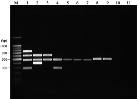
0.2 mM each deoxynucleoside triphosphate (Promega, Madison, WI), 10 pmol of each primer, and 2.5 U of HotStar Plus Taq polymerase (Qiagen, Gaithersburg, MD). After an initial denaturation step (8 min at 95°C) to activate the HotStar Plus Taq polymerase, 29 cycles of amplification were performed as follows: denaturation at 95°C for 60 s, annealing at 60°C for 40 s, and DNA extension at 72°C for 35 s. A final extension was performed at 72°C for 10 min. Amplification was carried out in a DNA 9700 thermocycler (TGradient; Biometra, Germany). After amplification, PCR products were analyzed on a 2.0% agarose gel, stained with ethidium bromide, and visualized on a UV transilluminator. Multiplex PCR products of 484, 398, 753, 608, and 296 bp resulted from amplification of the 16S rRNA gene, IS900, IS901, IS1311, and DT1 targets, respectively (Fig. 1). DNA isolated from M. avium subsp. paratuberculosis ATCC 19698, M. avium subsp.
avium ATCC 35712, M. intracellulare ATCC 13950, and M. terrae ATCC 15755
were used as controls for each primer set in each PCR run.
A priori MAC multiplex interpretation criteria, consistent with current MAC
nomenclature (39), are listed in Table 2. Simultaneous amplification of the three targets IS1311, the 16S rRNA gene, and IS900, was interpreted as corresponding to M. avium subsp. paratuberculosis; amplification of DT1, IS1311, the 16S rRNA gene, and IS901 was interpreted as corresponding to M. avium subsp. avium serotype 2 or 3 (“bird type”) or M. avium subsp. silvaticum. Amplification of both IS1311 and the 16S rRNA gene but neither IS900 nor IS901 was interpreted as corresponding to M. avium subsp. hominissuis. Amplification of only the DT1 and 16S rRNA gene targets indicated M. intracellulare. Mycobacterial species outside the MAC were indicated when 16S rRNA gene amplification alone was observed.
Bacterial strains, cultures, and preparation of mycobacterial single-cell sus-pensions.The origins, sources and/or strain names, descriptions, and reference identifications of mycobacterial strains used to evaluate the assay are listed in Table 3. Reference strains of each Mycobacterium species were obtained from the American Type Culture Collection (ATCC). All nonmycobacterial organisms were obtained from the ATCC or the State Laboratory of Hygiene, Madison, WI (WSLH). Mycobacteria were cultivated in 7H9 broth supplemented with 10% oleic acid-albumin-dextrose-catalase (OADC; Difco Laboratories, MD) supple-mented with 2g/ml of mycobactin J (Allied Monitor, Fayette, MO) for 2 weeks (rapid growers) or 4 weeks (slow growers) at 37°C. Nonmycobacterial organisms, such as Escherichia coli, were grown in Luria-Bertani (LB) liquid medium for 2
TABLE 1. Oligonucleotide primers used for MAC multiplex PCR
Genetic construct Product
size (bp) Orientation
a Oligonucleotide sequence Target organism(s)
IS900
398
F
5
⬘ TGGACAATGACGGTTACGGAGGTGG 3⬘
M. avium subsp. paratuberculosis
R
5
⬘ CGCAGAGGCTGCAAGTCGTGG 3⬘
IS901
753
F
5
⬘ GAACGCTGCTCTAAGGACCTGTTGG 3⬘
M. avium subsp. avium
R
5
⬘ GGAAGGGTGATTATCTGGCCTGC 3⬘
DT1
296
F
5
⬘ CGTTGGCTGGCCATTCACGAAGGAGT 3⬘
M. avium subsp. avium and
R
5
⬘ GCTAGTTGGATCGCGCCGAACACCGG 3⬘
M. intracellulare
IS1311
608
F
5
⬘ GCGTGAGGCTCTGTGGTGAA 3⬘
All MAC members
R
5
⬘ ATGACGACCGCTTGGGAGAC 3⬘
16S rRNA gene
484
F
5
⬘ ATAAGCCTGGGAAACTGGGT 3⬘
All mycobacterial species
R
5
⬘ CACGCTCACAGTTAAGCCGT 3⬘
days. All bacteria were washed 3 times with 10 mM phosphate-buffered saline (PBS; pH 7.4), bacterial cell pellets were collected after centrifugation, and small aliquots were stored at⫺80°C until use.
DNA extraction.To prepare the bacterial DNA from pure cultures, 1 ml of the bacterial pellet was resuspended in 10 mM PBS (pH 7.4) and transferred into a 1.5-ml Eppendorf tube. The DNA was extracted by using either a QIAamp DNA stool minikit (Qiagen Inc., Valencia, CA) or the conventional cetyltrimethyl-ammonium bromide (CTAB) method (18) following procedures described pre-viously (3) or as described by the manufacturer, with minor modifications.
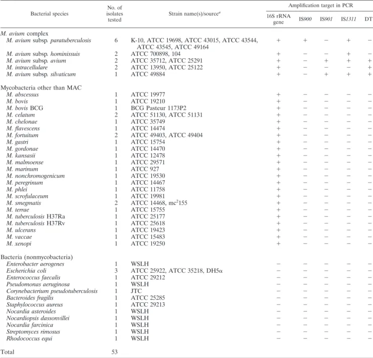
Multiplex specificity and sensitivity.To determine MAC multiplex specificity, a total of 53 reference bacterial strains consisting of 28 different mycobacterial species and 12 nonmycobacterial species were tested (Table 3). To evaluate assay sensitivity, purified DNA from M. avium subsp. avium ATCC 35712, M. avium subsp. paratuberculosis ATCC 19698, M. avium subsp. hominissuis 104, M.
intra-cellulare ATCC 13950, M. abscessus ATCC 19977, M. tuberculosis Ra, M. bovis
BCG, E. coli ATCC 35218, and M. terrae ATCC 15755 isolated from pure cultures were serially diluted from 100 ng to 5 fg, then assayed using the MAC multiplex.
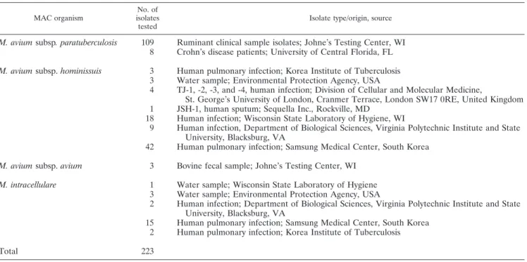
MAC multiplex PCR on clinical isolates from broth culture.A total of 223 MAC strains, consisting of 117 isolates of M. avium subsp. paratuberculosis, 80 isolates of M. avium subsp. hominissuis, 3 isolates of M. avium subsp. avium, and 23 isolates of M. intracellulare were obtained from human and veterinary diag-nostic laboratories. The origins and sources of these isolates are listed in Table
4. The M. avium subsp. paratuberculosis isolate identities had been confirmed by both analysis of L1 and L9 and single-target IS900 PCR detection (28). The human-origin M. avium subsp. paratuberculosis isolates were identified by using a previously described molecular typing method based on two IS900 integration loci (28), IS1311 restriction fragment length polymorphism (21), rpoB sequence analysis (5), and hsp65 code sequevar analysis (19, 40). All clinical isolates were tested by MAC multiplex PCR on a blind basis after their propagation in modified Bactec 12B or MGIT ParaTB culture media.
RESULTS
Specificity and sensitivity of MAC multiplex PCR.
The
MAC multiplex PCR’s five products were readily visualized on
agarose gels: these products were a 484-bp product specific for
the mycobacterial 16S rRNA gene, a 398-bp product from
IS900 specific for M. avium subsp. paratuberculosis, 753 bp
from IS901 specific for M. avium subsp. avium, 296 bp from DT
found in M. avium subsp. avium and M. intracellulare, and a
608-bp product from IS1311 found in all M. avium subspecies
members (Fig. 1). During testing of the 12 nonmycobacterial
FIG. 1. Representative gel resulting from the MAC multiplex. Lane M, molecular size marker; lane 1, M. avium subsp. avium ATCC 35712;
lane 2, M. avium subsp. paratuberculosis ATCC 19698; lane 3, M. avium subsp. hominissuis 104; lane 4, M. intracellulare ATCC 13950; lane 5, M.
terrae ATCC 15755; lane 6, M. phlei ATCC 11758; lane 7, Mycobacterium abscessus ATCC 19977; lane 8, M. tuberculosis Ra; lane 9, Mycobacterium
bovis (BCG); lane 10, E. coli ATCC 35218; lane 11, negative control.
TABLE 2. Interpretation criteria for MAC multiplex results
Identification
Criterion Panmycobacterium
16S rRNA gene IS900 IS901 IS1311 DT1
M. avium subsp. avium (serotypes 2 and 3) or
M. avium subsp. silvaticum
a⫹
⫺
⫹
⫹
⫹
M. avium subsp. avium (serotype 1)
⫹
⫺
⫹
⫹
⫺
M. avium subsp. paratuberculosis
⫹
⫹
⫺
⫹
⫺
M. avium subsp. hominissuis
⫹
⫺
⫺
⫹
⫺
M. intracellulare
⫹
⫺
⫺
⫺
⫹
Mycobacteria other than MAC
⫹
⫺
⫺
⫺
⫺
Nonmycobacterial species
⫺
⫺
⫺
⫺
⫺
a
species, no amplification was seen for any of the MAC
multi-plex targets. The MAC multimulti-plex correctly positioned the
ATCC mycobacterial species within or outside the M. avium
complex and identified all species within the MAC correctly
(Table 1).
Based on dilution trials with purified DNA from M. avium
subsp. paratuberculosis ATCC 19698, M. avium subsp. avium
ATCC 35712, M. intracellulare ATCC 13950, and M. bovis
(BCG), the analytical sensitivities of the MAC multiplex PCR
were approximately 2 to 5 fg for the 16S rRNA gene, 10 fg for
IS900, 10 fg for IS901, 5 fg for IS1311, and 50 fg for DT1 (data
not shown). This is roughly equivalent to 10
1to 10
2CFU/ml of
mycobacteria in liquid culture. The MAC multiplex was able to
detect M. avium subsp. paratuberculosis from a mixed pure
culture containing a hundredfold excess of non-MAC
myco-bacteria (data not shown).
Performance of MAC multiplex with clinical isolates.
Com-plete identity agreement was found between the MAC
multi-plex and the independent methods used by source institutions
providing the 223 different acid-fast strains isolated from
hu-mans and a wide variety of domestic and nondomestic animals
(Table 4).
TABLE 3. Type strains used in the specificity test for multiplex PCR
Bacterial species No. of isolates tested Strain name(s)/sourcea Amplification target in PCR 16S rRNA
gene IS900 IS901 IS1311 DT1
M. avium complex
M. avium subsp. paratuberculosis
6
K-10, ATCC 19698, ATCC 43015, ATCC 43544,
ATCC 43545, ATCC 49164
⫹
⫹
⫺
⫹
⫺
M. avium subsp. hominissuis
2
ATCC 700898, 104
⫹
⫺
⫺
⫹
⫺
M. avium subsp. avium
2
ATCC 35712, ATCC 25291
⫹
⫺
⫹
⫹
⫹
M. intracellulare
2
ATCC 13950, ATCC 25122
⫹
⫺
⫺
⫺
⫹
M. avium subsp. silvaticum
1
ATCC 49884
⫹
⫺
⫹
⫹
⫹
Mycobacteria other than MAC
M. abscessus
1
ATCC 19977
⫹
⫺
⫺
⫺
⫺
M. bovis
1
ATCC 19210
⫹
⫺
⫺
⫺
⫺
M. bovis BCG
1
BCG Pasteur 1173P2
⫹
⫺
⫺
⫺
⫺
M. celatum
2
ATCC 51130, ATCC 51131
⫹
⫺
⫺
⫺
⫺
M. chelonae
1
ATCC 35749
⫹
⫺
⫺
⫺
⫺
M. flavescens
1
ATCC 14474
⫹
⫺
⫺
⫺
⫺
M. fortuitum
2
ATCC 49403, ATCC 49404
⫹
⫺
⫺
⫺
⫺
M. gastri
1
ATCC 15754
⫹
⫺
⫺
⫺
⫺
M. gordonae
1
ATCC 14470
⫹
⫺
⫺
⫺
⫺
M. kansasii
1
ATCC 12478
⫹
⫺
⫺
⫺
⫺
M. malmoense
1
ATCC 29571
⫹
⫺
⫺
⫺
⫺
M. marinum
1
ATCC 927
⫹
⫺
⫺
⫺
⫺
M. nonchromogenicum
1
ATCC 19530
⫹
⫺
⫺
⫺
⫺
M. peregrinum
1
ATCC 14467
⫹
⫺
⫺
⫺
⫺
M. phlei
1
ATCC 11758
⫹
⫺
⫺
⫺
⫺
M. scrofulaceum
1
ATCC 19981
⫹
⫺
⫺
⫺
⫺
M. smegmatis
2
ATCC 14468, mc
2155
⫹
⫺
⫺
⫺
⫺
M. terrae
1
ATCC 15755
⫹
⫺
⫺
⫺
⫺
M. tuberculosis H37Ra
1
ATCC 25177
⫹
⫺
⫺
⫺
⫺
M. tuberculosis H37Rv
1
ATCC 25618
⫹
⫺
⫺
⫺
⫺
M. ulcerans
1
ATCC 19423
⫹
⫺
⫺
⫺
⫺
M. vaccae
1
ATCC 15483
⫹
⫺
⫺
⫺
⫺
M. xenopi
1
ATCC 19250
⫹
⫺
⫺
⫺
⫺
Bacteria (nonmycobacteria)
Enterobacter aerogenes
1
WSLH
⫺
⫺
⫺
⫺
⫺
Escherichia coli
3
ATCC 25922, ATCC 35218, DH5
␣
⫺
⫺
⫺
⫺
⫺
Enterococcus faecalis
1
ATCC 29212
⫺
⫺
⫺
⫺
⫺
Pseudomonas aeruginosa
1
WSLH
⫺
⫺
⫺
⫺
⫺
Corynebacterium pseudotuberculosis
1
JTC
⫺
⫺
⫺
⫺
⫺
Bacteroides fragilis
1
ATCC 25285
⫺
⫺
⫺
⫺
⫺
Staphylococcus aureus
1
ATCC 29213
⫺
⫺
⫺
⫺
⫺
Nocardia asteroides
1
WSLH
⫺
⫺
⫺
⫺
⫺
Nocardiopsis dassonvillei
1
WSLH
⫺
⫺
⫺
⫺
⫺
Nocardia farcinica
1
WSLH
⫺
⫺
⫺
⫺
⫺
Streptomyces rimosus
1
WSLH
⫺
⫺
⫺
⫺
⫺
Rhodococcus equi
1
WSLH
⫺
⫺
⫺
⫺
⫺
Total
53
aATCC, American Type Culture Collection, Manassas, VA; JTC, Johne’s Testing Center, Madison, WI; WSLH, Wisconsin State Laboratory of Hygiene, Madison, WI.
DISCUSSION
Insertion elements found in MAC subspecies provide useful
markers for their identification through genotyping (2, 4, 21,
39). Moreover, the markers appear to be associated with the
subspecies’ epidemiologies and pathogenicities, making the
use of these markers clinically relevant. With rare exceptions,
IS900 is a reliable genetic marker defining M. avium subsp.
paratuberculosis (4, 16), the cause of paratuberculosis (Johne’s
disease) in animals. IS901 is strongly associated with MAC
infections in avian species, and IS901-positive strains are thus
often called bird-type MAC members (7, 8, 39). IS1311 is
consistently associated with the MAC, with only rare
excep-tions reported (2, 21). Inclusion of primers for the 16S rRNA
gene target common to all mycobacteria in the MAC multiplex
serves as an internal control for confirmation of mycobacterial
identification (34).
Reliance on a single target for a collection of organisms so
closely related can be perilous and may result in inaccurate
diagnoses of mycobacterial infections (9). The MAC multiplex
has good analytical sensitivity (5 to 10 fg DNA), especially
considering that it simultaneously amplifies five separate
my-cobacterial genomic targets. The assay is designed specifically
to discriminate among MAC subspecies and is particularly
good at identifying M. avium subsp. paratuberculosis, aided in
all likelihood by the high copy number of IS900 (1). M. avium
subsp. paratuberculosis identification based solely on the
detec-tion of IS900 should be evaluated with caudetec-tion, since some
environmental mycobacteria have been reported to contain
IS900-like elements (1, 14, 42).
Despite the specificity of PCR primers and the full isolate
identification concordance shown in this study’s data set, the
presence of more than one mycobacterial species in a sample
can perturb the diagnosis. False-positive PCR results are not
unheard of (33). Primer inhibition due to sample components
or competition for primer targets in the assay also may occur
and should be monitored.
The data provided from multitarget PCR allow
discrimina-tion among MAC subspecies, increase diagnostic confidence in
the identity of isolates, and help resolve discrepancies. This
technique is not only very promising for clear characterization
of clinical MAC infections but is useful for epidemiologic
stud-ies as well.
ACKNOWLEDGMENTS
We acknowledge financial support by the Johne’s Testing Center,
the Basic Science Research Program through the National Research
Foundation of Korea (NRF) funded by the Ministry of Education,
Science and Technology (grant R01-2007-000-10702-0), and the
Johne’s Disease Integrated Program (award no. 2008-55620-18710)
from the USDA-NIFA program.
REFERENCES
1. Abubakar, I., D. Myhill, S. H. Aliyu, and P. R. Hunter. 2008. Detection of
Mycobacterium avium subspecies paratuberculosis from patients with Crohn’s
disease using nucleic acid-based techniques: a systematic review and meta-analysis. Inflamm. Bowel Dis. 14:401–410.
2. Alvarez, J., I. G. Garcia, A. Aranaz, J. Bezos, B. Romero, L. de Juan, A.
Mateos, E. Gomez-Mampaso, and L. Dominguez.2008. Genetic diversity of
Mycobacterium avium isolates recovered from clinical samples and from the
environment: molecular characterization for diagnostic purposes. J. Clin. Microbiol. 46:1246–1251.
3. Amaro, A., E. Duarte, A. Amado, H. Ferronha, and A. Botelho. 2008. Com-parison of three DNA extraction methods for Mycobacterium bovis,
Myco-bacterium tuberculosis and MycoMyco-bacterium avium subsp. avium. Lett. Appl.
Microbiol. 47:8–11.
4. Bartos, M., P. Hlozek, P. Svastova, L. Dvorska, T. Bull, L. Matlova, I.
Parmova, I. Kuhn, J. Stubbs, M. Moravkova, J. Kintr, V. Beran, I. Meli-charek, M. Ocepek, and I. Pavlik.2006. Identification of members of
Myco-bacterium avium species by Accu-Probes, serotyping, and single IS900, IS901,
IS1245 and IS901-flanking region PCR with internal standards. J. Microbiol. Methods 64:333–345.
5. Ben Salah, I., T. Adekambi, D. Raoult, and M. Drancourt. 2008. rpoB sequence-based identification of Mycobacterium avium complex species. Mi-crobiology 154:3715–3723.
TABLE 4. Identification of clinical isolates by MAC multiplex PCR (100% congruence with source methods)
MAC organism
No. of isolates tested
Isolate type/origin, source
M. avium subsp. paratuberculosis
109
Ruminant clinical sample isolates; Johne’s Testing Center, WI
8
Crohn’s disease patients; University of Central Florida, FL
M. avium subsp. hominissuis
3
Human pulmonary infection; Korea Institute of Tuberculosis
3
Water sample; Environmental Protection Agency, USA
4
TJ-1, -2, -3, and -4, human infection; Division of Cellular and Molecular Medicine,
St. George’s University of London, Cranmer Terrace, London SW17 0RE, United Kingdom
1
JSH-1, human sputum; Sequella Inc., Rockville, MD
18
Human infection; Wisconsin State Laboratory of Hygiene, WI
9
Human infection, Department of Biological Sciences, Virginia Polytechnic Institute and State
University, Blacksburg, VA
42
Human pulmonary infection; Samsung Medical Center, South Korea
M. avium subsp. avium
3
Bovine fecal sample; Johne’s Testing Center, WI
M. intracellulare
1
Water sample; Wisconsin State Laboratory of Hygiene
3
Water sample; Environmental Protection Agency, USA
2
Human infection; Department of Biological Sciences, Virginia Polytechnic Institute and State
University, Blacksburg, VA
15
Human pulmonary infection; Samsung Medical Center, South Korea
2
Human pulmonary infection; Korea Institute of Tuberculosis
6. Biet, F., M. L. Boschiroli, M. F. Thorel, and L. A. Guilloteau. 2005. Zoonotic aspects of Mycobacterium bovis and Mycobacterium avium-intracellulare com-plex (MAC). Vet. Res. 36:411–436.
7. Dvorska, L., T. J. Bull, M. Bartos, L. Matlova, P. Svastova, R. T. Weston, J.
Kintr, I. Parmova, D. Van Soolingen, and I. Pavlik.2003. A standardised restriction fragment length polymorphism (RFLP) method for typing
Myco-bacterium avium isolates links IS901 with virulence for birds. J. Microbiol.
Methods 55:11–27.
8. Dvorska, L., L. Matlova, W. Y. Ayele, O. A. Fischer, T. Amemori, R. T.
Weston, J. Alvarez, V. Beran, M. Moravkova, and I. Pavlik.2007. Avian tuberculosis in naturally infected captive water birds of the Ardeideae and Threskiornithidae families studied by serotyping, IS901 RFLP typing, and virulence for poultry. Vet. Microbiol. 119:366–374.
9. Falkinham, J. O., III. 1996. Epidemiology of infection by nontuberculous mycobacteria. Clin. Microbiol. Rev. 9:177–215.
10. Falkinham, J. O., III, C. D. Norton, and M. W. LeChevallier. 2001. Factors influencing numbers of Mycobacterium avium, Mycobacterium intracellulare, and other mycobacteria in drinking water distribution systems. Appl. Envi-ron. Microbiol. 67:1225–1231.
11. Feller, L., and J. Lemmer. 2007. Aspects of immunopathogenic mechanisms of HIV infection. SADJ 62:432–434, 436.
12. Field, S. K., and R. L. Cowie. 2006. Lung disease due to the more common nontuberculous mycobacteria. Chest 129:1653–1672.
13. Frothingham, R., and K. H. Wilson. 1994. Molecular phylogeny of the
Mycobacterium avium complex demonstrates clinically meaningful divisions.
J. Infect. Dis. 169:305–312.
14. Grant, I. R. 2005. Zoonotic potential of Mycobacterium avium ssp.
paratu-berculosis: the current position. J. Appl. Microbiol. 98:1282–1293.
15. Haist, V., F. Seehusen, I. Moser, H. Hotzel, U. Deschl, W. Baumgartner, and
P. Wohlsein.2008. Mycobacterium avium subsp. hominissuis infection in 2 pet dogs, Germany. Emerg. Infect. Dis. 14:988–990.
16. Harris, N. B., and R. G. Barletta. 2001. Mycobacterium avium subsp.
para-tuberculosis in veterinary medicine. Clin. Microbiol. Rev. 14:489–512.
17. Henry, M. T., L. Inamdar, D. O’Riordain, M. Schweiger, and J. P. Watson. 2004. Nontuberculous mycobacteria in non-HIV patients: epidemiology, treatment and response. Eur. Respir. J. 23:741–746.
18. Hurley, S. S., G. A. Splitter, and R. A. Welch. 1987. Rapid lysis technique for mycobacterial species. J. Clin. Microbiol. 25:2227–2229.
19. Ichikawa, K., T. Yagi, M. Moriyama, T. Inagaki, T. Nakagawa, K. Uchiya, T.
Nikai, and K. Ogawa.2009. Characterization of Mycobacterium avium clin-ical isolates in Japan using subspecies-specific insertion sequences, and iden-tification of a new insertion sequence, ISMav6. J. Med. Microbiol. 58:945– 950.
20. Jeong, Y. J., K. S. Lee, W. J. Koh, J. Han, T. S. Kim, and O. J. Kwon. 2004. Nontuberculous mycobacterial pulmonary infection in immunocompetent patients: comparison of thin-section CT and histopathologic findings. Radi-ology 231:880–886.
21. Johansen, T. B., B. Djonne, M. R. Jensen, and I. Olsen. 2005. Distribution of IS1311 and IS1245 in Mycobacterium avium subspecies revisited. J. Clin. Microbiol. 43:2500–2502.
22. Johnson-Ifearulundu, Y., J. B. Kaneene, and J. W. Lloyd. 1999. Herd-level economic analysis of the impact of paratuberculosis on dairy herds. J. Am. Vet. Med. Assoc. 214:822–825.
23. Koh, W. J., O. J. Kwon, and K. S. Lee. 2005. Diagnosis and treatment of nontuberculous mycobacterial pulmonary diseases: a Korean perspective. J. Korean Med. Sci. 20:913–925.
24. Lai, C. C., C. K. Tan, C. H. Chou, H. L. Hsu, C. H. Liao, Y. T. Huang, P. C.
Yang, K. T. Luh, and P. R. Hsueh.2010. Increasing incidence of nontuber-culous mycobacteria, Taiwan, 2000–2008. Emerg. Infect. Dis. 16:294–296. 25. Maekura, R., Y. Okuda, A. Hirotani, S. Kitada, T. Hiraga, K. Yoshimura, I.
Yano, K. Kobayashi, and M. Ito.2005. Clinical and prognostic importance of serotyping Mycobacterium avium-Mycobacterium intracellulare complex iso-lates in human immunodeficiency virus-negative patients. J. Clin. Microbiol.
43:3150–3158.
26. Matlova, L., L. Dvorska, K. Palecek, L. Maurenc, M. Bartos, and I. Pavlik. 2004. Impact of sawdust and wood shavings in bedding on pig tuberculous lesions in lymph nodes, and IS1245 RFLP analysis of Mycobacterium avium
subsp. hominissuis of serotypes 6 and 8 isolated from pigs and environment.
Vet. Microbiol. 102:227–236.
27. Miguez-Burbano, M. J., M. Flores, D. Ashkin, A. Rodriguez, A. M. Granada,
N. Quintero, and A. Pitchenik.2006. Non-tuberculous mycobacteria disease as a cause of hospitalization in HIV-infected subjects. Int. J. Infect. Dis.
10:47–55.
28. Motiwala, A. S., M. Strother, A. Amonsin, B. Byrum, S. A. Naser, J. R.
Stabel, W. P. Shulaw, J. P. Bannantine, V. Kapur, and S. Sreevatsan.2003. Molecular epidemiology of Mycobacterium avium subsp. paratuberculosis: evidence for limited strain diversity, strain sharing, and identification of unique targets for diagnosis. J. Clin. Microbiol. 41:2015–2026.
29. Nielsen, S. S., and N. Toft. 2009. A review of prevalences of paratuberculosis in farmed animals in Europe. Prev. Vet. Med. 88:1–14.
30. O’Toole, D., S. Tharp, B. V. Thomsen, E. Tan, and J. B. Payeur. 2005. Fatal mycobacteriosis with hepatosplenomegaly in a young dog due to
Mycobac-terium avium. J. Vet. Diagn. Invest. 17:200–204.
31. Ott, S. L., S. J. Wells, and B. A. Wagner. 1999. Herd-level economic losses associated with Johne’s disease on US dairy operations. Prev. Vet. Med.
40:179–192.
32. Petrini, B. 2006. Non-tuberculous mycobacterial infections. Scand. J. Infect. Dis. 38:246–255.
33. Pithua, P., S. J. Wells, S. M. Godden, S. Sreevatsan, and J. R. Stabel. 2010. Experimental validation of a nested polymerase chain reaction targeting the genetic element ISMAP02 for detection of Mycobacterium avium subspecies
paratuberculosis in bovine colostrum. J. Vet. Diagn. Invest. 22:253–256.
34. Rossi, M. C., A. Gori, G. Zehender, G. Marchetti, G. Ferrario, C. De
Madd-alena, L. Catozzi, A. Bandera, A. D. Esposti, and F. Franzetti.2000. A PCR-colorimetric microwell plate hybridization assay for detection of
My-cobacterium tuberculosis and M. avium from culture samples and
Ziehl-Neelsen-positive smears. J. Clin. Microbiol. 38:1772–1776.
35. Schweickert, B., O. Goldenberg, E. Richter, U. B. Gobel, A. Petrich, P.
Buchholz, and A. Moter.2008. Occurrence and clinical relevance of
Myco-bacterium chimaera sp. nov., Germany. Emerg. Infect. Dis. 14:1443–1446.
36. Steed, K. A., and J. O. Falkinham III. 2006. Effect of growth in biofilms on chlorine susceptibility of Mycobacterium avium and Mycobacterium
intracel-lulare. Appl. Environ. Microbiol. 72:4007–4011.
37. Tell, L. A., L. Woods, and R. L. Cromie. 2001. Mycobacteriosis in birds. Rev. Sci. Tech. 20:180–203.
38. Thorel, M. F., H. F. Huchzermeyer, and A. L. Michel. 2001. Mycobacterium
avium and Mycobacterium intracellulare infection in mammals. Rev. Sci.
Tech. 20:204–218.
39. Turenne, C. Y., D. M. Collins, D. C. Alexander, and M. A. Behr. 2008.
Mycobacterium avium subsp. paratuberculosis and M. avium subsp. avium are
independently evolved pathogenic clones of a much broader group of M.
avium organisms. J. Bacteriol. 190:2479–2487.
40. Turenne, C. Y., M. Semret, D. V. Cousins, D. M. Collins, and M. A. Behr. 2006. Sequencing of hsp65 distinguishes among subsets of the Mycobacterium
avium complex. J. Clin. Microbiol. 44:433–440.
41. Uppal, M., S. L. McLellan, M. T. Collins, and R. S. Lambrecht. 2002. A multiplex PCR assay that discriminates Mycobacterium avium subspecies
paratuberculosis from closely related Mycobacteria using primers which
de-tect specific insertion sequences, abstr. Z-58. Abstr. 102nd Annu. Meet. Am. Soc. Microbiol. American Society for Microbiology, Washington, DC. 42. Vansnick, E., P. De Rijk, F. Vercammen, D. Geysen, L. Rigouts, and F.
Portaels.2004. Newly developed primers for the detection of Mycobacterium
avium subspecies paratuberculosis. Vet. Microbiol. 100:197–204.
43. Wells, S. J., M. T. Collins, K. S. Faaberg, C. Wees, S. Tavornpanich, K. R.
Petrini, J. E. Collins, N. Cernicchiaro, and R. H. Whitlock.2006. Evaluation of a rapid fecal PCR test for detection of Mycobacterium avium subsp.