- 57 -
KISEP Original Articles J Rhinol 9(1,2 ), 2002
Clinical and Histologic Features of Antrochoanal Polyps
Jae Myeong Kim, M.D., Jun Yeon Hwang, M.D., Tae Young Kwon, M.D. and Sung Wan Kim, M.D.
ABSTRACT
Antrochoanal polyp (ACP) usually appears as a large, soft, gelatinous mass in the nasopharynx. Recently, ACP is considered as a separate entity among sinonasal polyps. Histologically, the antral part of the polyp can be cystic or polypoid with a fibrous and solid choanal part. However, the extent of inflammation of the paranasal sinuses has not well been demonstrated. In addition, pathologic patterns of ACP have not been well revealed. The aim of our study is to evaluate the disease patterns of ACPs by clinical and pathologic analysis. Thirty two patients with ACPs were evaluated by a retrospective study. We evaluated sinus involvement by operative findings and pathologic patterns were investigated in 28 cases by predominant infiltrating cells and types of polyp. In order to evaluate the histologic differences between the antral and choanal portion, each A CP was divided into proximal and distal portion. The involved sinuses were multiple and diverse and the most of them had a polyp. The most common pathologic type was chronic inflammatory polyp. Lymphocyte and neutrophil were visualized in many ACPs predo - minantly. The histologic differences between the proximal and distal portion was not observed significantly. Clinical analysis suggest that ACPs are not sole polyp but mixed disease with inflammation of the nose and sinuses in most cases. Histologic analysis suggest that ACPs are caused by chronic inflammation in many cases and each portion of ACPs is composed of the same histologic type.
KEY WORDS:Antrochoanal polyp・Histology.
INTRODUCTION
Antrochoanal polyp is a polyp that originates from the maxillary sinus mucosa and comes out through either the natural ostium or accessory ostium and pro- trudes toward the choana and nasopharynx. Since Ki- llian1) described the maxillary sinus as the origin of antrochoanal polyp, other researchers have reported the medial and posterior wall of the maxillary sinus mucosa as the most common origin site.2-4) Classified as a disorder from nasal polyps, this disease has an incidence rate of 4-6% of all polyps,5) and it espe- cially takes up 33% of all pediatric polyps.6) It is di- vided into a maxillary portion and a nasal cavity por-
tion. There have been reports that state they have a di- fferent histologic feature compared to general polyps,2) however, studies based on a difference among the two portions of antrochoanal polyps has still not been demonstrated.
Therefore, we plan to inquire the aspects of antrocho- anal polyps through its clinical and histologic findings.
MATERIAL AND METHOD
A retrospective study was carried out on 32 patients that have been diagnosed and surgically operated on for antrochoanal polyps from February, 1996 through February 2000. The age distribution was from 5 years old to 81 years old and the average age was 26 years old. There were 17 cases on the left, 10 cases on the right and 5 cases on both sides. Based on the surgical findings of the 37 cases of antrochoanal polyps, we carried out a study to inquire the extent of invaded areas of the paranasal sinus and whether or not a polyp exists in the paranasal sinus. Based on the 28 cases where histologic observation was possible, we studied each predominant infiltrating cell and the type of po- Department of Otolaryngology-Head and Neck Surgery, Kang-
Nam General Hospital, Public Corporation, Seoul, Korea Address correspondences and reprint requests to Sung Wan Kim, M.D., Department of Otolaryngology-Head and Neck Sur- gery KangNam General Hospital, Public Corporation, Seoul, Korea 171-1 Samsung-dong, Kangnam-ku, Seoul 135-740, Korea Tel:82-2-3430-0673, Fax:82-2-539-5256
E-mail:[email protected]
Accepted for publication on October 8, 2002
58 / J Rhinol 9(1,2), 2002
lyps in the paranasal sinus of the proximal portion which signifies the inner portion of the maxillary sinus and a distal portion which signifies the inner portion of the nasal cavity.
The polyps are classified into 4 types, which are, edematous eosinophilic polyps, chronic inflammatory polyps, polyps with hyperplasia of seromucinous glands and polyps with atypical stroma.7) The predominant infiltrating cells for each part was studied and all his- tologic studies were carried out by the same histologist (Fig. 1).
RESULTS
Surgical findings based on the condition of infiltr- ation of the paranasal sinus cavity, revealed the follo- wing results:15 cases where only the maxillary sinus
was invaded, 8 cases where the maxillary and ethmoid sinuses were both invaded, 1 case where the maxillary, ethmoid and sphenoid sinuses were invaded, 1 case where the frontal, maxillary and ethmoid sinuses were invaded, 1 case where only the frontal and maxillary sinuses were invaded and 11 cases where the frontal, maxillary, ethmoidal and sphenoidal sinuses were all invaded. There were 22 cases where the paranasal sinus, other than the maxillary sinus, was involved and 18 cases among them showed a polypous finding in the paranasal sinus (Table 1).
The predominant infiltrating cells revealed through histologic findings of the proximal and distal portions were each lymphocytes in 12 and 12 cases, neutrophils in 12 and 11 cases, eosinophils in 11 and 10 cases, and plasma cells in 7 and 8 cases, respectively. The lym- phocytes and eosinophils both revealed similar extents and there was no difference in infiltrating cells in the proximal and distal portion of the polyps (Table 2).
The most common pathologic type of polyps were
Table 1. Sinus involvement on antrochoanal polyp patients by operative findings
Involved sinus No. of patients No. of polyp
MS+ES 8 7
MS+FS 1 0
MS+ES+SS 1 1
MS+ES+FS 1 1
MS+ES+FS+SS 11 9
Total 22 18
MS:maxillary sinus ES:ethmoid sinus FS:frontal sinus SS:sphenoid sinus
Table 2. Predominant cells of proximal and distal portion of an- trochoanal polyp
Portion
Cell type Proximal portion Distal portion
Lymphocyte 12 (43%) 12 (43%)
Neutrophil 12 (43%) 11 (39%)
Eosinophil 11 (39%) 10 (36%)
Plasma cell 7 (25%) 8 (29%)
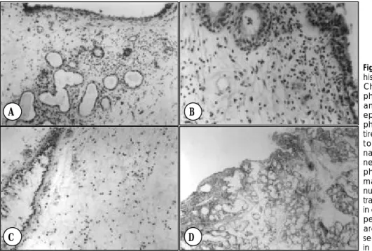
Fig. 1. Histologic features of different histologic types of nasal polyps. A:
Chronic inflammatory polyp with lym- phocyte predominant. There are squ- amous and cuboidal metaplasias of epithelium and diffuse numerous lym- phocyte infiltrations are seen in an en- tire specimen. B:Chronic inflamma- tory polyp with neutrophil predomi- nant. There are numerous segmented neutrophil infiltrations with a few lym- phocytes and plasma cells. C:Ede- matous eosinophilic polyp. There are numerous eosinophil and mast cell infil- trations with hyperplasia of goblet cells in edematous stroma. D:Polyp with hy- perplasia of seromucinous glands. There are numerous seromucinous glands seen with various inflammatory cells in loose and edematous stroma.
A B
C D
Kim et al:Antrochoanal Polyp / 59
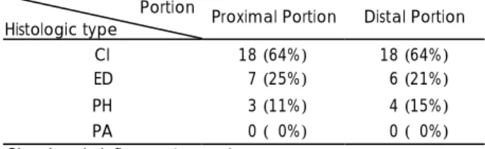
the chronic inflammatory polyps followed by edmatous eosinophilc polyps and polyps with hyperplasia of ser- omucinous glands, in order of occurrence. Polyps with atypical stroma were not found. In each maxillary sinus antrochoanal polyp, with an exception of 1 case among the proximal and distal portion, the type of polyps were almost all similar (Table 3).
DISCUSSION
Antrochoanal polyps are usually unilateral and they take up 3-6% of all polyps. Its origin is usually the mucosa of the maxillary sinus. It comes out through either the natural ostium or accessory ostium and pro- trudes toward the choana and nasopharynx. Differently from general polyps, antrochoanal polyps are more common in men than women and in children than adults.
It has almost no relation to allergies and is considered to be caused by chronic inflammations.2)
In this study, among all 32 patients, both men (19) and women (13) showed similar frequencies. There were 12 (48%) under the age 15, which showed a hi- gher distribution compared to general polyps. However, compared to studies by Kim et al.,8) and other resear- chers, the relative importance of children occupied a lower distribution in this study.
Surgical findings revealed 22 cases where the par - anasal sinus has been invaded. Among the invaded paranasal sinuses, 18 cases revealed polypous findings, which show that it is not merely a disease of the ma- xillary sinus but is a multiple disorder that accompanies chronic paranasal sinusitis.
The differences between antrochoanal polyps and simple polyps are that antrochoanal polyps originate from the posterior wall of the maxillary sinus, has no relation to allergies and has little eosinophils and mucinous glands.2)5) It is considered that antrochoanal
polyps are more related to chronic inflammations than allergies based on its infiltration of inflammatory cell and fibrosis.9)
Results of this test revealed that, among all 28 cases, the infiltrating extent of 2 types of cells were similar in 14 cases in the proximal portion and 13 cases in the distal portion based on a predominant infiltrating cell test and therefore was classified according to each in- filtrating cell. Based on this classification, lymphocytes and neutrophils were dominant over eosinophils and plasma cells which coincide with the fact that chronic inflammation may be the main cause of this disorder.
There have been reports that state that there is a difference between the nasal cavity area and maxillary sinus area of the antrochoanal polyp, histologically.
The nasal cavity has a thick wall and multiplication of fibrocytes, infiltration of various inflammatory cells such as neutrophils, lymphocytes and eosinophils and less vessels and mucinous glands compared to simple polyps, whereas, the maxillary sinus area carries a more cystic nature which has a thin wall filled with mucinous glands. This is caused by the polyp protruding through the natural ostium of the paranasal sinus that is ex- posed to the air current in the nasal cavity which sti- mulates fibroblast multiplication that forms a solid polyp by fibroblast multiplication and collagen sedi- mentation of the nasal cavity area. This causes a se- condary change that leads to purulent granuloma, mu- cinous liquefaction and so forth.5)10)
Based on the results of this study, chronic inflam- matory polyps were most commonly revealed in 18 cases in both the maxillary sinus and nasal cavity area of antrochoanal polyps of the maxillary sinus, which appropriates it as a chronic inflammation. However, there was almost no difference among the type of po- lyps and predominant infiltrating cells in the maxillary sinus area and nasal cavity area, which does not relate to reports that state the two portions as being different.5)
CONCLUSION
Antrochoanal polyp of the maxillary sinus is not just a simple polypous disorder but is a chronic inflam- matory disorder that invades numerous paranasal si- nuses. Histologically, between the nasal cavity and maxillary sinus area, a difference among the type of polyp and predominant infiltrating cells was not noticed.
Table 3. Histologic types of proximal and distal portion of antro- choanal polyp
Portion
Histologic type Proximal Portion Distal Portion
CI 18 (64%) 18 (64%)
ED 07 (25%) 06 (21%)
PH 03 (11%) 04 (15%)
PA 00 (00%) 00 (00%)
CI:chronic inflammatory polyp ED:edematous eosinophilic polyp
PH:polyp with hyperplasia of seromucinous gland PA:polyp with atypical stroma
60 / J Rhinol 9(1,2), 2002
REFERENCES
1) Killian G. The origin of choanal polyp. Lancet 1906;2:81-2.
2) Min YG, Chung JW, Shin JS, Chi JG. Histologic structure of an- trochoanal polyps. Acta Otolaryngol (Stock) 1995;115:543-7.
3) Hong SK. Endoscopic removal of the antral portion of antrochoanal polyp by powered instrumentation. Korean J Otolaryngol 2002;45:
41-6.
4) Jang CH, Wang WK, Kim ID, Cho JH, Lee JH, Yoon SW. Treatment of antrochoanal polyp by use of osteoplastic approach with sinos- copy. J Clin Otolaryngol Head Neck Surg 1992;3(l):76-82.
5) Heck WE, Hallberg OE, Williams HL. Antrochoanal polyps. Arch
Otolaryngol 1950;52:538-48.
6) Lee KC, Lee SC, Ban JH, Park SO, Jin SM, Lee LY. Outcomes of transnasal endoscopic sinus surgery in 62 cases of antrochoanal polyp. J Rhinol 1999;6:47-52.
7) Davidsson A, Hellquist HB. The so -called allergic nasal polyp. ORL J Otorhinolaryngol Relat Spec 1993;55(l):30-5.
8) Kim YD, Bai CH, Kim JW, Chung YS, Suh JS, Song KW. End- oscopic sinus surgery of antrochoanal polyp. Korean J Otolaryngol 1998;41:208-12.
9) Rhee CK, Jang YU. Arachidonic acid metabolites in antrochoanal polyp and the nasal polyp associated with chronic paranasal sinusitis.
Korean J Otolaryngol 1999;42:54-7.
10) Batsakis JG. Tumors of the Head and Neck. 2nd ed. Baltimore: The Williams & Wilkins Co;1979:514-30.