- 138 -
KISEP Original Articles J Rhinol 5((((2)))), 1998
Functional Differences of the Lymphocytes in Nasal Polyps between Allergic and Non-Allergic Patients
Yeon Gi Kim, M.D., Sung Hoon Baek, M.D., Jin Bok Park, M.D., Hyun Joo Joo, M.D., Jong Ok Kim, M.D. and Chong Ae Kim, M.D.
ABSTRACT
Lymphocytes can produce various kinds of cytokines which are responsible for the development of the infectious and allergic inflammation. We focused on the role of the lymphocyte in the pathogenesis of the nasal polyp. This study was designed to ev- aluate the functional differences of the lymphocytes between allergic and non-allergic polyp. Lymphocytes were obtained from peripheral blood and tissues of polyp in 12 patients with non-allergic polyp, 7 patients with allergic polyp and 5 normal subjects as control. Cytokines were measured with ELISA from each group of lymphocytes after stimulation with Concanavalin A (Con A). We compared the production of interleukin (IL)-2, IL-4, tumor necrosis factor (TNF)-α and interferon (IFN)-γ between the non-allergic and allergic groups. The levels of IL-4 and IL-6 from polyp tissue lymphocytes were higher in allergic group, while those of IL-2 and IFN-γ were higher in non-allergic group. The levels of IL-4, IL-6 and TNF-α from peripheral blood lymphocytes were higher in allergic group, and IFN-γ was higher in non-allergic group. These results suggest that cytokine productivity of the polyp tissue lymphocytes appear to be parallel to that of the peripheral blood lymphocytes in each group and shows distinct pattern of ytokine production between two groups.
KEY WORDS:Nasal polyp·Allergy·Lymphocyte·Cytokine·Concanavalin A.
INTRODUCTION
Numerous studies have been conducted on the pathogenesis of nasal polyposis. However the relationship of the polyp and allergy has not been proved yet. Nasal polyps are associated with numerous upper respiratory tract diseases including all- ergic rhinitis, recurrent sinusitis, asthma, aspirin intolerance, and cystic fibrosis. Allergy and bacterial infection have gene- rally been accepted as important etiologic factors in the for- mation of nasal polyps.
1)Nasal polyps have respiratory epithelium with infiltration of eosinophils, T and B lymphocytes, polymorphonuclear ce- lls (PMNs) and metachromatic cells. A number of studies have demonstrated that Th2-type T lymphocytes at sites of allergic inflammation produce granulocyte-macrophage colony stimulating factor (GM-CSF), interleukin (IL)-3, IL-4 and IL-5.
2-4)IL-4 is required for the production of IgE by inducing isotype switch in B cells.
5)And IL-3, IL-5 and GM-CSF sti-
mulate the growth and differentiation of eosinophils and in- crease the survival of eosinophils.
It has been suggested that the development and maintenance of the allergic inflammation may be due to T-lymphocyte ac- tivation with predominant production of the cytokines IL-4 and IL-5. However, the reports are not consistant with regard to the profile of cytokines in nasal polyp of allergic patients.
6-8)So we evaluated the functional differences of lymphocytes in nasal polyp and peripheral blood between allergic and non-allergic patients by measuring cytokines after stimulation with Con A.
MATERIALS AND METHODS Subjects
Nineteen patients with nasal polyp and chronic sinusitis were involved in this study. Among them, 7 patients had allergy to house dust mite, which was confirmed by positive skin test and/or RAST. The remaining 12 patients had no history of al- lergic diseases and showed negative skin prick test responses, and their serum IgE levels were less than 80 IU/ml. Five hea- lthy volunteers were also included in this study as controls.
None of study subjects had taken oral corticosteroids within 6 weeks before the study. Informed consent were obtained from all subjects.
Department of Otolaryngology, Wallace Memorial Baptial Ho- spital, Pusan, Korea
Address correspondence and reprint requests to Yeon Gi Kim, M.D., Department of Otolaryngology, Wallace Memorial Baptist Hospital, Choryang 3-dong, Dong-Ku, Pusan 601-013, Korea Tel:82-51-461-3272, Fax:82-51-466-2860
Accepted for publication on October 12, 1998
Kim et al:Functional Differences of Lymphocytes in Nasal Polyp / 139
Isolation of lymphocytes
Lymphocytes were isolated from nasal polyp specimens and peripheral blood of all patients.
And peripheral blood lymphocytes were isolated from control subjects. Polyp tissue was digested with trypsin and lymphocytes were isolated by using Ficoll-Conray density ce- ntrifugation. Peripheral blood lymphocytes of patient and co- ntrol groups were also isolated in a similar fashion by a Ficoll- Conray gradient. Isolated lymphocytes at a concentration of 1×10
6cells/ml were stimulated with Con A (10 μ/ml) for 48 hours. Cultured supernatants were kept at -20℃ until cy- tokine analysis.
Measurement of cytokines
Cytokine levels in cultured supernatants were analyzed by ELISA for IL-2, IL-4, IL-6, TNF-α and IFN-γ.
Statistical analyses
Statistical analyses were performed using unpaired t-test.
Values of p less than 0.01 were accepted as being statistically significant.
RESULTS
Cytokine production from polyp lymphocytes
Con A stimulated secretion of cytokine by lymphocytes isolated from polyp of allergic patients were compared with those from polyp of non-allergic patients. IL-4 and IL-6 were significantly higher in allergic group. And the levels of IL-2 and IFN-γ were significantly higher in non-allergic group.
But TNF-α was not significantly different between two gr- oups (Fig. 1).
Cytokine production from peripheral blood lymp- hocytes
Cytokine levels of peripheral blood lymphocytes were co-
mpared between allergic and non-allergic patients. The levels of IL-4, IL-6, and TNF-α were significantly higher in allergic group. In contrast, the levels of IFN-γ were significantly hi- gher in non-allergic group. But IL-2 was not significantly di- fferent between two groups.
Compared to control group, IL-4 was significantly higher in allergic group, whereas it was lower in non-allergic group.
IL-6 and TNF-α were significantly higher in allergic group than in control group, but not in non-allergic group. In contrast, IFN-γ was significantly higher in non-allergic group than in
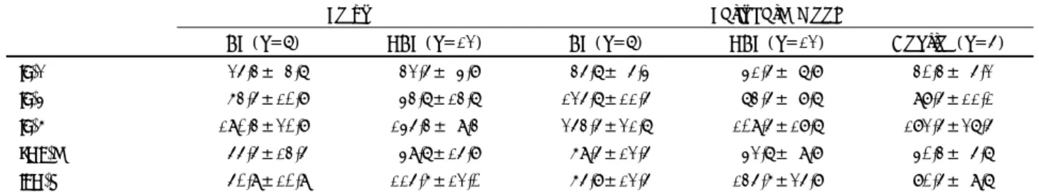
Table 1. Comparison of cytokine production of lymphocytes from nasal polyp and peripheral blood (unit:pg/ml)
Polyp Peripheral blood
AP (n=7) NAP (n=12) AP (n=7) NAP (n=12) Control (n=5) IL-2 25.3± 3.7 32.5± 4.8 35.7± 5.4 41.5± 7.8 30.3± 5.2 IL-4 63.5±10.8 43.7±13.7 125.7±11.5 73.5± 8.7 98.5±11.1 IL-6 190.3±20.8 145.3± 9.3 253.5±21.7 109.5±18.7 182.5±27.5 TNF-a 55.5±13.5 49.7±15.8 69.5±12.5 42.7± 9.8 40.3± 5.7 IFN-g 50.9±10.9 115.6±12.0 65.8±12.5 135.6±25.8 80.5± 9.7 Values are expressed as means±SD AP:Allergic polyp patient, NAP:Nonallergic polyp patient
Fig. 1. Comparison of cytokine productivity of polyp lymphoc- ytes. *p<0.01, **p<0.001
Fig. 2. Comparison of cytokine productivity of peripheral blood lymphocytes. *p<0.01, **p<0.001
140 / J Rhinol 5(2), 1998
control group, but not in allergic group (Fig. 2).
DISCUSSION
Some immunohistochemical studies about lymphocyte su- bpopulations in the nasal polyp revealed significantly more CD8
+than CD4
+cells.
9)10)They suggested that cellular im- munity of T lymphocytes rather than allergy may be involved in the formation of nasal polyp. We used Con A to stimulate T lymphocytes and this mitogen does not require the presence of accessory cells to activate T cells and dose not induce cyt- okine production from other cells.
11)The activated T cells in- clude subpopulations of both CD4
+and CD8
+cells. CD4
+T cell is divided into Th1 and Th2 subsets according to their cytokine profiles in tissue and peripheral blood of different disease. Th1 subset secretes IFN-γ and IL-2. IFN-γ is in- volved in delayed hypersensitivity, macrophage attraction and suppression of IgE secretion. Th2 type cytokines are IL-4, IL-5, IL-6, IL-10, and IL-13.
CD8
+cells are considered to have suppressor/cytotoxic fu- nction. But recently, distinct cytokine-secreting subsets of CD8
+T cell (Tc), similar to their CD4
+Th1 and Th2-type count- erparts, have been identified. Tc1 cells secrete predominantly IL-2 and IFN-γ, and Tc2 cells secrete IL-4 and IL-5.
12-14)As with CD4
+T cells, the presence of IL-4 in vitro favors the differentiation of CD8
+T cells toward Tc2-type immune re- sponse, resulting in production of IL-4 and IL-5, suppression of IFN-γ production, and facilitation in IgE synthesis.
15)16)The presence of IL-12 and IFN-γ is reported to favor the generation of Th1 cells in CD4
+T cells. And the effect of these cytokines on CD8
+T cells exhibits strong similarities with that on CD4
+T cell.
13)16)IL-4 induces the production of IgE and also stimulates the expression of VCAM-1 on endothelial cells, resulting in incr- eased binding of lymphocytes, monocytes, and especially eo- sinophils.
17)IL-6 helps the production of IgE. IL-5, which was not evaluated in this study, is the most specific eosinophil gr- owth and differentiation factor known.
18)Walker et al.
19)compared patients with intrinsic asthma versus patients with extrinsic asthma. Patient with intrinsic asthma had normal levels of IgE and negative results of skin tests for aeroallergens. Extrinsic asthma shows increased levels of IL-4 and IL-5 and low levels of IFN-γ and IL-2. However intrinsic asthma have elevated levels of IL-2, IFN-γ and IL-5 and low levels of IL-4.
Nasal polyp of non-allergic patients shows high levels of IFN-γ, IL-2 and tissue eosinophilia similar to intrinsic asthma.
If the level of IL-5 is high in nasal polyp of non-allergic pati- ent, it would indicate a similar immunologic picture to intrin- sic asthma.
8)But there are controversies about IL-5 levels in nasal polyp.
6)7)Localized nasal allergy could be an etiologic factor for the development of nasal polyp. Cho et al.
20)demonstrated local production of specific IgE in nasal polyp tissues. We found that cytokine productivity of lymphocytes in nasal polyp were parallel to that of peripheral blood lymphocytes. So it is likely that a certain systemic factor such as allergy or infection could be a cause of nasal polyp. In allergic patients, nasal polyp ly- mphocytes were predominantly Th2 type, whereas in non- allergic patients they were predominantly Th1 type in our study.
But this cannot be the direct evidence that Th2 type lymph- ocytes in nasal polyp play a major role in the formation of nasal polyp in allergic patients.
REFERENCES
1) Mygind N. Nasal polyps. In: Mygind N editor. Nasal allergy. 2nd ed. London: Blackwell Scientific Publication;1979. p.233-8.
2) Durham SR, Ying S, Varney VA, Jacobson MR, Sudderick RM, Mackay IS, et al. Cytokine messenger RNA expression for IL-3, L-4, IL-5, and granulocyte macrophage colony stimulating factor in the nasal mucosa after local allergen provocation: Relationship to tissue eosinophilia. J Immunol 1992;148:2390-4.
3) Wilson JW, Djukanovic R, Howarth PH, Holgate ST. Lymphocyte activation in bronchoalveolar lavage and peripheral blood in ato- pic asthma. Am Rev Respir Dis 1992;145:958-60.
4) Maggi E, Biswas P, Prete GD, Parronchi P, Macchia D, Simonelli C, et al. Accumulation of Th2-like helper T cell in the conjunc- tiva of patients with vernal conjunctivitis. J Immunol 1991;146:
1169-74.
5) Romagnani S. Regulation and deregulation of human IgE synthesis.
Immunol Today 1990;11:316-22.
6) Hamilos DL, Leung DY, Wood R, Cunningham L, Bean DK, Yas- ruel Z. et al. Evidence for distinct cytokine expression in allergic versus nonallergic chronic sinusitis. J Allergy Clin Immunol 1995;
96:537-44.
7) Min YG, Lee CH, Rhee CS, Kim KH, Kim CS, Koh YY. Inflam- matory cytokine expression on nasal polyps developed in allergic and infectious rhinitis. Acta Otolaryngol (Stockh) 1997;117:302-6.
8) Miller CH, Pudiak DR, Hatem F, Looney RJ. Accumulation of interferon gamma producing TH1 helper T cells in nasal polyps.
Otolaryngol Head Neck Surg 1994;111:51-8.
9) Stoop AE, Heijden HA, Biewenga J, Baan S. Lymphocytes and nonlymphoid cells in human nasal polyps. J Allergy Clin Immunol 1991;87:470-5.
10) Liu CM, Shun CT, Hsu MM. Lymphocyte subsets and antigen- specific IgE antibody in nasal polyps. Ann Allergy 1994;72:19-24.
11) Weiss A, Shields R, Newton M, Manger B, Imboden J. Ligandre- ceptor interaction required for commitment to the activation of the interleukin 2 gene. J Immunol 1987;138:2169-76.
12) Kemeny DM, Noble A, Holmes BJ, Diaz-Sanchez D. Immune re- gulation: A new role for the CD8+ T cells. Immunol Today 1994;
15:107-10.
13) Noble A, Kemeny DM. Interleukin-4 and interferon-γ regulate differentiation of CD8+ T cells into populations with divergent cytokine profiles. Int Arch Allergy Immunol 1995;107:186-8.
14) Stanciu LA, Shute J, Promwong C, Holgate ST, Djukanovic R.
Increased levels of IL-4 in CD8+ T cells in atopic asthma. J All- ergy Clin Immunol 1997;100:373-8.
Kim et al:Functional Differences of Lymphocytes in Nasal Polyp / 141
15) Erard F, Wild M, Garcia-Sanz J, Gros GL. Switch of CD8+ T cells to noncytolytic CD8- CD4- cells that make TH2 cytokines and help B cells. Science 1993;260:1802-5.
16) Croft M, Carter L, Swain SL, Dutton RW. Generation of polarized antigen-specific CD8 effector populations: Reciprocal action of interleukin (IL)-4 and IL-12 in promoting type 2 versus type 1 cytokine profiles. J Exp Med 1994;180:1715-28.
17) Bochner BS, Luskinskas FW, Gimbrone MA, Mewman W, Stervi- nsky SA, Derse-Anthony CD, et al. Adhesion of human basophils, eosinophils, and neutrophils to interleukin 1-activated human va- scular endothelial cells: Contribution of endothelial cell adhesion
molecules. J Exp Med 1991;173:1553-6.
18) Clutterbuck EJ, Hirst EM, Sanderson CJ. Human interleukin-5 (IL-5) regulates the produ- ction of eosinophils in human bone marrow cultures: Comparison and interaction with IL-1, IL-3, IL-6, and GM-CSF. Blood 1989;73:1504-12.
19) Walker C, Bode E, Bore L, Hansel TT, Blaser K, Virchow JC. Al- lergic and nonallergic asthmatics have distinct patterns of T-cell activation and cytokine production in peripheral blood and bronc- hoalveolar lavage. Am Rev Respir Dis 1992;146:109-15.
20) Cho CM, Jang TY, Yun YS, Jung DH. Local production of IgE in nasal polyp. J Rhinol 1997;4:126-8.