Received:May 7, 2019, Revised:June 9, 2019, Accepted:June 18, 2019 Corresponding to:Ki-Jo Kim http://orcid.org/0000-0002-3598-2396
Division of Rheumatology, Department of Internal Medicine, St. Vincent’s Hospital, College of Medicine, The Catholic University of Korea, 93 Jungbu-daero, Paldal-gu, Suwon 16247, Korea. E-mail:[email protected]
Copyright ⓒ 2019 by The Korean College of Rheumatology. All rights reserved.
This is an Open Access article, which permits unrestricted non-commerical use, distribution, and reproduction in any medium, provided the original work is properly cited.
Association of Uveitis with Radiographic Progression in Patients with Axial Spondyloarthritis: A Propensity Score Matching Analysis
Ki-Jo Kim, Young Bin Joo, Yune-Jung Park, Kyung-Su Park
Division of Rheumatology, Department of Internal Medicine, St. Vincent’s Hospital, College of Medicine, The Catholic University of Korea, Seoul, Korea
Objective. Acute anterior uveitis (AAU) is the most common extra-articular manifestation in patients with axial spondyloar- thritis (axSpA). However, the relationship between AAU and radiographic progression in axSpA remains unclear. Hence, we investigated whether the presence of AAU is associated with radiographic structural damage in patients with axSpA. Methods.
Clinical and radiographic data were obtained from 253 patients with axSpA. Radiographic progression over 2 years was as- sessed using the modified Stoke Ankylosing Spondylitis Spine Score (mSASSS). Progression was defined as mSASSS worsening by ≥two units. Using propensity score (PS) matching, differences between patients with and without AAU were analyzed.
Results. The proportion of progressors among patients with AAU was lower than that of patients without AAU (13.6% vs.
29.5%, p=0.058). The rate of increase in mSASSS and number of syndesmophytes were lower in patients with AAU than pa- tients without AAU (0.57±1.37 vs. 1.02±1.79, p=0.085 and 0.46±1.45 vs. 0.83±1.62, p=0.158). In multivariate regression analysis, presence of AAU was independently associated with slowed radiographic progression (odds ratio [95% confidence interval] 0.21 [0.07, 0.67], p=0.004). Conclusion. PS-matched axSpA patients with AAU showed significantly less radiographic progression than those without AAU. (J Rheum Dis 2019;26:248-256)
Key Words. Axial spondyloarthritis, Radiographic progression, Uveitis, Propensity score matching
INTRODUCTION
Axial spondyloarthritis (axSpA) is a chronic, pro- gressive disease characterized by inflammation of en- theses, leading to new bone formation and ankylosis of joints, primarily in the axial skeleton [1,2]. Functional disability resulting from progressive deformation and an- kylosis of the spine leads to decreases in physical activity and quality of life. Because axSpA occurs more commonly in economically active, young males, it can lead to pro- ductivity losses that result in substantial economic bur- den to society [3,4].
axSpA is an interesting rheumatic disease due to close associations with acute anterior uveitis (AAU), psoriasis,
and inflammatory bowel disease, so-called extra-articular manifestations [5,6]. The presence of extra-articular manifestations not only plays a critical role in the diag- nosis of axSpA [7], but also has an impact on health-re- lated quality of life and treatment choice [8]. AAU is the most common extra-articular manifestation, with a prev- alence of 20%∼30% [5,6,9]. Presence of AAU increases the probability of axSpA in patients presenting with chronic back pain [10] and is associated with higher dis- ease activity, poor functional ability, and advanced phys- ical impairment [11]. In axSpA patients with a history of AAU, infliximab and adalimumab are preferred over eta- nercept because of lower AAU flare rates [12].
Several parameters have been identified as risk factors
for radiographic progression in patients with axSpA: male sex, smoking, syndesmophyte(s) at baseline, high degree of sacroiliitis on magnetic resonance images, and in- creased level of C-reactive protein (CRP) [1]. High body mass index (BMI) and peripheral arthritis were also sug- gested as potential clinical predictors of radiographic pro- gression of the spine [13-17]. However, the relationship between AAU and radiographic progression in axSpA re- mains unclear. In a previous cohort study, history of AAU was associated with higher Bath Ankylosing Spondylitis Radiology Index (BASRI) score, but the association was found only in male patients and was not statistically sig- nificant [14].
The absence of randomization in observational studies can make inferences about factor or treatment effects sus- ceptible to indication [18]. Propensity score (PS) match- ing is a statistical technique that matches patients in two groups using a factor to balance observed characteristics, achieve exchangeability, and produce groups that are functionally randomized [18,19]. Therefore, once pa- tients have been matched based on PS, any observed dif- ference in outcomes is assumed to be a direct result of the factor or treatment dividing the two groups. To properly investigate the relationship between AAU and radio- graphic progression in axSpA, we used PS matching to match axSpA patients with and without AAU and com- pared the radiographic progression between the two groups.
MATERIALS AND METHODS
Patients
A total of 412 axSpA patients who fulfilled the Assessment of Spondyloarthritis International Society (ASAS) classification criteria for axSpA [7] and had re- ceived care at St. Vincent’s Hospital, The Catholic University of Korea (Suwon, Korea) between 2008 and 2017 was identified. Clinical and laboratory data and ra- diographic images were retrieved from medical records.
At baseline, sex, age at diagnosis, time since diagnosis, HLA-B27 status, smoking status, and history of extra- articular manifestations (uveitis, psoriasis, inflammatory bowel disease, peripheral arthritis, and enthesitis) were recorded. Disease activity was assessed using the Ankylosing Spondylitis Disease Activity Score (ASDAS) according to CRP level [20]. Of the total sample, 253 pa- tients who were followed for 2 years were assessed for ra- diographic progression. The study was carried out in ac-
cordance with the Helsinki Declaration and approved by the institutional review board of St. Vincent’s Hospital, The Catholic University of Korea (no. VC19RESI0090).
Radiographs and scoring
Radiographs of the sacroiliac joints and the cervical and lumbar spine were obtained by the local investigator at baseline and after 2 years of follow-up. All available radio- graphs for each patient were independently scored at the same time by two experienced readers who were blinded to all other data except radiograph chronology according to the modified Stoke Ankylosing Spondylitis Spine Score (mSASSS) [21]. Spinal radiographic progression was de- fined as worsening of the mean mSASSS by more than two units over 2 years, in conformity with previous stud- ies [22-24], and axSpA patients were categorized into two subgroups, progressors, and non-progressors. Radiographic sacroiliitis (SI) was scored according to the modified New York criteria [25], and radiological hip involvement was graded using the BASRI-hip scoring system [26].
Interobserver reliability was assessed by calculating the interclass correlation coefficient, which was 0.946 (95%
confidence interval [CI] 0.940 to 0.952).
Propensity score matching
Because we expected systematic differences in the base- line characteristics of subjects with or without AAU, we performed PS matching to adjust for these potential im- balances [18,19]. We used logistic regression to obtain the PS [27], and the variables included in PS estimation were sex, age at diagnosis, disease duration, BMI, HLA-B27 positivity, smoking status, history of peripheral arthritis, enthesitis, psoriasis, inflammatory bowel dis- ease, erythrocyte sedimentation rate at baseline, CRP lev- el at baseline, ASDAS-CRP at baseline, use of tumor ne- crosis factor (TNF) inhibitors, mSASSS, presence of syn- desmophyte(s), SI score, BASRI-hip score, and z-scores of bone mineral density at the lumbar spine and femur neck. Patients without AAU were matched based on PS using the nearest-neighbor matching algorithm without replacement and with a 1:3 ratio within 0.05 of the esti- mated PS [27].
Statistical analyses
For continuously distributed data, the results are shown as mean with standard deviation (SD); between-group comparisons were performed using Student’s t-test.
Categorical and dichotomous variables are expressed as
Table 1. Baseline characteristics of axSpA patients with and without AAU after propensity score matching
Variable Patients with AAU (n=44)
Patients without AAU After PS matching
(n=132) p-value† Before PS matching
(n=209) p-value‡
Male 34 (77.3) 107 (81.1) 0.744 153 (73.2) 0.712
Age at diagnosis (yr) 34.2±12.6 33.3±12.4 0.672 32.7±12.1 0.190 Disease duration (yr) 4.48±4.96 4.61±5.46 0.878 3.88±5.18 0.227
BMI (kg/m2) 0.794
Underweight, <18.5 0 (0.0) 1 (0.8) 9 (4.3)
Normal, 18.5∼25 24 (54.5) 80 (60.6) 133 (63.6)
Overweight, 25∼30 16 (36.4) 39 (29.5) 52 (24.9)
Obese, <30 4 (9.1) 12 (9.1) 15 (7.2)
HLA-B27 42 (95.5) 126 (95.5) 1.000 175 (83.7) 0.074
Smoking 0.861 0.578
Non-smoker 25 (56.8) 71 (53.8) 131 (62.7)
Smoker 19 (43.2) 61 (46.2) 78 (37.3)
Peripheral arthritis 8 (18.2) 27 (20.5) 0.913 77 (36.8) 0.027
Enthesitis 0 (0.0) 0 (0.0) 1.000 13 (6.2) 0.186
Psoriasis 1 (2.3) 3 (2.3) 1.000 3 (1.4) 1.000
Inflammatory bowel disease 1 (2.3) 1 (1.5) 1.000 5 (2.4) 1.000
ESR (mm/h) 30.2±21.4 31.5±25.1 0.768 36.1±27.6 0.122
CRP (mg/dL) 2.1±3.8 1.8±2.9 0.616 1.8±2.7 0.542
ASDAS-CRP 3.0±1.0 3.0±0.9 0.974 3.1±0.9 0.650
Use of TNF inhibitor* 25 (56.8) 76 (57.6) 1.000 114 (54.5) 0.913
Continuous use of NSAIDs 17 (61.4) 89 (67.4) 0.582 140 (67.0) 0.589
mSASSS, units 8.3±13.0 8.7±13.1 0.891 8.4±13.9 0.962
Presence of syndesmophyte(s) 19 (43.2) 52 (39.4) 0.790 82 (39.2) 0.751
Number of syndesmophyte(s) 2.3±4.5 2.5±4.3 0.835 2.6±4.8 0.781
Sacroiliac joint 0.859/0.746 0.552/0.560
Grade 0 (right/left) 7 (15.9)/7 (15.9) 16 (12.1)/18 (13.6) 39 (18.7)/46 (22.0) Grade 1 (right/left) 6 (13.6)/6 (13.6) 24 (18.2)/25 (18.9) 45 (21.5)/38 (18.2) Grade 2 (right/left) 12 (27.3)/13 (29.3) 42 (31.8)/35 (26.5) 59 (28.2)/53 (25.4) Grade 3 (right/left) 8 (18.2)/7 (15.9) 23 (17.4)/29 (22.0) 32 (15.3)/38 (18.2) Grade 4 (right/left) 11 (25.0)/11 (25.0) 27 (20.5)/25 (18.9) 34 (16.3)/34 (16.3)
Hip involvement 0.056/0.793 0.036/0.469
Grade 0 (right/left) 39 (88.6)/40 (90.9) 121 (91.7)/123 (93.2) 192 (91.9)/192 (91.9) Grade 1 (right/left) 2 (4.5)/1 (2.3) 3 (2.3)/4 (3.0) 7 (3.3)/9 (4.3) Grade 2 (right/left) 1 (2.3)/2 (4.5) 8 (6.1)/3 (2.3) 9 (4.3)/4 (1.9) Grade 3 (right/left) 0 (0.0)/0 (0.0) 0 (0.0)/1 (0.8) 1 (0.5)/3 (1.4) Grade 4 (right/left) 2 (4.5)/1 (2.3) 0 (0.0)/1 (0.8) 0 (0.0)/1 (0.5) BMD (g/cm2)
Lumbar spine 1.050±0.136 1.060±0.168 0.932 1.060±0.157 0.802
Femoral neck 0.862±0.096 0.856±0.131 0.733 0.847±0.117 0.384
Total hip 0.942±0.104 0.924±0.119 0.337 0.907±0.107 0.047
Z score
Lumbar spine −0.173±0.917 −0.227±1.210 0.754 −0.220±1.130 0.768
Femoral neck −0.118±0.900 −0.264±0.852 0.350 −0.386±0.804 0.073
Total hip −0.207±0.793 −0.304±0.817 0.488 −0.434±0.726 0.085
Values are presented as number (%) or mean±standard deviation. axSpA: axial spondyloarthritis, AAU: acute anterior uveitis, PS:
propensity score, BMI: body mass index, ESR, erythrocyte sedimentation rate, CRP: c-reactive protein, ASDAS: Ankylosing Spondylitis Disease Activity Score, TNF: tumor necrosis factor, NSAIDs: non-steroidal anti-inflammatory drugs, mSASSS: modified Stoke Ankylosing Spondylitis Spine Score, BMD: bone mineral density. *Counted if ever used. TNF inhibitors include etanercept, adalimumab, infliximab, and golimumab. †Comparison between patients with AAU and the matched patients without AAU.
‡Comparison between patients with AAU and the unmatched patients without AAU.
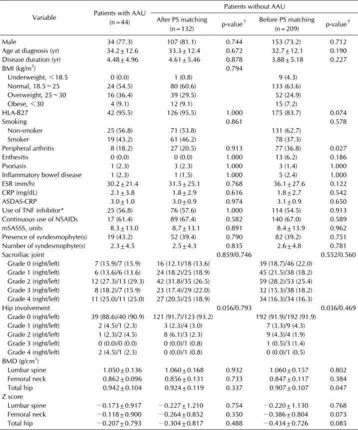
Figure 1. Radiographic progression over 2 years in axSpA patients with AAU and those without AAU. Upper panel indicates the result after PS matching and lower panel before PS matching. (A, D) Proportion of progressors. Comparison by chi-square test. (B, E) Change in mSASSS. Comparison by t-test. (C, F) Change in number of syndesmophytes. Comparison by t-test. axSpA: axial spon- dyloarthritis, AAU: acute anterior uveitis, PS: propensity score, mSASSS: modified Stoke Ankylosing Spondylitis Spine Score.
frequency and percentage and were compared using the chi-square test or Fisher’s exact test. Multivariable logis- tic regression analysis was performed to identify in- dependent predictors associated with radiographic progression. The effect size was computed by Cohen’s d method [28]. In our analysis, α was set at 5%, and a two-sided p-value less than 0.05 was considered statisti- cally significant. All statistical analyses were performed using R (version 3.5.3, The R Project for Statistical Computing; www.r-project.org).
RESULTS
Characteristics of the study sample
A total of 44 axSpA patients was identified to have a his- tory of AAU from the original sample of 253 patients who were followed for 2 years and assessed for radiographic progression. Through PS matching, 44 patients with AAU were matched to 209 patients without AAU at a 1:3 ratio and a comparator group of 132 patients without AAU was created. The baseline characteristics of the axSpA patients with AAU (n=44) and without AAU (n=132) did not differ and were well-balanced (Table 1).
Table 2. Multivariable logistic regression analysis for radiographic progression in patients with axial spondyloarthritis
Variables Crude OR (95% CI) Adjusted OR (95% CI) p-value
After PS matching
Male sex 3.18 (1.05, 9.57) 4.65 (1.02, 21.25) 0.047
Age at diagnosis 1.04 (1.01, 1.07) 1.04 (0.99, 1.09) 0.097
Disease duration 1.01 (0.96, 1.06) 1.06 (0.98, 1.14) 0.141
BMI 2.02 (1.23, 3.31) 2.89 (1.46 5.72) 0.002
HLA-B27 0.56 (0.13, 2.42) 0.20 (0.03, 1.53) 0.122
Smoking 1.73 (0.87, 3.42) 1.31 (0.51, 3.34) 0.576
CRP 1.04 (0.94, 1.15) 0.93 (0.76, 1.13) 0.454
ASDAS-CRP 1.30 (0.90, 1.88) 0.91 (0.47, 1.76) 0.790
Presence of syndesmophyte(s) at baseline 1.21 (1.11, 1.32) 1.16 (1.05, 1.28) 0.004
Use of TNF inhibitor 0.63 (0.32, 1.24) 0.39 (0.15, 1.01) 0.052
Continuous use of NSAIDs 1.85 (0.86, 3.97) 1.86 (0.70, 4.94) 0.211
Uveitis 0.38 (0.15, 0.96) 0.23 (0.07, 0.75) 0.015
Before PS matching
Male sex 5.62 (91.01, 1.06) 6.94 (2.08, 23.13) 0.002
Age at diagnosis 1.04 (1.01, 1.06) 1.04 (1.00, 1.08) 0.040
Disease duration 1.03 (0.99, 1.07) 1.06 (0.99, 1.13) 0.086
BMI 1.88 (1.25, 2.84) 2.44 (1.43, 4.18) 0.001
HLA-B27 1.02 (0.45, 2.30) 0.72 (0.24, 2.12) 0.549
Smoking 2.27 (1.27, 4.03) 1.62 (0.76, 3.47) 0.210
CRP 1.07 (0.98, 1.18) 0.97 (0.53, 1.56) 0.725
ASDAS-CRP 1.35 (0.99, 1.84) 0.91 (0.53, 1.56) 0.719
Presence of syndesmophyte(s) at baseline 1.19 (1.11, 1.27) 1.22 (1.02, 1.20) 0.011
Use of TNF inhibitor 0.70 (0.40, 1.24) 0.56 (0.27, 1.29) 0.134
Continuous use of NSAIDs 2.47 (1.26, 4.85) 2.14 (0.94, 4.91) 0.072
Uveitis 0.41 (0.17, 1.02) 0.23 (0.07, 0.69) 0.009
OR: odds ratio, CI: confidence interval, PS: propensity score, BMI: body mass index, CRP: c-reactive protein, ASDAS: Ankylosing Spondylitis Disease Activity Score, TNF: tumor necrosis factor, NSAIDs: non-steroidal anti-inflammatory drugs.
Before PS matching, in patients without AAU, HLA-B27 positivity was less frequent, peripheral arthritis was more common, and bone mineral density of total hip was lower compared with those with AAU.
Radiographic progression
We examined radiographic progression over 2 years in axSpA patients with AAU and those without AAU. The proportion of progressors among patients with AAU was lower than that among patients without AAU (13.6% vs.
29.5%, p=0.058; Figure 1A). The rate of increase in mSASSS was lower in patients with AAU compared with patients without AAU (0.57±1.37 vs. 1.02±1.79, p=
0.085; Figure 1B). The number of syndesmophytes showed a smaller increase among patients with AAU than patients without AAU (0.46±1.45 vs. 0.83±1.62, p=0.158; Figure 1C). Before PS matching, differences in the indices of radiographic progression (the proportion of progressors, the rate of increase in mSASSS, and change
in the number of syndesmophytes) were weak and not clear (Figure 1D∼F).
Risk factors for radiographic progression
To determine the relationship between presence of AAU and radiographic progression, we performed multivariable logistic regression analysis including known risk factors (Table 2). The presence of syndesmophyte(s) at baseline, male sex, and BMI had significant association with rapid radiographic progression (odds ratio [OR] [95% con- fidence interval, 95% CI] 1.16 [1.05, 1.28], 4.65 [1.02, 21.25], and 2.89 [1.46, 5.72], respectively, all p<0.05).
This agrees well with previous results [15,23,29]. The presence of AAU was independently associated with slowed radiographic progression (OR [95% CI] 0.23 [0.07, 0.75], p=0.015). Cumulative probability plots of spinal progression after stratification by AAU status at the onset of the next radiographic interval are presented in Figure 2. Difference of change in mSASSS between the
Figure 2. Cumulative probability plot of 2-year progression in the modified Stoke Ankylosing Spine Score (mSASSS), illustrating the change (Δ) in mSASSS values from baseline of each individual radiographic interval to 2 years in patients with acute anterior uveitis (AAU) and those without AAU. Radiographic progression was defined as an increase in mSASSS ≥2 in 2 years. The effect size was computed by Cohen’s d method. (A) After propensity score (PS) matching, (B) Before PS matching.
patients with AAU and without AAU were more evident and the effect size was moderate (Cohen’s d=0.532).
Before PS matching, the presence of AAU held the in- dependent association with retarded radiographic pro- gression (OR [95% CI] 0.23 [0.07, 0.69], p=0.009), but the effect size was small (Cohen’s d=0.203) (Table 2).
DISCUSSION
Our PS matching analysis matched axSpA patients with AAU with those having similar characteristics without AAU at a 1:3 ratio. This enabled the results from the two groups to be effectively and credibly compared in terms of radiographic progression (proportion of progressors, rate of mSASSS, and development of new syndesmophytes).
We confirmed a significant association between presence of AAU and delayed radiographic progression in axSpA patients in multivariable regression analysis.
Given the irreversibility of structural damage to the axial skeleton, the ability to effect early prediction of radio- graphic progression and aggressive treatment would be of great benefit for rheumatologists who wish to monitor patient risk [30]. The inverse association between AAU and radiographic progression is a novel finding and prob- ably was not captured in previous studies because of the wide phenotypic diversity and heterogeneity of patients with axSpA [31]. We addressed this limitation by PS matching [18,19,27].
In the previous reports, an association of AAU with ra- diographic progression in axSpA was not significant
[13,32]. Essers et al. [32] investigated whether the pres- ence of extra-articular manifestations is associated with more radiographic damage. In a multivariable model, AAU was not associated with mSASSS over time.
However, baseline mSASSS was much higher and periph- eral arthritis was more severe in the patients with AAU.
Baseline radiographic damage such as syndesmophyte(s) is well-known as the strongest predictor for further radio- graphic progression [30], which might countervail the ef- fect of uveitis. In addition, disease duration and age were longer and higher in patients with AAU and a long-term observation over 12 years was also a distinct difference from our study. Deminger et al. [13] investigated the pre- dictors of radiographic progression overall and by sex in the 166 patients with axSpA who were followed up over 5 years. Radiographic progression (either as an increase in mSASSS by ≥2 points or as development of new syn- desmophyte[s]) in this study was defined over the 5 years, which is quite different from the standard time frame, 2 years, to assess the radiographic progression [22-24]. In addition, the frequency of AAU (51%) was ab- normally high compared with the general prevalence (20%∼30%).
Smoking and CRP level was not captured as having a sig- nificant association with radiographic progression in our study, and it depends partly on the multivariable models, design of studies and/or cohorts [23,33]. Influence of smoking on radiographic progression was further de- termined by its dose and the interaction with baseline syndesmophyte(s) and inflammation [23,34]. Use of
TNF inhibitors can effectively suppress the inflammation, leading to the decrease in the radiographic progression regardless of baseline CRP level [33,35]. In this study, over a half of the patients (n=101, 57.4%) received TNF inhibitors and use of TNF inhibitors was marginally asso- ciated with the slowed progression (OR [95% CI] 0.39 [0.15, 1.01], p=0.052; Table 2). The robust use of TNF in- hibitors could mitigate against the effects of smoking and CRP in multivariable analysis.
Uveitis is the most common, clinically apparent, ex- tra-articular manifestation of axSpA, and one-third of axSpA patients experience uveitis at some point in the course of their disease [5,6]. From a clinical standpoint, uveitis usually presents as sudden onset episodes affect- ing only one eye at a time, but sacroiliitis and new bone formation insidiously progress at multiple levels [36].
This observation led us to hypothesize that there is an op- posing interaction between uveitis and radiographic progression. The uvea is the vascular middle layer of the eye, composed of the iris, ciliary body, and choroid [37].
The ciliary body includes the ciliary muscle and is con- nected to the lens through the zonular fiber, which is the suspensory ligament of the lens [38]. At a glance, the structure of the ciliary body looks like enthesis. This might be a clue to the intriguing association between uveitis and axSpA. However, to the best of our knowl- edge, there have been no studies addressing this potential relationship. Unraveling the intriguing coupling between uveitis and radiographic progression could help us better understand the etiopathogenesis of axSpA.
There are some limitations to be addressed in this study.
First, the study sample was relatively small and from a single center. Second, radiographic image evaluations were prospectively executed, but the data were retro- spectively collected. Retrospective data collection is in- herently susceptible to bias, including misclassification, information, and selection bias. Third, we cannot exclude the possibility of index event bias. When multiple risk factors contribute to the risk of an outcome, conditioning on the outcome induces dependence between the risk fac- tors, even when these risk factors are independently dis- tributed in the general population. This effect creates a spurious association among these risk factors with an in- dex event [39,40].
CONCLUSION
In the present study, we demonstrate that the presence
of AAU has not only a role in diagnosis and treatment choice, but also an association with radiographic out- come in axSpA. The association between AAU and radio- graphic progression in axSpA should be validated through further large, multi-center studies. Unraveling the mystery of this association could help us better un- derstand the pathoetiology of axSpA.
CONFLICT OF INTEREST
No potential conflict of interest relevant to this article was reported.
AUTHOR CONTRIBUTIONS
K.J.K. designed the study, and K.J.K. and Y.B.J. carried out data collection. K.J.K. performed statistical analysis and drafted the paper, and all authors (K.J.K., Y.B.J., Y.J.P., and K.S.P.) were involved in critically revising the final preparation. All authors approved the final version to be published.
REFERENCES
1. Sieper J, Braun J, Dougados M, Baeten D. Axial spondy- loarthritis. Nat Rev Dis Primers 2015;1:15013.
2. Sieper J, Poddubnyy D. Axial spondyloarthritis. Lancet 2017;390:73-84.
3. Boonen A, van der Linden SM. The burden of ankylosing spondylitis. J Rheumatol Suppl 2006;78:4-11.
4. Krüger K, von Hinüber U, Meier F, Tian H, Böhm K, Jugl SM, et al. Ankylosing spondylitis causes high burden to pa- tients and the healthcare system: results from a German claims database analysis. Rheumatol Int 2018;38:2121-31.
5. Stolwijk C, Essers I, van Tubergen A, Boonen A, Bazelier MT, De Bruin ML, et al. The epidemiology of extra-articular manifestations in ankylosing spondylitis: a population- based matched cohort study. Ann Rheum Dis 2015;74:
1373-8.
6. Stolwijk C, van Tubergen A, Castillo-Ortiz JD, Boonen A.
Prevalence of extra-articular manifestations in patients with ankylosing spondylitis: a systematic review and meta- analysis. Ann Rheum Dis 2015;74:65-73.
7. Rudwaleit M, van der Heijde D, Landewé R, Listing J, Akkoc N, Brandt J, et al. The development of Assessment of SpondyloArthritis International Society classification cri- teria for axial spondyloarthritis (part II): validation and final selection. Ann Rheum Dis 2009;68:777-83.
8. Gao X, Wendling D, Botteman MF, Carter JA, Rao S, Cifaldi M. Clinical and economic burden of extra-articular manifes- tations in ankylosing spondylitis patients treated with an- ti-tumor necrosis factor agents. J Med Econ 2012;15:1054-63.
9. Koo BS, Lim JW, Shin JH, Kim TH. Characteristics of uveitis in patients with ankylosing spondylitis in Korea: a sin- gle-center survey. J Rheum Dis 2018;25:28-33.
10. Moltó A, Paternotte S, Comet D, Thibout E, Rudwaleit M, Claudepierre P, et al. Performances of the Assessment of SpondyloArthritis International Society axial spondyloar- thritis criteria for diagnostic and classification purposes in patients visiting a rheumatologist because of chronic back pain: results from a multicenter, cross-sectional study.
Arthritis Care Res (Hoboken) 2013;65:1472-81.
11. Chen CH, Lin KC, Chen HA, Liao HT, Liang TH, Wang HP, et al. Association of Acute Anterior uveitis with disease ac- tivity, functional ability and physical mobility in patients with ankylosing spondylitis: a cross-sectional study of Chinese patients in Taiwan. Clin Rheumatol 2007;26:953-7.
12. Lie E, Lindström U, Zverkova-Sandström T, Olsen IC, Forsblad-d'Elia H, Askling J, et al. Tumour necrosis factor inhibitor treatment and occurrence of anterior uveitis in an- kylosing spondylitis: results from the Swedish biologics register. Ann Rheum Dis 2017;76:1515-21.
13. Deminger A, Klingberg E, Geijer M, Göthlin J, Hedberg M, Rehnberg E, et al. A five-year prospective study of spinal ra- diographic progression and its predictors in men and wom- en with ankylosing spondylitis. Arthritis Res Ther 2018;
20:162.
14. Atagunduz P, Aydin SZ, Bahadir C, Erer B, Direskeneli H.
Determinants of early radiographic progression in ankylos- ing spondylitis. J Rheumatol 2010;37:2356-61.
15. Jeong H, Bea EK, Lee J, Koh EM, Cha HS. Body mass index and estrogen predict radiographic progression in the spine in ankylosing spondylitis. Joint Bone Spine 2015;82:473-4.
16. Kim TJ, Lee S, Joo KB, Park DJ, Park YW, Lee SS, et al. The presence of peripheral arthritis delays spinal radiographic progression in ankylosing spondylitis: observation Study of the Korean Spondyloarthropathy Registry. Rheumatology (Oxford) 2014;53:1404-8.
17. Kim H, Lee J, Ahn JK, Hwang J, Park EJ, Jeong H, et al.
Predictive factors of radiographic progression in ankylosing spondylitis. Korean J Intern Med 2015;30:391-7.
18. Johnson SR, Tomlinson GA, Hawker GA, Granton JT, Feldman BM. Propensity score methods for bias reduction in observational studies of treatment effect. Rheum Dis Clin North Am 2018;44:203-13.
19. Luo Z, Gardiner JC, Bradley CJ. Applying propensity score methods in medical research: pitfalls and prospects. Med Care Res Rev 2010;67:528-54.
20. Molto A, Gossec L, Meghnathi B, Landewé RBM, van der Heijde D, Atagunduz P, et al. An Assessment in SpondyloArthritis International Society (ASAS)-endorsed definition of clinically important worsening in axial spondy- loarthritis based on ASDAS. Ann Rheum Dis 2018;77:
124-7.
21. Creemers MC, Franssen MJ, van't Hof MA, Gribnau FW, van de Putte LB, van Riel PL. Assessment of outcome in ankylos- ing spondylitis: an extended radiographic scoring system.
Ann Rheum Dis 2005;64:127-9.
22. Poddubnyy D, Conrad K, Haibel H, Syrbe U, Appel H, Braun J, et al. Elevated serum level of the vascular endothelial growth factor predicts radiographic spinal progression in patients with axial spondyloarthritis. Ann Rheum Dis 2014;
73:2137-43.
23. Poddubnyy D, Haibel H, Listing J, Märker-Hermann E, Zeidler H, Braun J, et al. Baseline radiographic damage, ele- vated acute-phase reactant levels, and cigarette smoking
status predict spinal radiographic progression in early axial spondylarthritis. Arthritis Rheum 2012;64:1388-98.
24. Poddubnyy D, Protopopov M, Haibel H, Braun J, Rudwaleit M, Sieper J. High disease activity according to the Ankylosing Spondylitis Disease Activity Score is associated with accelerated radiographic spinal progression in patients with early axial spondyloarthritis: results from the GErman SPondyloarthritis Inception Cohort. Ann Rheum Dis 2016;75:2114-8.
25. van der Linden S, Valkenburg HA, Cats A. Evaluation of di- agnostic criteria for ankylosing spondylitis. A proposal for modification of the New York criteria. Arthritis Rheum 1984;27:361-8.
26. MacKay K, Brophy S, Mack C, Doran M, Calin A. The devel- opment and validation of a radiographic grading system for the hip in ankylosing spondylitis: the bath ankylosing spon- dylitis radiology hip index. J Rheumatol 2000;27:2866-72.
27. Lee DK. An introduction to propensity score matching methods. Anesth Pain Med 2016;11:130-48.
28. Lee DK. Alternatives to P value: confidence interval and ef- fect size. Korean J Anesthesiol 2016;69:555-62.
29. van Tubergen A, Ramiro S, van der Heijde D, Dougados M, Mielants H, Landewé R. Development of new syndesmo- phytes and bridges in ankylosing spondylitis and their pre- dictors: a longitudinal study. Ann Rheum Dis 2012;71:
518-23.
30. Poddubnyy D, Sieper J. Radiographic progression in anky- losing spondylitis/axial spondyloarthritis: how fast and how clinically meaningful? Curr Opin Rheumatol 2012;24:
363-9.
31. Baeten D, Breban M, Lories R, Schett G, Sieper J. Are spon- dylarthritides related but distinct conditions or a single dis- ease with a heterogeneous phenotype? Arthritis Rheum 2013;65:12-20.
32. Essers I, Ramiro S, Stolwijk C, Blaauw M, Landewé R, van der Heijde D, et al. Do extra-articular manifestations influ- ence outcome in ankylosing spondylitis? 12-year results from OASIS. Clin Exp Rheumatol 2016;34:214-21.
33. Park JW, Kim MJ, Lee JS, Ha YJ, Park JK, Kang EH, et al.
Impact of tumor necrosis factor inhibitor versus non- steroidal antiinflammatory drug treatment on radiographic progression in early ankylosing spondylitis: its relationship to inflammation control during treatment. Arthritis Rheumatol 2019;71:82-90.
34. Villaverde-García V, Cobo-Ibáñez T, Candelas-Rodríguez G, Seoane-Mato D, Campo-Fontecha PDD, Guerra M, et al.
The effect of smoking on clinical and structural damage in patients with axial spondyloarthritis: a systematic literature review. Semin Arthritis Rheum 2017;46:569-83.
35. Molnar C, Scherer A, Baraliakos X, de Hooge M, Micheroli R, Exer P, et al.; Rheumatologists of the Swiss Clinical Quality Management Program. TNF blockers inhibit spinal radiographic progression in ankylosing spondylitis by re- ducing disease activity: results from the Swiss Clinical Quality Management Cohort. Ann Rheum Dis 2018;77:
63-9.
36. Rosenbaum JT, Rosenzweig HL. Spondyloarthritis: the eyes have it: uveitis in patients with spondyloarthritis. Nat Rev Rheumatol 2012;8:249-50.
37. Rosenbaum JT, Asquith M. The microbiome and HLA-B27- associated acute anterior uveitis. Nat Rev Rheumatol
2018;14:704-13.
38. Kanski JJ, Bowling B. Kanski’s clinical ophthalmology e-book: a systematic approach. 8th ed. Elsevier Health Sciences, 2015.
39. Choi HK, Nguyen US, Niu J, Danaei G, Zhang Y. Selection
bias in rheumatic disease research. Nat Rev Rheumatol 2014;10:403-12.
40. Dahabreh IJ, Kent DM. Index event bias as an explanation for the paradoxes of recurrence risk research. JAMA 2011;
305:822-3.