CASE REPORT
국소 진행성 위암 환자의 전이성 연수막 암종증
지정근, 정주원, 남승우, 최승규, 이동원, 김대인, 전병관, 신윤재
국립중앙의료원 내과 소화기센터
A Case of Isolated Leptomeningeal Carcinomatosis from Advanced Gastric Cancer
Jung Geun Ji, Joo Won Chung, Seung Woo Nam, Seung Kyu Choi, Dong Won Lee, Dae In Kim, Byung Gwan Jeon, and Yun Jae Shin Gastrointestinal Center, Department of Internal Medicine, National Medical Center, Seoul, Korea
Leptomeningeal carcinomatosis (LMC) is rare metastatic form of gastric cancer. Most cases are diagnosed in the final stage after multiple distant metastasis. An 84-year-old woman was admitted with melena, headache and vomiting. Esophagogastro- duodenoscopy showed an ulceroinfiltrating lesion at the stomach (Borrmann class III), and biopsy revealed a signet ring cell carcinoma. The abdominal-pelvic CT showed no evidence of metastasis. A sudden decrease of consciousness was noted, but the brain CT showed no active lesion while the brain MRI revealed enhancement of leptomeninges. A lumbar puncture was performed and the cerebrospinal fluid study revealed malignant neoplastic cells. With family consent, no further evaluation and treatment were administered and she died six weeks after the diagnosis of gastric cancer. We report an extremely rare case of a patient who initially presented with neurologic symptoms, and was diagnosed LMC from advanced gastric cancer without any evidence of metastasis in abdomen and pelvis. (Korean J Gastroenterol 2016;68:93-98)
Key Words: Gastric cancer; Leptomeningeal carcinomatosis; Neurologic manifestations
Received April 12, 2016. Revised May 13, 2016. Accepted May 14, 2016.
CC This is an open access article distributed under the terms of the Creative Commons Attribution Non-Commercial License (http://creativecommons.org/licenses/
by-nc/4.0) which permits unrestricted non-commercial use, distribution, and reproduction in any medium, provided the original work is properly cited.
Copyright © 2016. Korean Society of Gastroenterology.
교신저자: 정주원, 04564, 서울시 중구 을지로 245, 국립중앙의료원 내과 소화기센터
Correspondence to: Joo Won Chung, Gastrointestinal Center, Department of Internal Medicine, National Medical Center, 245 Eulji-ro, Jung-gu, Seoul 04564, Korea.
Tel: +82-2-2276-2267, Fax: +82-2-2267-8685, E-mail: [email protected] Financial support: None. Conflict of interest: None.
서 론
전이성 연수막 암종증(metastatic leptomeningeal carci- nomatosis)은 뇌연막과 지주막 사이의 공간으로 암세포가 전 이되어 광범위하게 침윤한 것으로 정의한다. 이러한 전이성 연수막 암종증은 전체 고형 종양의 약 3-8%에서 발생하는 것 으로 보고되고 있다.1 성인의 경우 유방암, 폐암, 림프종, 악성 흑색종에서 주로 발생하며 소아의 경우 백혈병이 흔한 원발 종양으로 알려져 있다.2 원발 종양에 관계없이 전이성 연수막 암종증은 대부분 그 예후가 매우 불량하다.3,4
위암은 갑상선암을 제외하고 우리나라에서 가장 많이 발생 하는 악성 종양이며, 위암의 5년 생존율은 73.1%로 비교적 길고 간, 폐, 복막 등의 부위에 주로 전이되는 것으로 알려져
있다.5 반면 위암에서 전이성 연수막 암종증이 발생하는 경우 는 약 0.06% 정도로 드물게 보고되고 있다.6 특히 전이성 연 수막 암종증으로 진단되는 시점에는 림프절이나 간의 전이, 복막의 암종증과 같이 원격 전이가 진행된 상태(final stage) 가 대부분이며 타 장기에 전이 없이 초기 단계에 진단되는 경우는 매우 드물다.4
저자들은 복부나 골반 내 전이의 소견이 없었던 진행성 위 암으로부터 전이성 연수막 암종증이 발생한 사례를 경험하여 문헌 고찰과 함께 보고하는 바이다.
증 례
25년 전 당뇨를 진단 받고 약물치료 중이던 84세 여자 환
Fig. 2. Pathologic findings (H&E). (A) The biopsy of stomach shows cellular infiltration with poorly differentiated adenocarcinoma, bizarre/
pleomorphic cells and few eosinophilic cells (×100). (B) Signet ring cells, characterized by a central optical clearing and globoid droplet of cytoplasmic mucin with an eccentrically placed nucleus, are observed (×400).
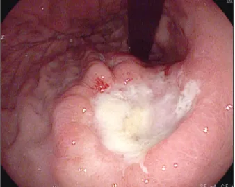
Fig. 1. Endoscopic finding. Endoscopy shows a 3 cm ulcero-infiltrative lesion with irregular base, nodular elevated margin, and abnor- malities of surrounding folds (Borrmann class III) at the posterior wall of the upper body.
각되는 경도의 고질소혈증 외에 이상 소견은 없었다.
위장관 출혈 여부를 확인하기 위해 시행한 상부소화관내시 경에서 흑색 변의 원인으로 추정되는 거대한 단일 궤양성 병
확인하기 위해 시행한 복부-골반 CT에서 상부 위벽의 비후 외에 림프절 비대나 타 장기의 침범 등 복부나 골반 내 전이를 시사할 만한 소견은 관찰되지 않았으며, 흉부 X선 검사에서도 폐 종괴 등 이상 소견은 보이지 않았다.
내원 2일째, 환자는 하루 종일 잠만 자려 하는 등의 의식 저하가 지속되었고 응급으로 시행한 뇌 CT에서 원인이 될 만 한 급성 병변은 보이지 않았다. 당시 활력 징후는 혈압 130/82 mmHg, 맥박 93회/분, 호흡수 20회/분, 체온 37.0oC 였다. 내원 4일째, 의식 저하가 지속되고 신경학적 검사에서 목 경직과 함께 Kernig sign 양성 소견을 보이는 등 수막 자 극 증상이 관찰되어 요추천자를 시행하였다. 뇌척수액 검사에 서 뇌척수압 12 cmH2O로 정상 범위를 보였고 백혈구 15/μL (호중구 52%, 림프구 22%), 단백질 222 mg/dL로 각각 증가 되었으며 포도당 11 mg/dL로 감소된 소견을 보였다. 뇌의 감염성 질환을 의심하여 시행한 뇌 MRI에서 뇌수막의 미만성 조영증강 소견과 함께 오른쪽 천막 하부에서 1 cm 크기의 국소적인 결절이 관찰되었다(Fig. 3). 뇌척수액에서 시행한 균 배양 검사에서 결핵, 박테리아, 진균 및 일부 바이러스에 대해 음성 결과를 보였고 세포 검사에서 종양세포는 발견되지 않았 다. 뇌척수액 및 뇌 MRI의 결과를 바탕으로 감염성 뇌수막염을
Fig. 3. MRI findings. (A) Brain MRI with gadolinium-enhanced, T1-weighted axial image shows multifocal diffuse leptomeningeal enhancement at cerebrum and cerebellum. (B) Focal enhancing nodule of 1 cm is noted on right infratentorium.
Fig. 4. Cytological findings (Wright’s stain). (A) The cerebrospinal fluid shows a few large malignant cells with high nuclear/cytoplasmic ratio, 2-3 nucleoli, and basophilic cytoplasms (×400). (B) Adenocarcinoma includes small vacuoles and protoplasmic projection (×400).
의심하여 경험적으로 항생제와 항결핵제를 시작하였다. 투약된 항생제는 ceftriaxone, ampicillin, vancomycin이었고 항결핵 제는 isoniazid, rifampin, ethambutol, pyrazinamide였다.
이후 4일간의 치료에도 불구하고 임상적인 호전은 보이지 않았으며 환자의 의식 수준은 점차 악화되어 반혼수 상태가 되었다. 또한 2차례의 발작이 발생하여 뇌파 검사를 시행하였 고 간질양 파형(epileptiform discharge)을 동반한 미만성 뇌 피질 손상 소견이 관찰되어 항간질제를 투여하였다. 임상적으 로 악화되는 소견을 보여 2번째 요추천자를 시행하였다. 뇌척 수액 검사에서 뇌척수압은 23 cmH2O로 증가되어 있었고 백 혈구 13/μL (호중구 96%, 림프구 2%), 단백질 457 mg/dL로 각각 증가되었으며 포도당은 16 mg/dL로 감소된 소견을 보였 다. 2번째 뇌척수액에서 시행한 세포 검사에서 전이성 선암의
특징을 보이는 악성 종양세포가 관찰되었다(Fig. 4). 이에 진행 성 위암에서 기원한 전이성 연수막 암종증으로 진단하였다.
투약 중이던 항생제와 항결핵제는 모두 중단하였고 연수막 외 위암의 원격 전이 여부를 확인하기 위해 PET-CT를 계획 하였으나, 보호자가 환자의 연령 및 병의 진행 상태를 고려하 여 적극적인 검사나 항암 화학 약물 치료를 하는 것을 원치 않아 검사는 시행하지 못하였다. 내원 15일째, 환자는 호흡부 전이 발생하였고 기관 삽관 후 인공호흡기 치료를 시작하였 다. 이후 보존적인 치료를 하면서 경과 관찰하였고 환자의 임 상 징후는 점차 악화되었다. 결국 환자는 위암으로 진단된 지 6주 만에 전이성 연수막 암종증으로 인한 다발성 장기 부전으 로 사망하였다.
Table 2. Cases of Leptomeningeal Carcinomatosis from Gastric Cancer without Involvement of Any Other Sites
Reference Age (yr)/
sex
Initial presenting
symptom
Endoscopic & histologic finding (site/Bormann
type/differentiation)
MRI finding Treatment
Survival time from diagnosis
of LMC Deeb et al., 199713 53/M Nausea,
vomiting, dizziness
EG junction/ – / PDAC–
Normal Intrathecal CTx
(MTX)
>6 mo
Yamada et al., 200812 53/M Anorexia, headache, diplopia
Antrum/Bormann II/
PDAC
Slight enhancement of cerebellar sulcus
Whole brain RTx
4.2 mo
Guo et al., 20148 40/F Headache, cervical pain
Antrum/Bormann I/
PDAC
Hydrocephalus, enhancement along ventral surface of brain stem, cerebellum and spinal cord (C1-T4)
No
4 mo
Present case 81/F Melena, headache, vomiting
Upper body/Bormann III/PDAC with signet ring cell
Multifocal enhancement along ventral surface of cerebrum and cerebellum
No
6 wk
M, male; F, female; EG, esophago-gastric; PDAC, poorly differentiated adenocarcinoma; CTx, chemotherapy; MTX, methotrexate; RTx, radiotherapy;
LMC, leptomeningeal carcinomatosis.
Table 1. Summary of Six Retrospective Studies of Leptomeningeal Carcinomatosis in Gastric Cancer
Referencce Patient
Bormann classification
III or IV
Poorly differentiated adenocarcinoma
Signet ring cell
Cancer stage III or IV
Median duration from diagnosis of gastric cancer to diagnosis of
LMC (mo)
Median survival time from diagnosis of
LMC (wk)
Lisenko et al., 20032 8 5/8 (62.5) 8/8 (100) 6/8 (75) 7/8 (87.5) 12.0 6.0
Lee et al., 20041 19 18/19 (94.7) 15/19 (79.0) 8/19 (42.1) 19/19 (100) 6.6 4.0
Oh et al., 20094,a 54 38/45 (84.4) 44/47 (93.6) 19/47 (40.4) 49/51 (96.1) 6.3 6.7
Emoto et al., 20115 7 7/7 (100) 6/7 (85.7) 5/7 (71.4) 7/7 (100) 13.0 4.7
Tomita et al., 20129 12 NA NA NA 9/12 (75.0) 15.6 8.6
Kim et al., 20143 9 8/9 (88.9) 5/9 (55.6) 2/9 (22.2) 7/8 (87.5) 10.0 4.0
Values are presented as n only or n (%).
LMC, leptomeningeal carcinomatosis; NA, not available.
aMulti-center study at four institutions from 1994 to 2007.
후향성 연구들을 종합해보면 Table 11-5,9과 같다. 위암에서 전 이성 연수막 암종증의 진단 시기는 대부분(75-100%) stage III-IV이며 간이나 폐, 뼈 등에 원격 전이가 있는 경우도 11-63%의 빈도를 보였다. 위암의 첫 진단에서 연수막 암종증 이 진단될 때까지 걸리는 중앙 간격은 6.3-15.6개월이었다. 조 기 위암에서 발생한 전이성 연수막 암종증은 오직 부검 증례 보고만 있으며,10 조기 위암 수술 후 29개월이 지나 연수막 암종증이 발생한 최근 보고가 있으나11 이 또한 원격 림프절
Table 1의 연구들에 따르면, 위암의 내시경 소견은 Bormann 3형 혹은 4형이 주로 관찰되었고, 병리학적으로 대 부분 저분화성 선암을 보이면서 반지 모양 세포의 성상을 띠 는 경우가 많았다. 이번 증례도 Bormann 3형의 소견을 보이 면서 반지 모양 세포를 동반한 저분화성 선암으로 진단되었던 경우이다.
연수막 암종증의 임상적인 증상은 비특이적이며 침범한 부 위에 따라 크게 뇌, 척수, 뇌신경에 의한 것으로 분류할 수
있다. 뇌압 상승으로 인해 두통, 어지러움, 구역 및 구토, 의식 변화가 발생할 수 있고, 척수 압박으로 인해 목 및 허리 통증, 하지 위약을 보일 수 있다.8 뇌신경과 관련된 증상으로는 복 시,12 시력 저하, 청력 저하,14 안면마비 등이 있다. 이 중 두통 이 가장 흔한 증상으로, 이번 증례의 경우 두통과 함께 다른 뇌 관련 증상들을 보였다. 하지만 원발 종양이 진단되기 전이 나 암의 초기 단계에 상기 증상들이 관찰되었을 경우 연수막 암종증을 의심하고 진단하는 것은 어려운 일이다. 특히 암세 포로 인한 응고성 항진(hypercoagulability) 상태에서 뇌경색 이 동반된 경우 진단에 혼선을 가져올 수 있다.15
전이성 연수막 암종증의 확진은 뇌척수액에서 종양 세포를 확인하는 것이며 영상 검사로 gadolinium-enhanced MRI가 진단에 도움을 줄 수 있다. MRI의 경우 진단에 약 76%의 민 감도와 77%의 특이도를 보이며 이는 CT에 비해 2배 이상 높 은 수치이다.16 이번 증례에서도 볼 수 있듯이 뇌나 척수 연수 막의 여러 부위에 걸친 미만성 조영 증강이 전형적인 소견이 며16 뇌수막염 같은 뇌의 감염성 질환과의 감별이 필요하다.
한 번의 요추천자를 시행할 경우 그 민감도가 54%로 낮지 만 추가적인 검사를 통해 91%까지 증가시킬 수 있는 것으로 보고하고 있다.17 이번 증례에서는 2번째 시행한 검사에서 종 양세포가 발견되었으며 따라서 연수막 암종증이 의심될 경우 반복적인 요추천자의 시행이 반드시 필요하다. 뇌척수액 검사 에서 상승된 뇌척수압, 증가된 백혈구 수, 단백질의 증가, 포 도당의 감소를 보일 시 전이성 연수막 암종증을 의심할 수 있다.2 또한 위암에서 기원한 연수암 암종증의 경우 암표지자 인 발암배아성 항원(CEA)이 상승되어 있는 경우가 많으나,8 이번 증례에서는 척수액에서 CEA 검사를 시행하지 않아 확 인할 수 없었다.
전이성 연수막 암종증은 이미 타 장기에 다발성 전이가 진 행된 말기의 환자에서 발생하는 경우가 많고 신경학적 합병증 으로 인해 예후는 매우 불량하다. 진단 후 중앙 생존기간은 4-8.6주이고 적극적인 치료를 시행한 경우에도 2-4개월 정도 로 보고되고 있다.1 현재까지 확립된 표준 치료는 없으나 경막 내 항암 화학요법이 주로 시행되고 있다.2,6 최근 irinotecan, 5-fluorouracil을 이용한 고용량 전신적 항암 화학요법을 시행 한 환자의 경우 생존기간이 13개월이었으며 이는 생존율에 큰 향상을 보여준 사례이다.18 또한 경막내 항암 화학요법에 추가하여 전뇌 방사선치료, 뇌실-복강간 션트를 시행한 일부 환자들에게서 생존율의 향상과 함께 연수막 암종증으로 인한 신경학적 증상의 완화를 가져왔다는 결과가 보고된 바 있다.9
저자들은 복부나 골반 내 전이 소견이 없는 진행성 위암의 진단 직후 신경학적 증상을 보여 전이성 연수막 암종증이 확 인된 매우 드문 증례를 경험하였다. 이 환자는 진단 직후 급격 한 임상 증상의 악화로 치료를 받지 못하고 진단 후 6주 만에
사망하여 매우 불량한 예후를 보였다. 위암의 연수막 암종증 의 경우 아직까지 적절한 치료 방법과 치료 효과가 입증되지 는 않았다. 하지만 다양한 치료를 통해 환자의 중앙 생존기간 의 연장과 증상 완화를 통한 삶의 질 향상을 기대할 수 있다는 최근 보고들이 이어지고 있어, 이를 조기에 진단하고 적극적 인 치료를 시도해 볼 필요가 있다. 따라서 암이 의심되거나 초기 단계의 암으로 진단된 환자에게서도 비특이적인 신경학 적 증상들을 보이는 경우 전이성 연수막 암종증의 가능성을 항상 염두에 두고 접근하는 것이 필요하다.
REFERENCES
1. Lee JL, Kang YK, Kim TW, et al. Leptomeningeal carcinomatosis in gastric cancer. J Neurooncol 2004;66:167-174.
2. Lisenko Y, Kumar AJ, Yao J, Ajani J, Ho L. Leptomeningeal carcino- matosis originating from gastric cancer: report of eight cases and review of the literature. Am J Clin Oncol 2003;26:165-170.
3. Kim NH, Kim JH, Chin HM, Jun KH. Leptomeningeal carcinoma- tosis from gastric cancer: single institute retrospective analysis of 9 cases. Ann Surg Treat Res 2014;86:16-21.
4. Oh SY, Lee SJ, Lee J, et al. Gastric leptomeningeal carcinoma- tosis: multi-center retrospective analysis of 54 cases. World J Gastroenterol 2009;15:5086-5090.
5. Emoto S, Ishigami H, Yamaguchi H, Yamashita H, Kaisaki S, Kitayama J. Frequent development of leptomeningeal carcino- matosis in patients with peritoneal dissemination of gastric cancer. Gastric Cancer 2011;14:390-395.
6. Kim KW, Kim SM, Kim JS. Clinical features and prognosis of lep- tomeningeal carcinomatosis. J Korean Neurol Assoc 1989;7:
210-217.
7. Lee HG, Lee B, Kim SM, Suh BJ, Yu HJ. A case of gastric ad- enocarcinoma presenting as meningeal carcinomatosis.
Korean J Intern Med 2007;22:304-307.
8. Guo JW, Zhang XT, Chen XS, et al. Leptomeningeal carcinoma- tosis as the initial manifestation of gastric adenocarcinoma: a case report. World J Gastroenterol 2014;20:2120-2126.
9. Tomita H, Yasui H, Boku N, et al. Leptomeningeal carcinomatosis associated with gastric cancer. Int J Clin Oncol 2012;17:361- 366.
10. Grove A. Meningeal carcinomatosis from a clinically undiag- nosed early gastric cancer. Pathol Res Pract 1991;187:341- 345.
11. Park KK, Yang SI, Seo KW, Kim YO, Yoon KY. A case of metastatic leptomeningeal carcinomatosis from early gastric carcinoma.
World J Surg Oncol 2012;10:74.
12. Yamada T, Furukawa K, Yokoi K, Ohaki Y, Okada S, Tajiri T. Case of meningeal carcinomatosis with gastric cancer which man- ifested meningeal signs as the initial symptom; the palliative benefit of radiotherapy. J Nippon Med Sch 2008;75:216-220.
13. Deeb LS, Yamout BI, Shamseddine AI, Shabb NS, Uthman SM.
Meningeal carcinomatosis as the presenting manifestation of gastric adenocarcinoma. Am J Gastroenterol 1997;92:329-