pISSN 1598-9992 eISSN 2233-6869
ORIGINAL ARTICLE
이차 병원 건강검진자의 바렛식도 유병률 및 분문부 장상피화생과의 비교 연구
최철영, 서승철, 박재석, 이현정, 이종섭, 최효선, 박현성, 홍승권
샘안양병원 내과
The Prevalence of Barrett’s Esophagus and the Comparison of Barrett’s Esophagus with Cardiac Intestinal Metaplasia in the Health Screening at a Secondary Care Hospital
Cheul Young Choi, Seungchul Suh, Jae Serk Park, Hyun Jeong Lee, Jong Sup Lee, Hyo Sun Choi, Hyun Sung Park and Seung Goun Hong
Department of Internal Medicine, SAM Anyang Hospital, Anyang, Korea
Background/Aims: The purpose of this study was to estimate the prevalence of Barrett’s esophagus (BE) and its association with reflux esophagitis (RE) and peptic ulcer disease detected by free charge endoscopy which was covered by the National Health Insurance at a secondary care hospital, and to compare the results of the biopsy of BE with that of cardiac intestinal metaplasia (CIM).
Methods: A total of 4,002 patients underwent endoscopy from March 2010 to December 2012. BE was diagnosed if there was histologically proven specialized intestinal metaplasia, and CIM was diagnosed if intestinal metaplasia was accompanied with chronic gastritis.
Results: Four hundred twenty four patients underwent endoscopic biopsy, and the prevalence of BE was 1.0% (42/4,002).
The mean age and the proportion of males in BE were significantly higher than those of the rest of study population, and BE had slight tendency related to RE than the rest of study population. CIM was observed in 34 patients and BE and CIM showed similar results, regarding age, sex and association with RE. The mean length of endoscopic Barrett’s mucosa of BE group was 9.2±5.1 mm, and it was similar to that of CIM.
Conclusions: The prevalence of BE in the secondary care hospital was not low, and old age and male sex were significantly associated with BE. Because BE was observed in about 10% of biopsied patients and CIM was observed in a similar percentage with BE, the precise targeted biopsy is warranted and the biopsy method should be reestablished through the large prospective study of multiple secondary care hospitals. (Korean J Gastroenterol 2012;60:219-223)
Key Words: Barrett; Prevalence; Cardia; Metaplasia; Free
Received March 19, 2012. Revised April 25, 2012. Accepted May 7, 2012.
CC This is an open access article distributed under the terms of the Creative Commons Attribution Non-Commercial License (http://creativecommons.org/licenses/
by-nc/3.0) which permits unrestricted non-commercial use, distribution, and reproduction in any medium, provided the original work is properly cited.
교신저자: 홍승권, 430-733, 안양시 만안구 삼덕로 9, 샘안양병원 내과
Correspondence to: Seung Goun Hong, Department of Internal Medicine, SAM Anyang Hospital, 9 Samdeok-ro, Manan-gu, Anyang 430-733, Korea. Tel: +82-31-467- 9114, Fax: +82-31-449-0151, E-mail: [email protected]
Financial support: None. Conflict of interest: None.
서 론
우리 나라는 현재 위암의 선별검사로 40세 이상 남녀에게 보험 공단에서 1-2년에 한번씩 무료로 위내시경검사 시행을 권장하고 있다.1 이러한 건강검진자에서 위내시경검사를 시행
하다 보면 종종 바렛식도가 의심되는 병변을 만나게 된다. 내 시경에서 바렛식도가 의심되는 병변은 편평상피-원주상피접 합부가 위식도접합부보다 근위부로 이동되어 있고, 사이는 연 어빛 붉은 오렌지색을 띄는 점막 부위로 원주상피식도(colu- mnar-lined esophagus)로 불리운다. 바렛식도는 전통적으로
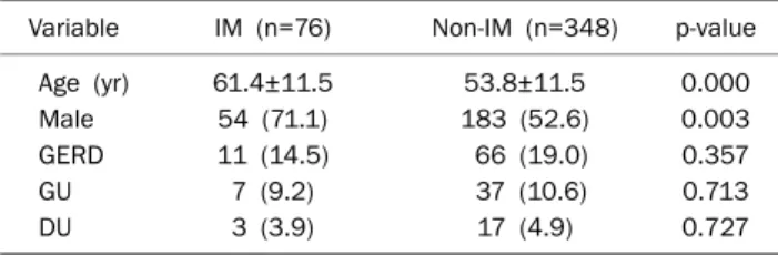
Table 1. Comparison between the Intestinal Metaplasia Group and the Non-Intestinal Metaplasia Group
Variable IM (n=76) Non-IM (n=348) p-value
Age (yr) 61.4±11.5 53.8±11.5 0.000
Male 54 (71.1) 183 (52.6) 0.003
GERD 11 (14.5) 66 (19.0) 0.357
GU 7 (9.2) 37 (10.6) 0.713
DU 3 (3.9) 17 (4.9) 0.727
Values are presented as mean±SD or n (%).
IM, intestinal metaplasia; GERD, gastroesophageal reflux disease;
GU, gastric ulcer; DU, duodenal ulcer.
배세포(goblet cell)를 포함한 특수 장상피화생이 조직학적으 로 증명된 경우에 정의하는 것이 일반적이다. 바렛식도는 식 도선암의 전구병변으로 알려져 있고, 내시경적 원주상피식도 인 원위부 식도에서 조직검사를 할 경우 바렛식도 외에 위식 도접합부인 분문부에 존재하는 정상 위점막 및 위염조직 외에 위염과 동반된 장상피화생이 검출될 수 있다. 따라서 조직검 사 시에 정확하게 하부 식도에서 시행되어야 하는데 전통적으 로 1-2 cm 간격으로 네 방향에서 다수의 조직 채취방법이 권유되고 있다.2 그러나, 아시아의 경우 서양의 유병률 및 이 형성의 발생에 비해 이전보다 증가하고 있기는 하지만 낮은 수준이기 때문에 조직검사는 육안적으로 점막에 특이한 변화 가 없을 경우 잘 이루어지지 않고 다수의 조직검사 방법의 번거로움 및 출혈 등의 합병증으로 인하여 일정기간을 두고 추적검사가 이루지는 것이 보통이다.3 이에 저자들은 한 이차 병원에 방문한 건강검진자를 대상으로 위내시경 검사에서 발 견되는 바렛식도가 의심되는 곳에서 조직검사를 시행하여 바 렛식도의 유병률 및 역류성 식도염, 위궤양의 동반 등의 임상 학적 차이를 분문부의 위염과 동반된 장상피화생군과 전향적 으로 비교 분석하고자 하였다.
대상 및 방법
1. 대상
2010년 5월부터 2012년 12월까지 위내시경 검사를 위하여 본원에 방문한 건강검진 수진자 4,002명을 대상으로 하였다.
2. 방법
한 명의 소화기내과 내시경 세부 전문의에 의해 위식도접 합부 상방에 편평상피접합부 직하방의 바렛식도가 의심되는 병변에서 1-3차례의 조직검사를 시행하여 한 명의 병리의사 가 hematoxylin and eosin (H&E) 염색에 배세포가 관찰되 는 특수 장상피화생(specialized intestinal metaplasia)이 증 명된 경우 조직학적 바렛식도로 여겼고, 내시경적 길이가 1 cm 미만인 경우 초단분절(ultrashort segment) 바렛식도, 1 cm 이상 3 cm 미만인 경우 단분절(short segment) 바렛식 도, 3 cm 이상인 경우 장분절(long segment) 바렛식도로 정 의하였다.4 단분절 내지 초단분절이 의심되는 곳에서 조직검 사를 시행했으나, 만성 위염에서 보이는 소와 과증식(foveolar hyperplasia) 등이 장상피화생과 동반된 경우는 위식도접합 부의 분문부(cardia) 장상피화생으로 여겼다.5-7 내시경적으로 발견된 위식도 역류질환의 진단은 로스 엔젤레스 분류법을 따 랐으며8 미세변화 위식도 역류질환은 포함시키지 않았고 위, 십이지장 궤양의 동반 여부를 비교하였다. 이 연구는 본원의 기관윤리위원회의 승인을 받았다.
3. 통계분석
SPSS Program version 12.0 (SPSS Inc., Chicago, IL, USA)을 이용하여 성비 및 역류성 식도염, 위궤양, 십이지장 궤양 동반 여부의 범주형 변수의 분석에는 chi-square test 및 Fisher's exact test를 이용하였고, 나이, 바렛식도의 길이 등의 연속형 변수의 분석에는 independent t-test 및 ANOVA 를 이용하였으며, 연령대별 바렛식도의 유병률의 차이는 로지 스틱 회귀분석을 이용하였다. 통계적 유의수준은 p값이 0.05 미만일 경우로 하였다.
결 과
1. 대상군의 역학적 특징
전체 수진자 4,002명 중에서 내시경적으로 바렛식도의 소 견을 보여 조직검사를 시행한 수진자는 424명(10.5%)이었다.
조직검사를 받은 군은 남자가 237명(55.9%)으로 평균 나이는 55.1±11.8세였고, 비조직검사군은 남자가 1,653명(46.2%)으 로 평균 나이는 54.6±9.9세였다. 조직검사에서 장상피화생이 보인 군은 76명(1.9%)으로 평균 나이는 61.4±11.5세였고 남 자가 54명(71.1%)이었다. 장상피화생이 보이지 않는 정상 위 점막 내지 위염소견을 보인 348명의 평균 나이는 53.8±11.5 세로 남자가 183명(52.6%)이었다. 장상피화생군과 비장상피 화생군의 비교에서는 장상피화생군에서 평균 나이 및 남성의 비율이 통계적으로 높았고, 역류성 식도염과 소화성 궤양의 동반율은 차이를 보이지 않았다(Table 1).
2. 바렛식도의 유병률 및 역류성 식도염과 소화성 궤양의 동반율 장상피화생이 보인 군 중 위염과 동반된 분문부 병변을 제 외한 최종 바렛식도는 42명으로 기간 유병률은 1.0% (42/
4,002)로 나타났다. 42명의 평균 나이는 60.6±13.2세였고 이 중 남자는 28명(66.7%)이었다. 바렛식도군은 분문부 장상피 화생군과 성별과 나이에서 통계적 차이를 보이지 않았으나,
Table 2. Comparison between the Barrett’s Esophagus Group and the Cardiac Intestinal Metaplasia Group
Variable BE (n=42) CIM (n=34) p-value
Age (yr) 60.6±13.2 62.3±9.2 0.526
Male 28 (66.7) 26 (76.5) 0.349
GERD LA-A 6 (14.3) 1 (2.9)
LA-B 0 2 (5.9)
LA-C 0 2 (5.9)
Total 6 (14.3) 5 (14.7) 0.158
GU 2 (4.8) 5 (14.7) 0.530
DU 2 (4.8) 1 (2.9) 0.738
Values are presented as mean±SD or n (%).
BE, Barrett’s esophagus; CIM, cardiac intestinal metaplasia; GERD, gastroesphageal reflux disease; LA, Los Angeles Classification;
GU, gastric ulcer; DU, duodenal ulcer.
Table 3. Prevalence of Barrett’s Esophagus in Relation to Age Distribution
Age (yr) Total (n=4,002)
People biopsied (n=424)
Barrett’s esophagus
(n=42) 40-49 1,287 (32.2) 126 (29.7) 3 (7.1) 50-59 1,500 (37.5) 140 (33.0) 16 (38.1) 60-69 787 (19.7) 107 (25.2) 13 (31.0) 70-79 362 (9.0) 39 (9.2) 7 (16.7)
80-89 66 (1.6) 12 (2.8) 3 (7.1)
Values are presented as n (%).
Table 4. Comparison between the Intestinal Metaplasia Group and the Non-intestinal Metaplasia Group according to the Length of the Endoscopic Barrett’s Mucosa
IM (n=76)
Non-IM (n=348) p-value BE
(n=42)
CIM (n=34)
Ultrashort segment 26 19 233
Short segment 15 14 115
Long segment 1 1 0
Length (mm) 9.2±5.1 9.5±5.0 0.827a
9.3±5.0 8.3±3.4 0.029b
9.2±5.1 8.4±3.6 0.180c
Values are presented as number or mean±SD.
IM, intestinal metaplasia; BE, Barrett’s esophagus; CIM, cardiac intestinal metaplasia.
Comparison abetween BE with CIM, bbetween IM with non-IM,
cbetween BE with CIM and non-IM.
조직검사자 중 바렛식도를 제외한 382명의 평균 나이는 54.5±11.5세로 바렛식도군이 의미있게 높았고(p=0.002), 성 별에선 차이를 보이지 않았다. 전체 수진자 중 비조직검사군 인 3,578명에서 위식도 역류질환은 286명(8.0%)으로 바렛식 도군의 14.3%와 분문부 장상피화생군의 14.7%가 높은 경향 을 보였으나 통계적인 차이는 보이지 않았다. 비조직검사군의 위 궤양 및 십이지장 궤양은 각각 10.6%, 3.6%에서 동반되었 고 이 역시 바렛식도군 및 분문부 장상피화생군과 의미있는 차이를 보이지 않았다. 바렛식도군은 전체 수진자중 바렛식도 를 제외한 군의 평균 나이인 54.6±10.0세에 비해 고령이었 고, 연령대별 유병률은 40대 0.2% (3/1,287), 50대 1.1%
(16/1,500), 60대 1.7% (13/787), 70대 1.9% (7/362), 80대 4.5% (3/66)로 연령대가 증가함에 따른 유의한 차이를 보였으 며(p=0.003), 남성의 비율은 바렛식도군에서 비바렛식도군의 46.9%에 비해 의미있게 높았다(p=0.011). 바렛식도군과 분 문부 장상피화생군과의 비교는 나이, 성별 및 역류성 식도염 등에서 유의한 차이를 보이지 않았다(Tables 2, 3).
3. 바렛식도의 내시경적 길이에 따른 비교
전체 조직검사군에서 장분절은 2명, 단분절은 144명, 초단
분절은 278명이었고, 바렛식도군에서는 장분절이 1명, 단분 절이 15명, 초단분절이 26명이었다. 바렛식도군의 그 길이의 평균은 9.2±5.1 mm였고, 분문부 장상피화생군의 그 길이의 평균은 9.5±5.0 mm로 통계적인 차이를 보이지 않았다. 조직 검사자 중 바렛식도를 제외한 군의 그 길이의 평균은 8.4±
3.6 mm로 바렛식도군과 차이를 보이지 않았다. 장상피화생 군과 비장상피화생군에서 그 길이의 평균은 각각 9.3±5.0 mm, 8.3±3.4 mm로 통계적인 차이를 보였다(Table 4).
고 찰
우리 나라 바렛식도의 유병률은 최근에 대학병원 및 3차 의료기관을 대상으로 한 대규모 연구의 보고에 의하면 아직까 지 서구에 비해 낮은 수준으로 유지되고 있다.9-13 이번 연구는 지방의 한 이차 의료기관 건강검진센터에서 약 2년의 기간 동안 한 명의 소화기내과 내시경 세부 전문의사에 의해, 원주 상피식도(columnar-lined esophagus)가 보이는 곳에서 일반 적인 1-2 cm 간격으로 네 방향 조직검사가 아닌 2-3개의 조 직검사를 바렛변화가 의심되는 식도점막에서 시행하여, 바렛 식도의 유병률 및 바렛식도 유무에 따른 역류성 식도질환 등 을 비교한 연구이다.
이번 연구의 대상은 이전의 대규모 3차 병원 기반의 바렛 식도 유병률 조사의 구성원과 사회경제적 상태 및 질병의 중 등도 등에 차이가 있을 것으로 생각되었으나, 약 2년 기간 동 안 유병률은 1%로 생각보다 높게 확인되었다. 내시경적으로 바렛식도가 보이는 424명 중 76명에서 장상피화생이 확인되 었고, 이 중 34명의 분문부 장상피화생을 제외한 42명에서 바렛식도가 진단되었다. 분문부 장상피화생은 우리나라와 같 이 만성위염이 상대적으로 흔한 곳에서 동반되는 소견이
다.5,6,14-16 분문부 장상피화생과 바렛식도의 구분은 단지 조직 학적 소견으로는 구분이 힘들어, 동반된 임상양상이나 H&E 염색법 외에 alcian blue 염색, 장상피화생의 완전형과 불완 전형에 따른 구분, cytokeratin 7/20의 면역학적 반응정도, p53 돌연변이의 발현, Helicobacter pylori 양성 유무 등의 소견을 종합하여 판단하는 것으로 알려져 있다.5,6,17-19
본원에서 확인된 수진자의 내시경적 바렛식도는 초단분절 과 단분절이 대부분을 차지하고 약 반수가 분문부 장상피화생 이어서 식도 말단 부위의 망상 혈관 등의 존재가 교란 인자로 작용할 수 있어 정확한 식도조직의 채취가 중요함을 보여주었 고,20 실제 임상적으로 내시경적 바렛식도의 길이가 짧은 경우 에 조직검사만으로 두 소견의 구분은 거의 불가능할 수도 있 을 것으로 생각된다. 또한 대부분이 국민건강보험공단에서 1-2년에 한번씩 무료로 제공하는 위내시경검사를 시행하러 오는 사람들인 데다가 이차 병원 특성상 보다 전문적인 특수 염색의 추가가 힘든 점도 더욱 그러하다고 할 수 있다. 장분절 바렛식도는 내시경적으로 2명에서 의심되었으나, 한 명이었 다. 이번 연구에서 바렛식도의 평균 길이는 9.2 mm이고 위식 도 역류질환은 42명 중 6명(14.3%)에서 확인되었고, 모두 로 스 앤젤레스 분류 A에 해당되었다. 분문부 장상피화생을 소 견을 보인 34명에서 위식도역류질환은 5명(14.7%)으로 로스 앤젤레스 분류 A가 1명, B가 2명, C가 2명이었다. 관찰 기간 동안 바렛식도군은 비바렛식도군에 비해 고령이고 남자의 비 율이 높았으며, 위식도 역류질환은 약간 높은 경향을 보여 이 전 연구결과와 비슷하였다. 이전의 대규모 연구에서 바렛식도 의 위식도 역류질환 동반율이 10-40% 정도로 다양하게 보고 되어, 추후 이차 병원에서 시행되는 무료 위내시경 수진자를 대상으로 바렛식도에 동반된 위식도 역류질환에 대한 보다 큰 규모의 연구가 필요할 것으로 보인다.9-11,13
이번 연구에서 식도 이형성이나 선암은 발견되지 않았고, 이전 연구에서도 매우 드물거나 없는 것으로 보고되어 있
다.9,11,12 분문부 장상피화생의 이형성 내지 악성 변화여부에
대해서는 아직까지 논란이 많으나, 그것으로 인한 선암 발생 은 지속적으로 증가 추세에 있고, 바렛식도보다는 H. pylori와 의 관련성이 있으며, 위전정부의 장상피화생의 발생과도 상관 관계가 높은 것으로 알려져 있다.5,6,21,22 이번 연구는 무료 건 강검진 위내시경 수진자들을 대상으로 했기 때문에 H. pylori 감염여부 확인을 위한 조직검사 또는 신속 요소분해효소 검 사, 그리고 전정부의 장상피화생을 알기 위한 조직검사 등이 일률적으로 이루어지지 못해서 비조직검사군의 대조군 연구 가 이루어지지 못한 취약점이 있다. 또한 바렛식도와 분문부 장상피화생과의 비교에서는 특별히 임상적 차이를 보이지 않 았는데 이는 원위부 식도에서 원주상피식도 수진자에서만 조 직검사가 이루어져서 전체 수진자의 분문부 장상피화생을 대
표하지 못하기 때문에 추후 이런 점을 추가해서 연구가 필요 할 것으로 생각된다. 아울러 최근에 협대역 내시경의 활발한 도입으로 하부 식도의 바렛 변성이 의심되는 혈관 및 점막이 상을 보다 자세히 관찰하여 네 방향 조직검사 등 임상적으로 비실용적인 방법 없이도 정확성을 높일 수 있어, 이차 병원에 서도 이를 도입하여 검사가 이루어진다면 더욱 효과적일 것이 다.
요약하면 한 이차 병원에서 무료 건강검진 위내시경검사 수진자를 대상으로 시행한 바렛식도의 유병률은 약 1%로 이 전 3차 병원 규모의 다기관 연구에 비해 매우 낮지 않았고 대부분이 초단분절이었으며, 고령 및 남성의 비율이 높았다.
또한 내시경적 원주상피식도군에서 분문부 장상피화생군도 비슷한 수로 관찰되어 보다 주의깊은 조직검사가 필요할 것으 로 생각되며 추후 이차 병원 규모의 대단위 연구를 바탕으로 조직검사 방법의 새로운 정립이 필요할 것이다.
요 약
목적: 한 이차 병원에서 국민건강보험에서 제공하는 무료 위 내시경에서 바렛식도가 의심되는 병변에서 조직검사를 시행 하여 그것의 유병률 및 역류성 식도염과 소화성 궤양의 동반 정도를 분문부 장상피화생과 비교 연구하고자 하였다.
대상 및 방법: 2010년 3월부터 2012년 12월까지 총 4,002명 의 수진자가 위내시경을 받았다. 바렛식도는 조직학적으로 특 수 장상피화생이 동반되는 것으로 정의하였고 장상피화생이 만성위염을 동반한 것은 분문부 장상피화생으로 여겼다.
결과: 내시경상 424명에서 조직검사가 이루어졌고, 바렛식도 의 유병률은 1% (42/4,002)이었다. 바렛식도군은 전체 수진자 중 바렛식도를 제외한 군에 비해 나이가 많았고, 남자의 비율 이 의미있게 높았으며, 역류성 식도염이 약간 많이 동반되는 경향을 보였다. 분문부 장상피화생은 34명에서 관찰되었고, 바렛식도군과의 나이, 성별, 역류성 식도염의 비교에서 특별 한 차이를 보이지 않았다. 바렛식도군의 내시경적 바렛점막의 평균 길이는 9.2±5.1 mm였고, 분문부 장상피화생의 그것과 비슷하였다.
결론: 한 이차 병원에서 바렛식도의 유병률은 낮지 않았고, 고령, 남성이 비바렛식도군에 비해 의미있게 관련되어 있었 다. 조직검사자 중에서 약 10% 정도가 바렛식도에 합당하였 고, 분문부 장상피화생군도 바렛식도군과 비슷한 숫자로 확인 되어 내시경적 바렛식도가 보이는 경우 더욱 정확한 조직검사 와 다수의 이차 병원의 대규모 전향적 연구를 통한 그 방법의 정립이 필요할 것이다.
색인단어: 바렛; 유병률; 분문부; 화생; 무료
REFERENCES
1. Choi KS, Kwak MS, Lee HY, Jun JK, Hahm MI, Park EC. Screening for gastric cancer in Korea: population-based preferences for endoscopy versus upper gastrointestinal series. Cancer Epide- miol Biomarkers Prev 2009;18:1390-1398.
2. Ishimura N, Amano Y, Appelman HD, et al. Barrett's esophagus:
endoscopic diagnosis. Ann N Y Acad Sci 2011;1232:53-75.
3. Chang CY, Cook MB, Lee YC, et al.; Asian Barrett's Consortium.
Current status of Barrett's esophagus research in Asia. J Gas- troenterol Hepatol 2011;26:240-246.
4. Mueller J, Werner M, Stolte M. Barrett's esophagus: histopatho- logic definitions and diagnostic criteria. World J Surg 2004;28:
148-154.
5. Liu GS, Gong J, Cheng P, Zhang J, Chang Y, Qiang L. Distinction between short-segment Barrett's esophageal and cardiac in- testinal metaplasia. World J Gastroenterol 2005;11:6360- 6365.
6. Miao Q, Peng YS, Cai GH, Chen XY. Comparison of intestinal met- aplasia in gastric cardia and Barrett's esophagus. J Dig Dis 2011;12:272-278.
7. Voutilainen M, Juhola M, Färkkilä M, Sipponen P. Foveolar hyper- plasia at the gastric cardia: prevalence and associations. J Clin Pathol 2002;55:352-354.
8. Lundell LR, Dent J, Bennett JR, et al. Endoscopic assessment of oesophagitis: clinical and functional correlates and further vali- dation of the Los Angeles classification. Gut 1999;45:172-180.
9. Kim JH, Rhee PL, Lee JH, et al. Prevalence and risk factors of Barrett's esophagus in Korea. J Gastroenterol Hepatol 2007;22:
908-912.
10. Kim JY, Kim YS, Jung MK, et al. Prevalence of Barrett's esoph- agus in Korea. J Gastroenterol Hepatol 2005;20:633-636.
11. Lee IS, Choi SC, Shim KN, et al. Prevalence of Barrett's esoph- agus remains low in the Korean population: nationwide cross-sectional prospective multicenter study. Dig Dis Sci 2010;
55:1932-1939.
12. Lee JI, Park H, Jung HY, Rhee PL, Song CW, Choi MG. Prevalence
of Barrett's esophagus in an urban Korean population: a multi- center study. J Gastroenterol 2003;38:23-27.
13. Park JJ, Kim JW, Kim HJ, et al.; H. pylori and GERD Study Group of Korean College of Helicobacter and Upper Gastrointestinal Research. The prevalence of and risk factors for Barrett's esoph- agus in a Korean population: A nationwide multicenter pro- spective study. J Clin Gastroenterol 2009;43:907-914.
14. Abdo-Francis JM, Sobrino-Cossío S, Bernal-Sahagún F, Hernán- dez-Guerrero A. Prevalence of intestinal metaplasia of the gas- tric cardia and its relation with Helicobacter pylori strains cagA and vacA. Cir Cir 2010;78:315-321.
15. Bak YT, Jung GM, Yeon JE, et al. Validity of the specialized colum- nar epithelium as a diagnostic criterion of the short segment Barrett's esophagus. Korean J Intern Med 1998;13:99-103.
16. Lee JS, Kim HW, Lee JH, Youn HS, Jung WT, Ko GH. Relationships between types of proximal gastric mucosa and clinicopatholo- gical features. Korean J Pathol 2003;37:15-18.
17. Kim CW, Lee BI, Kim BW, et al. Immunohistochemical expression of the p53 and Ki-67 proteins in Barrett's esophagus in Korea.
Korean J Gastroenterol 2005;46:189-195.
18. Ormsby AH, Vaezi MF, Richter JE, et al. Cytokeratin immunor- eactivity patterns in the diagnosis of short-segment Barrett's esophagus. Gastroenterology 2000;119:683-690.
19. Rothery GA, Patterson JE, Stoddard CJ, Day DW. Histological and histochemical changes in the columnar lined (Barrett's) oeso- phagus. Gut 1986;27:1062-1068.
20. Chung JW, Lee GH, Choi KD, et al. Prevalence of the endoscopic Barrett's esophagus determined by palisading vessel and in- ter-observer variation. Korean J Gastrointest Endosc 2007;34:
239-243.
21. Chandrasoma P, Wickramasinghe K, Ma Y, DeMeester T. Is in- testinal metaplasia a necessary precursor lesion for adenocar- cinomas of the distal esophagus, gastroesophageal junction and gastric cardia? Dis Esophagus 2007;20:36-41.
22. Chandrasoma P, Wijetunge S, DeMeester S, et al. Columnar- lined esophagus without intestinal metaplasia has no proven risk of adenocarcinoma. Am J Surg Pathol 2012;36:1-7.