大韓放射線훌훌學會誌 第 26 卷 第1 號 pp. 148 - 150, 1990 Journal 01 Korean Radiological Society, 26 (J) 148 - 150, 1990
〈국문초록〉
Cystic Lymphangioma of the Pancreas
- A Case Report -
In Mook Cho, M.D., 8ang Wook Lee, M.D., Kyung 800 Kim, M.D., Yong Hyun Kim
,
M.D.Department of Radiology, Masan Korea General Hospial
춰|장에 발생한 낭형 임파관종 -1 례 보고-
마산고려영원 진단땅사선파
조인묵·이상욱·김경수·김용현
임파판종은임파계의 양성종양으로 주로 경부에 발생하며, 취장에 발생한임파판종은애우드물 어, 수예가 운헌상 보고되었다.
저자들은 취 장에 발생 한 임파판종을 경험 하였기 에 운헌고찰파 함께 보고하는 바이 다. CT 소 견으로는 훼장에 경계가 분영한 낭성종괴였고 수솔로 다엽성의 낭성종괴를 적출하였다.
- Abstract-
Cystic lymphangioma of the pancreas is very rare tumor and only few cases have been reported in the literature. CT findings has been described in only one case report. Authors present a case of cystic lymphangioma of the pancreas in 47 year old women showing a well defined huge cystic leacion on CT
Index Words: Pancreas, neoplasms 77.31 Pancreas, CT 77.1211
phangioma, in par ticular, is very rare. Only a few Introduction cases have been reported in the literature3- 7
) and not studied by CT except for pand이f03-8). We Lymphangiomas are benign congenital mal- report a case of cystic lymphangioma of the pan- formations of the lymphatic system usually affect- creas with a brief review of the literature. ing the neck and axilla 1). Involvement of the
visceral organs is less common2). Pancreatic lym- Case history
이 논운은 1989 년 11월 18 일 접 수하여 1989년 11월 24 일
에 채 택 되 었 음 A 47-year-old women admitted with lO-year- Received November 18, accepted November 24, 1989 history of postprandial epigastric discomfort and
- 148 -
- ln Mook Cho‘ et al.: Cystic Lymphangioma 01 the Pancreas -
indigestion. Physical examination reveald minimal tenderness over the right upper quadrant and epi- gastrium. No definite mass was palpated. CT de monstrated huge cystic lesion in the body and tail of the pancreas compressing the stomach anterior ly(Fig. 1
,
2). The lesion was well defined and clear- ly separated from the adjacent structures. Atte- nuation value was 10 HU. The administration of contrast agent showed enhancement of a hyper- vascular capsule. The patient was explored with provisional diagnosis of cystic tumor of the pan-Fig. 1. (a) CT scan at the region of body of the pan- creas reveals huge oval shaped cystic lesion in gastro- hepatic space. The stomach is compressed
까혁鍵鍵鍵혔熱熱; ‘ E;전
X훨찮 캔앓τ::;:S;헛상
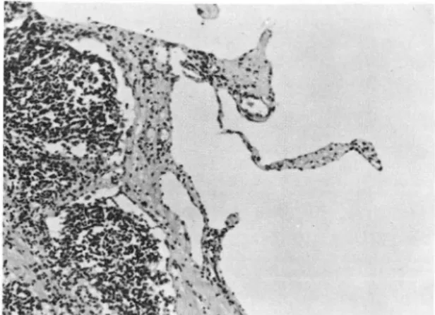
Fig. 2. photomicrograph of the tumor shows hugely di- lated cystic spaces lined by a single layer of flattened endothelial cells and separated by a scant fibrous con- nective tissue with adjacent aggregations of lymphoid cells (H-E, X 100)
creas.
At surgery, multilocular mass originating from the anterior and superior surface of the body and tail of the pancreas was resected. Cystic masses consist of three different size: largest - 14 X 10 X 10 cm, medium-l0X8X 10 cm, small- 3X2X3 cm.
Pathological diagnoisis was cystic lymphangioma of the pancreas(Fig. 3).
Discussion
Although the origin of cystic lymphangioma is not known 1)
,
they are developmental abnormali- ties (hamartomas) rather than true neoplasms9).Of cystic lymphangiomas, 75 % occur in the neck (cystic hygroma); 20 % are found in the axillary region; the other 5 % arise in the mediastinum, retroperitoneum, mesentery (mesenteric cyst), olJ1entum (omental cyst) , pelvis, groin, spleen, bone, and skin 10.11). Cystic lymphangioma may at times develop from cavernous lymphangioma.
When symptomatic, these tumors usually present in the first two years of life as a palpable mass in Fig. l.(b) CT scan at the region of tail of the pancreas the abdomen of flank.
reveals huge cystic lesion in the body and tail of pan-
creas. -N-~-~~id~~~~ ~f-;~~a~io~ in~~-;u :r~~n-di~g-s~:~- Pancreatic lymphangioma, in particular, is a
ture is seen. very rare tumor. Only a few cases have been re-
- 149 -
-大韓放射線훌훌學會註 : 第26卷 第1 號 1990-
ported in the literature and none studied by CT except for PandolfoS- S). In our case the lesion appeared as well encapsulated water density, mul tilocular mass indistinguishable from cystadenoma6). Although our patient was unusual in that she presented with epigastric pain in the adulthod
,
the correct diagnosis was made preoper- atively. CT was valuable because the low attenua- tion value of 10 HU suggested the presence of fat containing fluid.In summary, a preoperative diagnosis of cystic lymphangioma can be suggested by appearance of CT
4. Hanelin LG, Schimmel DH. Lymphangioma of the pancreas exhibiting an unusual pattern of calcifica- tion. Radiology 1977: 122:636
5. Dubois JD. Lymphangioma of the pancreas mas- querading as acute appendicitis and torsion of the left testis. Br ] Surg 1981. 68.799-800
6. deSantos LA, Bernardino ME, Paulus DD, Martin RE‘ Computed tomography of cystadenoma of the pancreas. ] Comput Assist Tomogr 1978: 2:222-5 7. Wolfman NT, Ramquist NA, Karstaedt N, Hopkins
MB. Cystic neoplasms of the pancreas: CT and sonography. A]R 1982: 138:37-41
8. Pandolfo 1, Seribano E, Gaeta M et al. Cystic lymphangioma of the pancreas.' CT demonstration ]CAT 9.209, 1985
REFERENCES 9. Willis RA. the borderland of embryology and pathology, 2d ed. London: Butterworth, 1962:358 1. Bill AH, Sumner DS. A unified concept of lymphan- 10. Rekhi BM, Esselstyn CB J r, Levy
r
, et al. Re- gioma and cystic hygroma. Surg Gynecol Obstet troperitoneal cystic lymphangioma. Report of two1965: 120:79-86 cases and review of the literature. Cleve Clin Q
2. Peradotto F, Zannetti PP, Obialero M, et al. Lym- 1972. 39:125-128
phangioma of the spleen. Remarks on two obserbed 11. Singh S, Baboo ML, Pathank
r c.
Cystic lymphan- cases. Minerva 1νed 1986: 75:2295-2300 gioma in children: reprt of 32 cases including le- 3. Dodds WJ. Margolin FR, Goldberg HL. Cavernous sions at rare sites. Surgery 1971: 69:947-951lymphangioma of the pancreas. Radiol Clin (Basel) 12. Harrow BR. Retroperitoneal lymphatic cyst (cystic
1969: 38.-267-70 lymphangioma). ] Urol 1957: 77.'82-89
n U
표