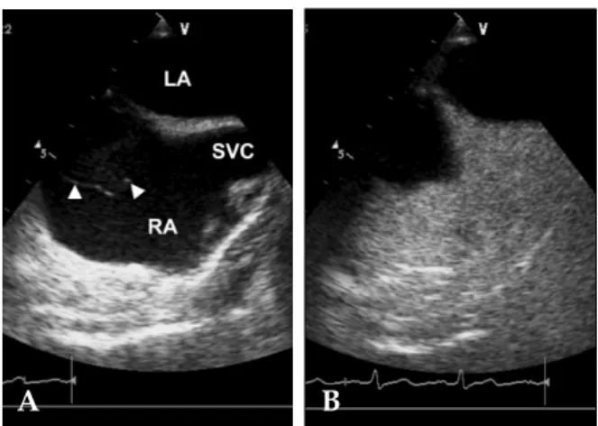
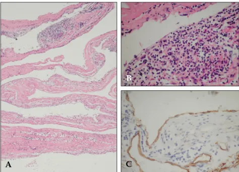
A Case of Cardiac Lymphangioma Presenting as a Cystic Mass in the Right Atrium
5
0
0
전체 글
(2)
(3)
(4)
(5)
수치
관련 문서
