INTRODUCTION
Lymphangiomas are rare benign tumors, and are located preferentially in head, neck, and axilla in children. However, lymphangiomas in abdomen are extremely rare, particularly in adults. Of all the lymphangiomas in the peritoneal cavi- ty, about 70% of them have been found in the mesentery of small intestine. This report describes a case of omental cystic lymphangioma of the stomach associated with earlier subto- tal gastrectomy for gastric cancer.
CASE REPORT
A 76-yr-old man who presented with an 8-month history of postprandial abdominal discomfort and bowel habits change was referred from a local clinic, because of a huge intraab- dominal cystic lesion on abdominal computed tomographic (CT) scan. According to his medical history, he underwent subtotal gastrectomy (Billroth II operation) 10 yr ago for early gastric cancer at St. Mary’s Hospital, The Catholic Universi- ty of Korea, Seoul, Korea. A non-tender left upper quadrant abdominal mass was palpated on physical examination. Lab- oratory data, including tumor markers, were unremarkable.
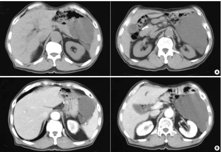
Gastrointestinal endoscopic examination revealed no local recurrence at the previous gastrojejunostomy anastomosis or intraluminal pathologic lesion. An abdominal computed tomography showed about 12×15 cm sized non-enhancing
cystic mass, suspicious of a cystic neoplasm of pancreas, mesen- teric cyst, or omental lymphangioma (Fig. 1).
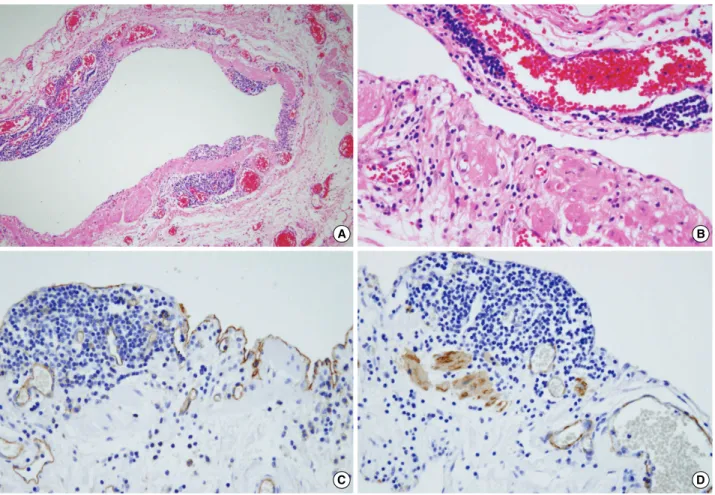
On October 31, 2003, the patient underwent exploration which showed no evidence of ascites, peritoneal dissemination, or metastasis in abdominal cavity. During exploration and resec- tion, the cystic tumor was found not to involve mesentery of the colon, remnant stomach and retroperitoneum, however, technically complete removal of the tumor without resection of distal pancreas and spleen was not feasible. Therefore, En- bloc resection of the cystic tumor with the greater omentum, distal pancreas and spleen with no spillage of cystic fluid was performed. After resection, cystic tumor was grossly measured 16.5×12.0×3.5 cm, but the attached distal pancreas and spleen were grossly unremarkable (Fig. 2). The encapsulated cystic mass had a pinkish smooth external surface attached with distal pancreas and spleen in continuity, and cut sections showed grayish white, trabeculated, and glistening in the inner cut surface of multilocular cyst. The cyst was filled with straw- berry colored serous fluid, the thickness of cystic wall was measured less than 0.1 cm, and there was no solid area. His- tologically, the cystic wall consisted of a fibroconnective tis- sue accompanied by dilated lymphatic spaces of varying sizes and lymphoid cell aggregations in the endothelial lining of lymphatic vessels, and there was no evidence of recurrence or metastasis from gastric cancer. Immunohistochemical stainings showed positive for CD31 and smooth muscle actin (SMA) (Fig. 3). These findings were consistent with a diagnosis of cystic lymphangioma. The patient had an uneventful post-
1212
Jong Han Kim1, Woo Sang Ryu1, Byung Wook Min1, Tae Jin Song1, Gil Soo Son1, Seung Joo Kim1, Young Sik Kim2, and Jun Won Um1
Departments of Surgery1and Pathology2, Korea University College of Medicine, Seoul, Korea
Address for correspondence Jun Won Um, M.D.
Department of Surgery, Korea University Ansan Hospital, Korea University College of Medicine, 516 Gojan-dong, Danwon-gu, Ansan 425-707, Korea Tel : +82.31-412-5952, Fax : +82.31-413-4829 E-mail : [email protected]
J Korean Med Sci 2009; 24: 1212-5 ISSN 1011-8934
DOI: 10.3346/jkms.2009.24.6.1212
Copyright � The Korean Academy of Medical Sciences
Acquired Omental Cystic Lymphangioma after Subtotal Gastrectomy:
A Case Report
We herein describe a case of cystic lymphangioma in the greater omentum of the remnant stomach, which is thought it to be related with subtotal gastrectomy 10 yr ago for early gastric cancer. A 76-yr-old man was admitted to our department with postprandial abdominal discomfort and bowel habit change. Intraabdominal multi- locular cystic mass was detected by ultrasonography and computed tomography.
We performed a complete En-bloc tumor resection including spleen and distal pan- creas, and histological examination confirmed cystic lymphangioma originated from the greater omentum of the remnant stomach. Although the etiology of omental lym- phangioma remains largely unclear, these findings suggested strongly that obstruc- tion of the lymphatic vessels after gastric resection for gastric carcinoma might be the most plausible cause. The surgical extirpation with resection of organs involved appears to be a treatment of choice for such unusual case.
Key Words : Abdominal Neoplasms; Surgery; Lymphangioma; Omentum; Gastrectomy; Adult
Received : 22 November 2007 Accepted : 29 June 2008
Acquired Omental Cystic Lymphangioma 1213
Fig. 2. Gross specimen showed an encapsulated cystic tumor combined with distal pancreas and spleen (A, Anterior view; B, Posterior view).
A B
Fig. 1. An abdominal CT scan show- ed about 12.0×15.0 cm sized non-enhancing cystic mass in the left ab- dominal cavity. This mass was atta- ched to pancreas body and tail su- peromedially, and displaced a remnant of stomach medially, spleen laterally, and adjacent bowel loops inferomedially. This cystic mass contained internal septum, like structure in the upper aspect of tumor.
A
B
1214 J.H. Kim, W.S. Ryu, B.W. Min, et al.
operative course, and no evidence of recurrence had also been found in a 3-yr follow-up period.
DISCUSSION
Cystic lymphangioma is a rare congenital malformation of the lymphatics which is found predominantly in children, and abdominal lymphangioma is far more extremely rare, especially in adult population (1-5). Although the etiology remains unclear, lymphangioma is thought to be due to a developmental failure and possible lymphatic obstruction followed by the inflammation of lymphatic channels (1-5).
Cystic lymphangiomas can be located in any part of the body except the brain, and they are most frequently a single cyst and also affect a single organ (2, 5). The tumors are usu- ally located in the neck (75%), axilla (20%), and other part of the body (4-5%) (2, 5), and less than 1% of lymphangio- mas affect the mesentery, greater omentum, and retroperi- toneum (5).
Abdominal lymphangiomas can occur at any age and most
are asymptomatic. In adults, symptoms and signs are com- monly insidious in onset rather than acute (1, 2, 4-6).
It is important to distinguish cystic lymphangioma from other cystic neoplasms such as mesenteric cyst, peritoneal mesothelioma, and retroperitoneal cystic neoplasm. Histo- logically, lymphangioma is characterized by a flat epithelial endothelium and a wall containing alternatively lymphoid tissue, small lymphatic spaces, smooth muscle, and foam cells (5-7). Furthermore, this endothelial lining and stroma cells are usually stained positively with anti-factor VIII-related antigen or anti-CD 31 by immunohistochemistry (7, 8).
The treatment of choice is complete resection to yield a cure for this benign lesion, but resectability depends on its size, location, and adhesions or invasions to vital structures (1-6). In the present case, En-bloc resection of cystic tumor with distal pancreatectomy and splenectomy was indicated because this huge neoplasm could be a malignant tumor and a possible local recurrence if the resection was incomplete.
In English-written literatures, cystic lymphangiomas aris- ing from the omentum have mostly been described as con- genital primary lesions, and some secondary lesions concur-
Fig. 3. Histology of lymphangioma. (A, B) Cystic wall was consisted of fibroconnective tissue accompanied by dilated lymphatic spaces and lymphoid cell aggregations in the endothelial lining of lymphatic vessels (H&E ×100, ×400). Immunohistochemical staining. (C) Posi- tive for CD31 in the lining cells of dilated lymphatic vessels (×400). (D) Positive for SMA in the cystic wall, and they were smooth muscle bundles surrounding lymphatic vascular endothelium (×400).
A B
C D
Acquired Omental Cystic Lymphangioma 1215
rently with other primary tumors have also been reported.
To the best of our knowledge, this could be the first case report of an acquired intraabdominal cystic lymphangioma arising from the greater omentum, associated with earlier gastric resection for gastric cancer.
In general, during subtotal gastrectomy for distal gastric cancer, the greater omentum at the high body and around the spleen is not indicated for dissection and resection. In the present case, as described above in the operative findings, cystic lymphangioma was thought to originated from the remnant greater omentum after incomplete omentectomy in the previous subtotal gastrectomy for distal gastric carci- noma. Long standing lymphatic obstruction and stasis dur- ing the past 10 yr after subtotal gastrectomy is strongly sug- gested as the pathophysiologic predisposing factor to the development of this unusual case.
REFERENCES
1. Takiff H, Calabria R, Yin L, Stabile BE. Mesenteric cysts and intra-
abdominal cystic lymphangiomas. Arch Surg 1985; 120: 1266-9.
2. Roisman I, Manny J, Fields S, Shiloni E. Intra-abdominal lymphan- gioma. Br J Surg 1989; 76: 485-9.
3. Fonkalsrud EW. Congenital malformations of the lymphatic system.
Semin Pediatr Surg 1994; 3: 62-9.
4. de Perrot M, Rostan O, Morel P, Le Coultre C. Abdominal lymphan- gioma in adults and children. Br J Surg 1998; 85: 395-7.
5. Losanoff JE, Richman BW, El-Sherif A, Rider KD, Jones JW. Mesen- teric cystic lymphangioma. J Am Coll Surg 2003; 196: 598-603.
6. Allen JG, Riall TS, Cameron JL, Askin FB, Hruban RH, Campbell KA. Abdominal lymphangiomas in adults. J Gastrointest Surg 2006;
10: 746-51.
7. Chung JH, Suh YL, Park IA, Jang JJ, Chi JG, Kim YI, Kim WH. A pathologic study of abdominal lymphangiomas. J Korean Med Sci 1999; 14: 257-62.
8. Miettinen M, Lindenmayer AE, Chaubal A. Endothelial cell markers cd31, cd34, and bnh9 antibody to h- and y-antigens--evaluation of their specificity and sensitivity in the diagnosis of vascular tumors and comparison with von willebrand factor. Mod Pathol 1994; 7:
82-90.