2013 EDISON 계산화학 경진대회 13 Molecular Modeling of the Subtype Dopamine Receptor-ligand Interactions
Molecular Modeling of the Subtype Dopamine Receptor-ligand Interactions
Minkyung Baek and Woong-Hee Shin
Department of Chemistry, SNU,1 Gwanak-ro, Gwanak-gu, Seoul, 157-742, Korea.
Tel: 010-6354-7994, E-mail: [email protected]
Chemworks (or Chemworks2) ID: ecc13_SNU_minkyung
ABSTRACT
Dopamine agonists and antagonists and its receptor play a critical role in the information transfer in the nervous system, and dopamine receptor-ligands interactions are deeply related to Parkinson’s disease, schizophrenia and some other mental diseases. However, the only experimental 3D structure available for dopamine receptors is human D3 dopamine receptor. Therefore, it is important to create model of subtype dopamine receptor-ligands interactions. We report here the 3D structures of the human D1 and D2 dopamine receptor predicted by using GalaxyTBM, and its predicted binding site determined by using GalaxyDock. The highly conserved Asp on TM 3 and Phe on TM 6 have critical role in ligand binding.
Also, highly conserved serines on TM 5 are essential for binding agonists and some kinds of antagonists.
We identify differences between binding sites of agonists and antagonists of human D1 and D2 dopamine receptor, and find the reasons of selective binding of antagonists.
Key Words : human D1 dopamine receptor, human D2 dopamine receptor, receptor-ligands interactions
2013 EDISON 계산화학 경진대회 15 Molecular Modeling of the Subtype Dopamine Receptor-ligand Interactions
Introduction
Dopamine modulates diverse biological functions, including movement, endocrine function, and memory formation through activation of five distinct subtype dopamine receptors1. The subtype dopamine receptors are classified into two subfamilies, D1-like dopamine receptors and D2-like dopamine receptors, based on their biochemical and pharmacological properties2. While the D1-like subfamily consists of D1 and D5 dopamine receptor, the D2-like includes D2, D3, and D4 dopamine receptor. The D1 and D2 dopamine receptors are the most abundant subtype dopamine receptors2. D1 and D2 receptor-selective agonists and antagonists are potential drugs for treatment of Parkinson’s disease, schizophrenia, and other neuropsychiatric disorders3. Despite the importance of solving the 3D structure of the subtype dopamine receptors in the structure-based drug-design, the only experimental 3D structure available for a dopamine receptor is human D3 dopamine receptor. Therefore, it is important to create model of subtype dopamine receptor-ligands interactions. Some mutagenesis studies on D1 dopamine receptor(D1DR) and D2 dopamine receptor(D2DR) found the Asp 3.32 has critical role in binding ligands, and serines on TM 5 are essential of agonists binding4,5. However, other receptor-ligands interactions and selectivity of ligand binding were unclear in these mutagenesis studies. Here, we predicted the 3D structures of D1DR and D2DR by using GalaxyTBM. Furthermore, based on predicted 3D structures, we predicted the binding site of agonists and antagonists in D1DR and D2DR, and analyzed the receptor- ligand interactions. The predicted binding sites contain the critical residues for binding ligands that have been identified by mutagenesis studies.
Theory and Computational Method
Prediction of the D1DR and D2DR 3D Structures
The global structures and spatial arrangement of the main secondary structure elements are highly conserved in GPCRs6. Therefore, we presumed that we are able to create reasonable models for D1 and D2 dopamine receptor by homology modeling. We predicted the 3D structures of D1DR and D2DR using GalaxyTBM7. Because second extracellular loop (ECL2) is too difficult to predict well, we removed the ECL2 region for docking process.
201 Mo
Fig cla Pr
rec fro
13 EDISON olecular Mode
gure 1. A, ass I antago rediction of
We used th ceptors8. Th om PubChe
계산화학 경진 eling of the Sub
a) Agonists nists, and c f the Ligan
he GLIDA(G he selected em compou
진대회 btype Dopami
s, b) class I ) class II an d Binding S
GPCR-ligan ligands are und databas
ine Receptor-li
antagonists ntagonists fo
Sites
nd Database e shown in se9. Depend
igand Interact
s, and c) cl or D2DR.
e) to select a Figure 1. T ding on th
tions
ass II antag
agonists and The coordin he structure
gonists for D
d antagonist nates of the
s of antago
D1DR. B. a
t of D1 and ese ligands onists, we
a) Agonists,
d D2 dopam were obtain
classified
17
, b)
mine ned the
antagonists into two classes: (i) class I, bulky antagonists and (ii) class II antagonists that have two aromatic or ring moieties connected by a flexible linker10. Hydrogen atoms were added, and Gasteiger charges were assigned to all of the ligands using the Dock Prep module in Chimera11. To predict binding sites, all of the agonists and antagonists were docked into dopamine receptor using GalaxyDock12. The center of a grid box was set on Cγ atoms of ASP3.32 which is thought to play a critical role in ligand binding5. Binding site residues were defined as those residues with at least one heavy atom within a 5Å from any heavy atom of the ligand.
Results and Discussion
Comparison of Binding Site between Agonists and Antagonists of D1DR
Figure 2 shows the 3D structures of the human D1DR predicted by using GalaxyTBM, and the predicted binding site of agonists and antagonists determined by using GalaxyDock. We found the following residues to be essential for binding of ligands in the D1 dopamine receptor.
1. Val-100, Asp-103, Ile-104, Ser-107, Tyr-194, Phe-288, Asn-292 and Trp-321 are essential for binding agonists, antagonists class I and II. The carboxyl group of the Asp-103 forms hydrogen bonds with the protonated amine group of the ligands. Ser-107, Tyr-194, and Asn-292 interacts with polar groups of ligands. The other residues, Val-100, Ile-104, Phe-288, Trp-321 form hydrophobic pocket for ligands.
2. Ser-198, Ser-199, Ser-202, and Trp-285 are necessary for binding agonists and antagonists class II. The polar functional group of Ser-198, Ser-199 and Ser-202 interacts with polar groups of agonists and antagonists class II, but there are some differences between agonists and antagonists
2013 EDISON 계산화학 경진대회 19 Molecular Modeling of the Subtype Dopamine Receptor-ligand Interactions
class II. While oxygen or nitrogen atoms of agonists interact with three serines, sulfur or halide atoms of antagonists class II interact with three serines. The Trp-285 interacts with ligands by hydrophobic interaction.
3. Val-317 interacts with both of antagonist class I and class II, but it doesn’t contact with agonists.
Ala-199 and Phe-313 contacts with only antagonist class II through hydrophobic interaction.
Figure 2. The binding site of D1DR. The residues colored in orange interact with all agonists, class I antagonists, class II antagonists. The residues colored in cornflower blue contact with agonists and class II antagonists. The residue colored in orchid interacts with class I and II antagonists, and the residues colored in medium purple contact with only class II antagonists.
Comparison of Binding Site between Agonists and Antagonists of D2DR
Figure 3. The binding site of D2DR. The residues colored in orange interact with all agonists, class I antagonists, class II antagonists. The residue colored in yellow interacts with only agonists. The residues colored in cornflower blue contact with agonists and class II antagonists. The residues colored in orchid interact with class I and II antagonists, and the residue colored in medium purple contacts with only class II antagonists
Figure 3 shows the predicted 3D structures of the human D2DR, and the predicted binding site of agonists and antagonists. We found the following residues to be essential for binding of ligands in the D2 dopamine receptor.
1. Asp-114, Val-115, Phe-389, and Tyr-410 are essential for binding agonists, antagonists class I and II. The Asp-114 forms salt bridge with the protonated amine group of the ligands. The OH group of Tyr-410 also helps form salt bridge with ligand by hydrogen bond. The other residues, Val-115, and Phe-389 form hydrophobic pocket for ligands.
2. Ser-193, Ser-194, and His-393 are necessary for binding agonists and antagonists class II. The polar functional groups of these residues interact with polar groups of agonists and antagonists class II. His-393 sometimes interacts with aromatic ring of ligands.
3. Trp-386 interacts with only agonists through hydrophobic interactions. Val-91, Leu-94, Phe-110,
2013 EDISON 계산화학 경진대회 21 Molecular Modeling of the Subtype Dopamine Receptor-ligand Interactions
Val-111, Tyr-408, and Thr-412 interacts with both of antagonist class I and class II, but it doesn’t contact with agonists. Cys-118 contacts with only antagonist class II through polar interaction.
Comparison of Receptor-ligands Interactions Between D1DR and D2DR
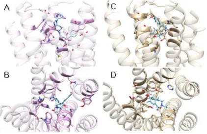
Figure 4. A, top view and B, side view of the SCH-23390 binding pose in D1DR. C, top view and D, side view of the SCH-23390 binding pose in D2DR.
D1 dopamine receptor and D2 dopamine receptor have different pharmacological properties. The binding affinity of SCH-23390 for D1 dopamine receptor is higher than that for D2 dopamine receptor (Ki
value for D1DR 0.11~0.35nM and Ki value for D2DR 270~1100nM)13. Figure. 4 shows the binding pose of SCH-23390 in D1DR and D2DR. While the SCH-23390 in DRD1 occupies the region between TM3, TM5, TM6, and TM7, the SCH-23390 in DRD2 occupies the region between TM2, TM3, TM6, and TM7.
These differences between occupying regions come from the smaller binding pocket of D2DR because of the inward shift of TM6 (Figure 5).
Figure 5. The top view of D1DR(orchid) and D2DR(cyan). There is the inward shift of TM 6 of D2DR.
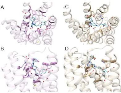
Figure 6. A, top view and B, side view of the spiperone binding pose in D1DR. C, top view and D, side view of the spiperone binding pose in D2DR.
The binding affinity of spiperone for D2 dopamine receptor is higher than that for D1 dopamine receptor (Ki value for D1DR 99~350nM and Ki value for D2DR 0.06~0.37nM)13. Figure 6 shows the binding pose of spiperone in D1DR and D2DR. The key interaction residue of D1DR and D2DR is different. Non-conservative Cys-118 of D2DR interacts with F atom of spiperone, and non-conservative Tyr-416 of D2DR interacts with the carbonyl group of spiperone. These non-conservative amino acids are essential for selective binding.
2013 EDISON 계산화학 경진대회 23 Molecular Modeling of the Subtype Dopamine Receptor-ligand Interactions
Conclusion
From the results of the binding site prediction, the Asp of TM3 (Asp-103 of D1DR and Asp-114 of D2DR) plays a critical role in ligand binding of dopamine receptors by forming the salt bridge. Phe of TM 6 (Phe-288 of D1DR and Phe-389 of D2DR) are also essential for ligand binding. Highly conservative serines of TM 5 are necessary for agonists and antagonists class II binding. These serines interact with polar groups of ligands. The Phe-313, Val-317 of D1DR and Tyr-408, Thr-412 of D2DR are necessary for antagonist binding. And some other hydrophobic residues contact with ligands by hydrophobic interactions. Also, we found the different binding poses of selective antagonist of D1DR and D2DR. The SCH-23390 which has higher affinity for D1DR occupies the region between TM 3, TM 5, TM6, and TM 7 in D1DR, but it occupies the region between TM 2, TM3, TM6, and TM 7 in D2DR because of the inward shift of the TM 6 of D2DR. The spiperone which has higher affinity for D2DR interacts non-conservative amino acids between D1DR and D2DR, and these non-conservative amino acids are the key residues for interaction.
Acknowledgments. This work has been supported by the project EDISON (EDucation-research Integration through Simulation On the Net), Chemistry.
References
1. Gingrich, J. A.; Caron, M. G. Annu. Rev. Neurosci. 1993, 16, 299.
2. Kebabian, J. W.; Calne, D. B. Nature, 1979, 277, 93.
3. Neve, K. A.; DuRand, C. J.; Teeter, M. M. Neurological Disease and Therapy, 2003, 56, 77.
4. Tomic M. et. al. Biochemical and Biophsical Research Communications, 1993, 191, 1020.
5. Mansour A. et. al. European Journal of Pharmacology – Molecular Pharmacology Section, 1992, 227, 205.
6. Congreve, M; Langmead, C; Marshall F. H. Adv. Pharmacol, 2011, 62, 1.
7. Ko, J.; Park, H.; Seok, C. BMC Bioinformatics, 2012, 13, 198.
8. Okuno, Y.; Yang, J.; Taneishi, K.; Yabuuchi, H.; Tsujimoto, G. Nucleic acids research, 2006, 34, 673.
9. Bolton, E. E.; Wang, Y.; Thiessen, P. A.; Bryant, S. H. Annual reports in computational chemistry, 2008, 4, 217.
10. Kalani, M. Y. S. et. al. PNAS, 2004, 101, 3815.
11. Pettersen, E. F.; Goddard, T. D. et. al., Journal of computational chemistry, 2004, 25, 1605.
12. Shin, W. H.; Seok, C. Journal of chemical information and modeling, 2012. 52, 3225.
13. Vallone, D.; Picetti, R.; Borrelli, E. Neuroscience & Biobehavioral Reviews, 2000, 24, 125.